. 2018 Aug 7:9:1826.
doi: 10.3389/fmicb.2018.01826. eCollection 2018.

The Cranberry Extract Oximacro® Exerts in vitro Virucidal Activity Against Influenza Virus by Interfering With Hemagglutinin

Affiliations

The Cranberry Extract Oximacro® Exerts in vitro Virucidal Activity Against Influenza Virus by Interfering With Hemagglutinin

Anna Luganini et al. Front Microbiol. .

Abstract

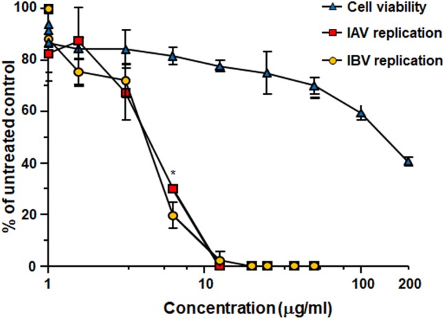
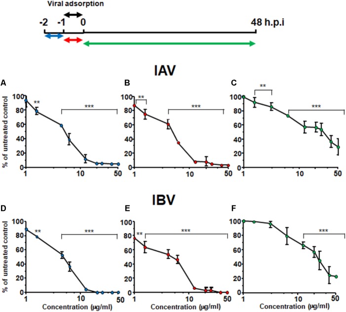
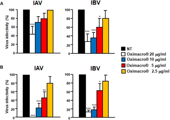
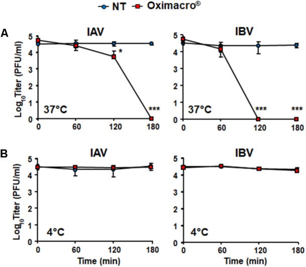
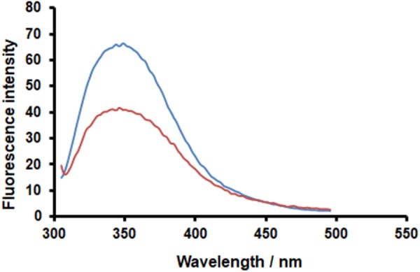
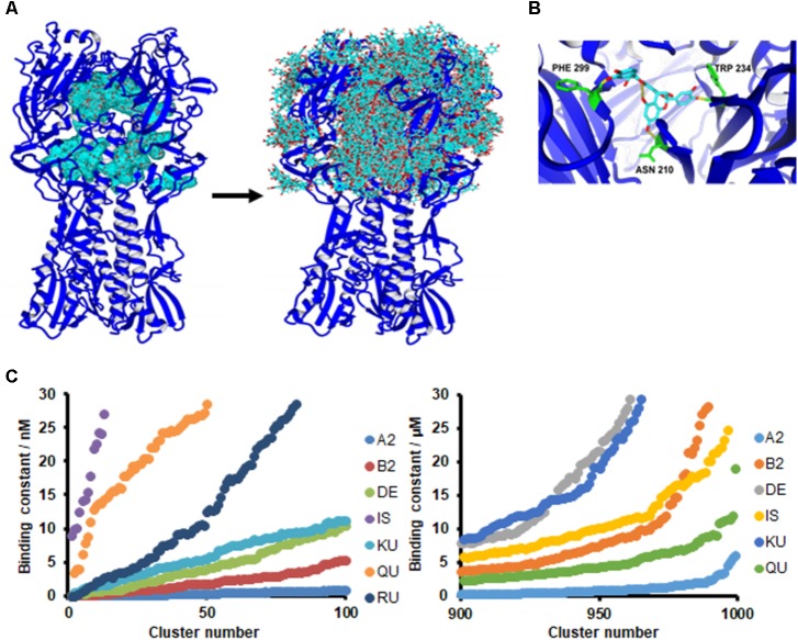
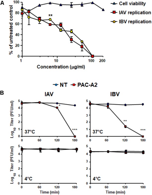
The defense against influenza virus (IV) infections still poses a series of challenges. The current antiviral arsenal against influenza viruses is in fact limited; therefore, the development of new anti-influenza strategies effective against antigenically different viruses is an urgent priority. Bioactive compounds derived from medicinal plants and fruits may provide a natural source of candidates for such broad-spectrum antivirals. In this regard, cranberry (Vaccinium macrocarpon Aiton) extracts on the basis of their recognized anti-adhesive activities against bacteria, may provide potential compounds able to prevent viral attachment to target cells. Nevertheless, only few studies have so far investigated the possible use of cranberry extracts as an antiviral tool. This study focuses on the suitability of a cranberry extract as a direct-acting anti-influenza compound. We show that the novel cranberry extract Oximacro® inhibits influenza A and B viruses (IAV, IBV) replication in vitro because of its high content of A-type proanthocyanidins (PAC-A) dimers and trimers. Mechanistic studies revealed that Oximacro® prevents attachment and entry of IAV and IBV into target cells and exerts a virucidal activity. Oximacro® was observed to interact with the ectodomain of viral hemagglutinin (HA) glycoprotein, thus suggesting the interference with HA functions and a consequent loss of infectivity of IV particles. Fluorescence spectroscopy revealed a reduction in the intrinsic fluorescence of HA protein after incubation with purified dimeric PAC-A (PAC-A2), thus confirming a direct interaction between HA and Oximacro® PAC-A2. In silico docking simulations further supported the in vitro results and indicated that among the different components of the Oximacro® chemical profile, PAC-A2 exhibited the best binding propensity with an affinity below 10 nM. The role of PAC-A2 in the anti-IV activity of Oximacro® was eventually confirmed by the observation that it prevented IAV and IVB replication and caused the loss of infectivity of IV particles, thus indicating PAC-A2 as the major active component of Oximacro®. As a whole, these results suggest Oximacro® as a potential candidate to create novel antiviral agents of natural origin for the prevention of IV infections.

Keywords: Oximacro®; PAC-A2; antiviral and virucidal activities; cranberry extract; dimeric A-type proanthocyanidins; hemagglutinin; influenza virus.


Figures

FIGURE 1
FIGURE 1
Antiviral activity of Oximacro® against influenza A and B viruses. MDCK cell monolayers were infected with either IAV or IBV (40 PFU/well), and, where indicated, the cells were treated with increasing concentrations of Oximacro® 1 h before, during virus adsorption, and after adsorption throughout the experiment. At 48 h p.i., plaques were stained and microscopically counted. The mean plaque counts for each concentration are expressed as a percentage of the mean plaque count for the control virus. The number of plaques was plotted as a function of Oximacro® concentration; concentrations producing 50 and 90% reductions in plaque formation (IC50 and IC90) were thus determined. Data represent means ± SD (error bars) of three independent experiments performed in duplicate. Statistical analysis was performed by comparing IAV and IBV replication curves. ∗∗∗p < 0.001, ∗∗p < 0.01, and p < 0.05. To determine cell viability, MDCK cells were exposed to increasing concentrations of Oximacro®. After 3 days of incubation, the number of viable cells was determined by the 3-(4,5-imethylthiazol-2-yl)-2,5-diphenyltetrazolium bromide (MTT) method.
FIGURE 2
FIGURE 2
Oximacro® acts in an early stage of IV-replicative cycle. A schematic summary of the time-of-addition experiment is shown at the top of the figure. Accordingly, MDCK cell monolayers were left untreated or treated with different concentrations of Oximacro® from -2 to -1 h (pre-adsorption) (A,D) or from -1 to 0 h (adsorption) (B,E) before infection with IAV or IBV (40 PFU/well). In the post-adsorption treatment (from 0 to 48 h p.i.) (C,F), untreated and infected cells were exposed to the same concentrations of Oximacro® as indicated in the scheme on the top and were infected with IAV or IBV (40 PFU/well) at -1 h p.i. At 48 h p.i., plaques were stained and microscopically counted. Data represent means ± SD (error bars) of three independent experiments performed in duplicate. For each experimental condition, statistical analysis was carried out by comparing treated samples with the untreated control (100% infection). ∗∗∗p < 0.001, ∗∗p < 0.01, and p < 0.05.
FIGURE 3
FIGURE 3
IV attachment and entry is prevented by Oximacro®. (A) Oximacro® affects IV attachment. Prechilled MDCK cells were infected with precooled IAV or IBV (40 PFU/well) in the presence of different concentrations of Oximacro® (20-10-5-2.5 g/ml) at 4°C for 2 h. After viral adsorption, the cells were washed and overlaid with 0.7% Avicel. At 48 h p.i., viral plaques were stained and microscopically counted. The results shown are means ± SD (error bars) from three independent experiments performed in duplicate. (B) Oximacro® inhibits IV entry. Prechilled MDCK cells were infected with IAV or IBV (40 PFU/well) for 2 h at 4°C to allow virion attachment to the cells. After adsorption, the cells were treated with different concentrations of Oximacro® (20-10-5-2.5 μg/ml) for 2 h at 37°C, prior to inactivation of extracellular virus with acidic glycine buffer for 30 s at RT. After further washing, the cells were incubated with medium-containing 0.7% Avicel. At 48 h p.i., viral plaques were stained and microscopically counted. The results shown are means ± SD (error bars) of three independent experiments performed in duplicate. ∗∗∗p < 0.001, ∗∗p < 0.01, and p < 0.05 compared with the 100% virus infectivity of untreated controls.
FIGURE 4
FIGURE 4
Incubation of IV particles with Oximacro® abrogates infectivity. IAV or IBV (2 × 10⁴ PFU) were incubated at 37°C (A) or at 4°C (B) for various lengths of time in the absence of Oximacro® (NT) (closed circles) or with 25 g of the Oximacro® (closed squares). After incubation, the samples were diluted to reduce Oximacro® concentration below that which inhibits IV attachment (0.25 μg/ml). Plaques were microscopically counted, and the mean plaque counts was expressed as PFU/ml on a log10 scale. The data shown represent means ± SD (error bars) of three independent experiments performed in duplicate. ∗∗∗p < 0.001, ∗∗p < 0.01, and p < 0.05 compared with the titer of untreated controls (NT).
FIGURE 5
FIGURE 5
Oximacro® interacts with the recombinant extracellular domain of IAV HA. (A) SDS–PAGE and immunoblot analysis of recombinant IAV-HA glycoprotein. Purified HA protein was analyzed on 10% SDS-polyacrylamide gels. Gel was either stained with Coomassie blue or analyzed by immunoblotting with an anti-HA IAV mAb. Left panel: Coomassie blue-stained gel of 1 g of purified recombinant HA-IAV. Right panel: Immunoblot analysis with 200 ng of the same sample as in the left panel. (B,C) Oximacro® interacts with recombinant HA in a concentration- (B) and time-dependent (C) manner. (B) Purified recombinant HA-IAV (1 g) was incubated at 37°C with medium or increasing amounts of Oximacro® for 3 h, and then mixtures were analyzed by SDS–PAGE. (C) Purified recombinant HA-IAV (1 g) was incubated at 37°C for various lengths of time with medium or 6 g of Oximacro®. At 30 min, 1, 2, and 3 h of incubation, mixtures were analyzed by SDS–PAGE. Gels were then stained with Coomassie blue. Sizes are indicated in kilodaltons.
FIGURE 6
FIGURE 6
PAC-A2 determines quenching of HA-IAV fluorescence. Recombinant HA-IAV was resuspended in PBS buffer (pH 7.4) at a concentration of 1.2 μM. Fluorescence of HA-IAV aliquots were measured in the absence (blue curve) and presence (orange curve) of 25 M PAC-A2 after incubation for 1 h at 25°C. Fluorescence emission spectra were monitored in the 295–500 nm range using a scan-rate of 200 nm/min upon excitation at 290 nm. Data reported were normalized by subtracting the PAC-A2 fluorescence contribution.
FIGURE 7
FIGURE 7
PAC-A2 interacts with HA in an in silico model. (A) Binding of PACA2 to HA-IAV. Crystal structures of HA-IAV (A/Puerto Rico/8/34-PDB ID: 1RU7) and PAC-A2 were docked using the AutoDock algorithm, embedded in YASARA. Docking analysis revealed that PAC-A2 binds within the internal grooves of the HA structure first (left panel) and subsequently to the surface of the structure (righ panel). (B) Best pose of PAC-A2 to HA-IAV. This represents the best complex obtained after 999 docking runs. The HA-IAV structure is represented in blue; PAC-A2 is in cyan; residues forming hydrogen bonds to the ligand are in green; and hydrogen bonds are in yellow. (C) PAC-A2 is the Oximacro® component that binds best to HA. Chemical structures of different components of Oximacro® were subjected to docking analysis. PAC-A2 (A2), PAC-B2 (B2), Delphinidin 3-glucoside (DE), Isorhamnetin (IS) Kuromanin (KU), Quercetin (QU), and Rutin (obtained from Pubchem) were docked with HA. In the left panel, the distribution of the binding constants of the top 100 scoring complex clusters is shown; in the right panel, the distribution of the worst 100 scoring complexes is shown. In the right panel, Rutin is not shown because its binding constant was >30 μM.
FIGURE 8
FIGURE 8
Purified PAC-A2 inhibits IV replication by causing loss of virion infectivity. (A) PAC-A2 inhibits replication of IAV and IBV. MDCK cell were infected with either IAV or IBV (40 PFU/well), and, where indicated, the cells were treated with increasing concentrations of PAC-A2 1 h before, during virus adsorption, and after adsorption throughout the experiment. At 48 h p.i., plaques were stained and microscopically counted. To determine cell viability, MDCK cells were exposed to increasing concentrations of PAC-A2, and the number of viable cells was determined by the MTT method. Statistical analysis was accomplished by comparing IAV and IBV replication curves. ∗∗∗p < 0.001, ∗∗p < 0.01, and p < 0.05. (B) Treatment with PAC-A2 abolishes the infectivity of IV particles. IAV or IBV (2 × 10⁴ PFU) were incubated at 37°C (A) or at 4°C (B) for various lengths of time in the absence (closed circles) or in the presence of 50 μg of PAC-A2 (closed squares). After incubation, the samples were diluted to reduce PAC-A2 below that which inhibits IV replication and the residual infectivity assessed by plaque assay. The mean plaque counts were expressed as PFU/ml on a log10 scale. The data shown represent means ± SD (error bars) of three independent experiments performed in duplicate. ∗∗∗p < 0.001, ∗∗p < 0.01, and p < 0.05 compared with the titer of untreated controls (NT).

Similar articles

Cited by

References

    1. Alame M. M., Massaad E., Zakaret H. (2016). Peramivir: a novel intravenous neuraminidase inhibitor for treatment of acute influenza infections. Front. Microbiol. 7:450. 10.3389/fmicb.2016.00450 - DOI - PMC - PubMed
    1. Bahramsoltani R., Sodagari H. R., Farzaei M. H., Abdolghaffari A. H., Gooshe M., Rezaei N. (2016). The preventive and therapeutic potential of natural polyphenols on influenza. Expert Rev. Anti Infect. Ther. 14 57–80. 10.1586/14787210.2016.1120670 - DOI - PubMed
    1. Blumberg J. B., Camesano T. A., Cassidy A., Kris-Etherton P., Howell A., Manach C., et al. (2013). Cranberries and their bioactive constituents in human health. Adv. Nutr. 4 618–632. 10.3945/an.113.004473 - DOI - PMC - PubMed
    1. Catucci G., Gilardi G., Jeuken L., Sadeghi S. J. (2012). In vitro drug metabolism by C-terminally truncated human flavin-containing monooxygenase 3. Biochem. Pharmacol. 83 551–558. 10.1016/j.bcp.2011.11.029 - DOI - PubMed
    1. Charlton A. J., Baxter N. J., Khan M. L., Moir A. J., Haslam E., Davies A. P., et al. (2002). Polyphenol/peptide binding and precipitation. J. Agric. Food Chem. 50 1593–1601. 10.1021/jf010897z - DOI - PubMed