Review
. 2018 Jul 30:9:1770.
doi: 10.3389/fimmu.2018.01770. eCollection 2018.

Armed and Ready: Transcriptional Regulation of Tissue-Resident Memory CD8 T Cells

Affiliations
Review

Armed and Ready: Transcriptional Regulation of Tissue-Resident Memory CD8 T Cells

Felix M Behr et al. Front Immunol. .

Abstract

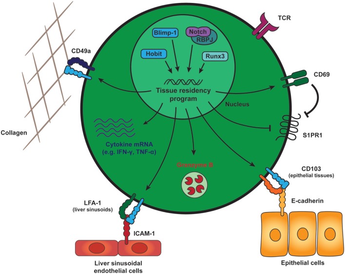
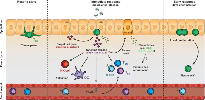
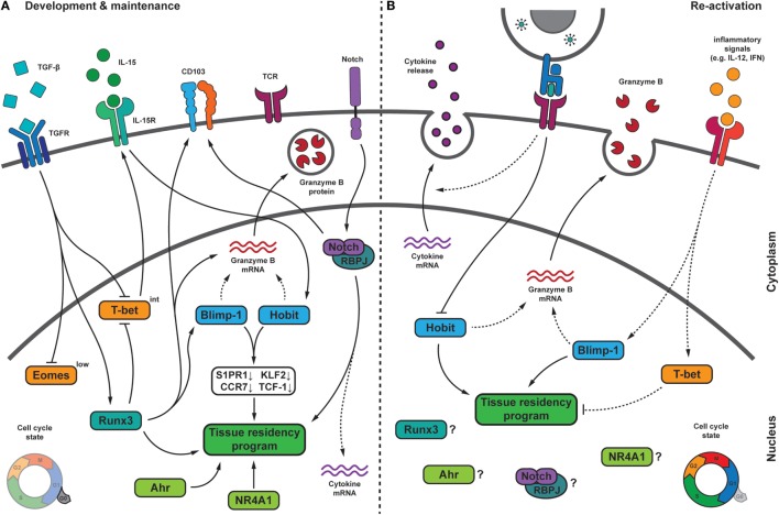
A fundamental benefit of immunological memory is the ability to respond in an enhanced manner upon secondary encounter with the same pathogen. Tissue-resident memory CD8 T (TRM) cells contribute to improved protection against reinfection through the generation of immediate effector responses at the site of pathogen entry. Key to the potential of TRM cells to develop rapid recall responses is their location within the epithelia of the skin, lungs, and intestines at prime entry sites of pathogens. TRM cells are among the first immune cells to respond to pathogens that have been previously encountered in an antigen-specific manner. Upon recognition of invading pathogens, TRM cells release IFN-γ and other pro-inflammatory cytokines and chemokines. These effector molecules activate the surrounding epithelial tissue and recruit other immune cells including natural killer (NK) cells, B cells, and circulating memory CD8 T cells to the site of infection. The repertoire of TRM effector functions also includes the direct lysis of infected cells through the release of cytotoxic molecules such as perforin and granzymes. The mechanisms enabling TRM cells to respond in such a rapid manner are gradually being uncovered. In this review, we will address the signals that instruct TRM generation and maintenance as well as the underlying transcriptional network that keeps TRM cells in a deployment-ready modus. Furthermore, we will discuss how TRM cells respond to reinfection of the tissue and how transcription factors may control immediate and proliferative TRM responses.

Keywords: BLIMP-1; Notch; RUNX3; T cell diferentiation; homolog of Blimp-1 in T cells; secondary responses; tissue-resident memory T cells; transcription factors.


Figures

Figure 1
Figure 1
General features of tissue-resident memory CD8 T (TRM) cells. Generation and maintenance of TRM cells is regulated by a distinct set of transcription factors, including Runx3, Blimp-1, and its homolog of Blimp-1 in T cells as well as the transcriptional activator Notch. These transcription factors instruct a tissue-residency program that allows for the long-term retention and maintenance of TRM cells within peripheral tissues. TRM cells across tissues maintain expression of CD69, which promotes tissue residency by interfering with spingosine-1 phosphate receptor (S1PR1) function. S1PR1 mediates egress of T cells into the circulation and its downregulation is a core characteristic of TRM cells. In many tissues, TRM cells also express high levels of CD49a, an adhesion molecule binding to collagen (in complex with β1 integrin) to establish tissue residency. The αE integrin CD103 is expressed by mucosal TRM cells and may contribute to tissue retention by interaction with E-cadherin on the surrounding epithelial cells. In liver sinusoids, local TRM cells upregulate LFA-1, which supports their tissue residence by binding to ICAM-1 on liver sinusoidal endothelial cells. In addition to these adhesion molecules, TRM cells in many tissues are characterized by elevated transcript levels encoding for pro-inflammatory cytokines, e.g., IFN-γ and TNF-α, and protein expression of the cytotoxic serine protease granzyme B. Abbreviations: Runx3, Runt-related transcription factor 3; Blimp-1, B lymphocyte-induced maturation protein-1; Hobit, homolog of Blimp-1 in T cells; RBPJ, recombining binding protein suppressor of hairless; LFA-1, lymphocyte function-associated antigen-1; ICAM-1, intercellular adhesion molecule 1; IFN-γ, interferon γ; TNF-α, tumor necrosis factor α.
Figure 2
Figure 2
Protective effector responses of epithelial TRM cells upon secondary infection. TRM cells in the epithelia continually patrol their local environment, projecting dendritic extensions in multiple directions. Upon pathogen challenge and antigen re-encounter, TRM cells rapidly release pro-inflammatory cytokines, including IFN-γ, TNF-α, and IL-2, which induce several immune cell- and tissue-specific effects. Local cytokine release by TRM cells results in recruitment and activation of natural killer (NK) cells and dendritic cells (DCs), as well as upregulation of VCAM-1 on endothelial cells in local blood vessels, which may enhance the recruitment of TCM, TEM, and B cells from the circulation. TRM cell reactivation and cytokine release also induces a tissue-wide state of alert, resulting in upregulation of many innate immune response genes, including interferon-induced transmembrane protein 3 (IFITM3), and the increased local expression of inflammatory chemokines. The protective capacity of TRM cells may also rely on perforin-mediated killing of target cells. One to two days after antigen re-encounter, TRM cells undergo local proliferation. Further investigation is required to determine whether TRM cells exit their local environment after reactivation. Abbreviations: IFN-γ, interferon γ; TNF-α, tumor necrosis factor α; IL-2, interleukin 2; ICAM-1, vascular cell adhesion molecule 1; TCM cell, central memory T cell; TEM cell, effector memory T cell; CCL, C-C motif chemokine; CXCL9, C-X-C motif chemokine 9.
Figure 3
Figure 3
Transcriptional regulation of TRM cells during development, maintenance, and upon pathogen re-challenge. (A) During their formation, TRM cells receive multiple signals from the tissue microenvironment that integrate into a transcriptional program, which drives TRM differentiation and maintenance. In several tissues, transforming growth factor β (TGF-β) signaling drives the downregulation of the T-box transcription factors Eomesodermin (Eomes) and T-bet. Residual T-bet expression is required for IL-15Rβ expression. The transcription factor homolog of Blimp-1 in T cells (Hobit) can be induced in an IL-15-dependent manner and, together with its homolog Blimp-1, represses the expression of S1PR1, CCR7, Krϋpple-like factor 2 (KLF2), and TCF-1, which is crucial for tissue residency. Blimp-1 and Hobit may also contribute to granzyme B maintenance in TRM cells. The transcription factor Runx3, which can be induced by TGF-β signaling, is crucial for the establishment and maintenance of many aspects of TRM cells, including granzyme B and CD103 expression. Runx3 has been shown to induce Blimp-1. Notch may regulate expression of the adhesion molecule CD103, is essential for maintenance of TRM cells and might contribute to the elevated transcript levels encoding for pro-inflammatory cytokines in TRM cells. Other factors regulating TRM cells include the nuclear receptor NR4A1, and the aryl hydrocarbon receptor (AhR). During quiescence, TRM cells show low proliferative activity. (B) Following pathogen re-encounter, TRM cells are exposed to antigen-dependent T cell receptor (TCR) triggering and a variety of inflammatory signals. TCR triggering in TRM cells may result in downregulation of Hobit, thereby weakening its contribution to maintenance of tissue residency. Inflammatory signals, such as IL-12 and type I interferons (IFN), can induce expression of Blimp-1 and T-bet. While increased Blimp-1 expression might fortify TRM features, elevated levels of T-bet could interfere with tissue residency. TRM cells rapidly release pro-inflammatory cytokines upon reactivation in an antigen-dependent manner. Upon re-infection, TRM cells undergo local proliferation. Dashed lines indicate relations that require further investigation. Abbreviations: Blimp-1, homolog of B lymphocyte-induced maturation protein 1; IL, interleukin; S1PR1, sphingosine-1-phosphate receptor 1; CCR7, C-C chemokine receptor type 7; TCF-1, T cell factor 1; Runx3, Runt-related transcription factor 3; RBPJ, recombining binding protein suppressor of hairless; NR4A1, nuclear receptor subfamily 4 group A 1.

References

    1. Blattman JN, Antia R, Sourdive DJD, Wang X, Kaech SM, Murali-Krishna K, et al. Estimating the precursor frequency of naive antigen-specific CD8 T cells. J Exp Med (2002) 195(5):657–64.10.1084/jem.20001021 - DOI - PMC - PubMed
    1. Bousso P, Casrouge A, Altman JD, Haury M, Kanellopoulos J, Abastado J-P, et al. Individual variations in the murine T cell response to a specific peptide reflect variability in naive repertoires. Immunity (1998) 9(2):169–78.10.1016/S1074-7613(00)80599-3 - DOI - PubMed
    1. Casrouge A, Beaudoing E, Dalle S, Pannetier C, Kanellopoulos J, Kourilsky P. Size estimate of the αβ TCR repertoire of naive mouse splenocytes. J Immunol (2000) 164(11):5782–7.10.4049/jimmunol.164.11.5782 - DOI - PubMed
    1. van Heijst JWJ, Gerlach C, Swart E, Sie D, Nunes-Alves C, Kerkhoven RM, et al. Recruitment of antigen-specific CD8+ T cells in response to infection is markedly efficient. Science (2009) 325(5945):1265–9.10.1126/science.1175455 - DOI - PubMed
    1. Butz EA, Bevan MJ. Massive expansion of antigen-specific CD8+ T cells during an acute virus infection. Immunity (1998) 8(2):167–75.10.1016/S1074-7613(00)80469-0 - DOI - PMC - PubMed

Publication types