

Review of the Variations of the Superficial Veins of the Neck
- PMID: 30131919
- PMCID: PMC6101467
- DOI: 10.7759/cureus.2826
Review of the Variations of the Superficial Veins of the Neck
Abstract
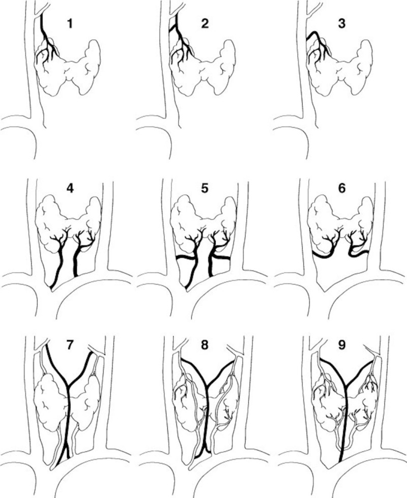
The venous drainage of the neck can be characterized into superficial or deep. Superficial drainage refers to the venous drainage of the subcutaneous tissues, which are drained by the anterior and external jugular veins (EJVs). The brain, face, and neck structures are mainly drained by the internal jugular vein (IJV). The superficial veins are found deep to the platysma muscle while the deep veins are found encased in the carotid sheath. The junction of the retromandibular vein and the posterior auricular vein usually form the EJV, which continues along to drain into the subclavian vein. The anterior jugular vein is usually formed by the submandibular veins, travels downward anterior to the sternocleidomastoid muscle (SCM), and drains either into the EJV or the subclavian vein. Other superficial veins of the neck to consider are the superior, middle, and inferior thyroid veins. The superior thyroid and middle thyroid veins drain into the IJV whereas the inferior thyroid vein usually drains into the brachiocephalic veins.
Keywords: external jugular; internal jugular; superficial; thyroid vein; vein.
Conflict of interest statement
The authors have declared that no competing interests exist.
Figures


References
-
- The anterior jugular venous system: variability and clinical impact. Schummer W, Schummer C, Bredle D, Frober R. Anesth Analg. 2004;99:1625–1629. - PubMed
-
- Extraordinary cerebral venous drainage pathway with mastoid emissary and posterior external jugular veins detected by contrast-enhanced neck computed tomography. Bulbul E, Yanik B, Demirpolat G, Koksal V. Surg Radiol Anat. 2015;37:1191–1194. - PubMed
-
- Facial vein draining into external jugular vein in humans: its variations, phylogenetic retention and clinical relevance. Gupta V, Tuli A, Choudhry R, Agarwal S, Mangal A. Surg Radiol Anat. 2003;25:36–41. - PubMed
-
- Williams PL, Warwick R, Dyson M, Bannister LH. Vol. 37. Edinburgh: Churchill Livingstone; 1989. Gray’s Textbook of Anatomy; pp. 777–781.
-
- Review of venous anatomy for venographic interpretation in chronic cerebrospinal venous insufficiency. Werner JD, Siskin GP, Mandato K, Englander M, Herr A. J Vasc Interv Radiol. 2011;22:1681–1690. - PubMed
Publication types
LinkOut - more resources
Full Text Sources
Other Literature Sources