

Craniotomy Improves Traumatic Optic Neuropathy
- PMID: 30131928
- PMCID: PMC6101456
- DOI: 10.7759/cureus.2835
Craniotomy Improves Traumatic Optic Neuropathy
Abstract
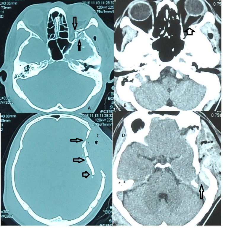
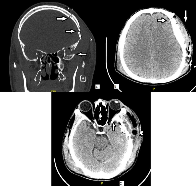
Traumatic optic neuropathy (TON) is a rare devastating complication of traumatic head injury and is an ophthalmic emergency. Herein, we report a rare case of a 46-year-old gentleman who experienced severe blurring of vision, binocular diplopia, and pain over his left eye following a fall from a tree about three meters in height. Examinations revealed the visual acuity was 6/60 with a marked relative afferent pupillary defect and generalized ophthalmoplegia over his left eye. Emergency computed tomography (CT) brain and orbit showed a left frontotemporoparietal extradural hemorrhage, comminuted frontotemporoparietal and greater wing of sphenoid fracture with a bony spur impinging the lateral rectus and indirectly on the optic nerve. A diagnosis of left frontotemporoparietal bone fracture with traumatic optic neuropathy was made. An emergency left craniotomy, elevation of depressed skull fracture, and evacuation of clot was done. Postoperatively, his visual acuity showed marked improvement with visual acuity of 6/6 and all optic nerve functions were normal.
Keywords: bony spur; emergency craniotomy; traumatic optic neuropathy.
Conflict of interest statement
The authors have declared that no competing interests exist.
Figures
References
-
- Optic nerve trauma: clinical, electrophysiological and histological remarks. Nau HE, Gerhard L, Foerster M, Nahser HC, Reinhardt V, Joka T. https://link.springer.com/article/10.1007/BF01406662. Acta Neurochir. 1987;89:16–27. - PubMed
-
- Traumatic optic neuropathy. Steinsapir KD, RA Goldberg. Surv Ophthalmol. 1994;38:487–518. - PubMed
-
- High dose corticosteroids for treatment of vision loss due to indirect injury to the optic nerve. Seiff SR. https://www.healio.com/ophthalmology/journals/osli/1990-6-21-6/%7B1c9b49.... Ophthalmic Surg. 1990;21:389–395. - PubMed
-
- Sphenoethmoid approach to the optic nerve. Sofferman RA. Laryngoscope. 1981;91:184–196. - PubMed
-
- Intrachiasmal hemorrhage: a cause of delayed post‐traumatic blindness. Crowe NW, Nickles TP, Troost BT, Elster AD. Neurology. 1989;39:863. - PubMed
Publication types
LinkOut - more resources
Full Text Sources
Other Literature Sources