Review
. 2018 Aug 7:6:82.
doi: 10.3389/fcell.2018.00082. eCollection 2018.

Endothelial Cell Metabolism in Atherosclerosis

Affiliations
Review

Endothelial Cell Metabolism in Atherosclerosis

Kosta Theodorou et al. Front Cell Dev Biol. .

Abstract

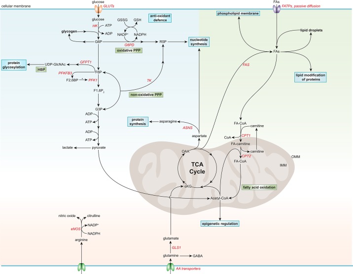
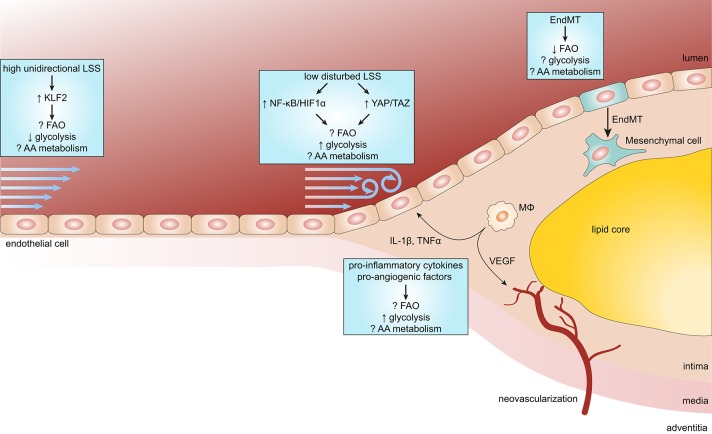
Atherosclerosis and its sequelae, such as myocardial infarction and stroke, are the leading cause of death worldwide. Vascular endothelial cells (EC) play a critical role in vascular homeostasis and disease. Atherosclerosis as well as its independent risk factors including diabetes, obesity, and aging, are hallmarked by endothelial activation and dysfunction. Metabolic pathways have emerged as key regulators of many EC functions, including angiogenesis, inflammation, and barrier function, processes which are deregulated during atherogenesis. In this review, we highlight the role of glucose, fatty acid, and amino acid metabolism in EC functions during physiological and pathological states, specifically atherosclerosis, diabetes, obesity and aging.

Keywords: aging; atherosclerosis; endothelial cells; hyperglycemia; hyperlipidemia; inflammation; metabolism.


Figures

Figure 1
Figure 1
Simplified general overview of glucose, fatty acid, and amino acid metabolism in healthy endothelial cells. Glucose enters ECs via glucose transporters (GLUTs), which is converted to glucose-6-phosphate (G6P) by hexokinase (HK) at the expense of adenosine triphosphate (ATP). G6P can be converted to glycogen for storage or used in the oxidative pentose phosphate pathway (PPP). The oxidative PPP generates reduced glutathione (GSH) and ribose-5-phosphate (R5P), which are used in the anti-oxidant defense and nucleotide synthesis, respectively. G6P can be further metabolized to fructose-6-phosphate (F6P) which is converted to uridine diphosphate N-acetylglucosamine (UDP-GlcNAc), a substrate used for protein glycosylation, in the hexosamine biosynthesis pathway (HBP). F6P can also be converted to fructose-2,6-bisphophate (F2,6BP) by 6-phosphofructo-2-kinase/fructose-2,6-bisphosphatase 3 (PFKF3B), the main isoform in ECs. F2,6BP is a positive allosteric regulator of phosphofructokinase 1 (PFK1), which converts F6P to fructose-1,6-bisphophate (F1,6P2), which is further metabolized to glyceraldehyde-3-phosphate (G3P). G3P together with F6P can be used in the non-oxidative PPP by transketolase (TK) to eventually produce R5P. G3P can also undergo several conversion steps leading to the production of ATP from adenosine diphosphate (ADP) and pyruvate, which can be further metabolized to lactate. After being taken up by fatty acid (FA) transporters (FATPs) or through passive diffusion, FAs become metabolically activated by coupling to coenzyme A (CoA). For FA oxidation (FAO) to occur, FAs have to be imported into the mitochondria, which is carried out by the acyl-carnitine shuttle system. At the outer mitochondrial membrane (OMM), carnitine palmitoyltransferase 1 (CPT1) converts FA-CoAs into FA-carnitines which facilitates the transport of FAs across the inner mitochondrial membrane (IMM) where they are converted back to FA-CoA by CPT2 (located in the IMM). FAO generates ATP and acetyl-CoA which is used in the tricarboxylic acid cycle (TCA). TCA intermediates are used for nucleotide synthesis or FA production, facilitated by fatty acid synthase (FAS). FAs can be stored in lipid droplets, converted to phospholipids to maintain the cell membrane or used for lipid modification of proteins. The amino acid (AA) glutamine is the most consumed AA in ECs. Once inside the cell, glutamine is converted to glutamate by glutaminase 1 (GLS1), which is further metabolized to α-ketoglutarate (αKG), a key intermediate in the TCA cycle. Through several metabolic steps, αKG is converted to oxaloacetic acid (OAA), which can be used for the generation of the AA aspartate. Aspartate can be used as a precursor for nucleotide synthesis or converted to the AA asparagine by asparagine synthetase (ASNS) using an ammonia group from glutamine thereby generating glutamate. Another important AA in ECs is arginine, which is used for the generation of the anti-atherogenic gaseous molecule nitric oxide using nicotinamide adenine dinucleotide phosphate (NADPH) as a cofactor. G6PD, glucose-6-phosphate dehydrogenase; GFPT1, glutamine:fructose-6-phosphate amidotransferase 1; GSSG, oxidized glutathione; NADP+, nicotinamide adenine dinucleotide.
Figure 2
Figure 2
Endothelial metabolism in atherosclerosis. Endothelial cells (ECs) exposed to high unidirectional laminar shear stress (LSS) activate atheroprotective signaling via the transcription factor Krüppel-like factor 2 (KLF2) which reduces glycolysis and maintains ECs in a quiescent state. In contrast, atheroprone regions are subjected to low disturbed LSS enhances EC glycolysis via the nuclear factor κB (NF-κB)/hypoxia inducible factor 1α (HIF1α) axis and mechanotransducers Yes-associated protein (YAP) and transcriptional coactivator with PDZ-binding motif (TAZ). During the process of atherosclerosis, ECs are exposed to a pro-inflammatory milieu [e.g., cytokines interleukin 1β (IL-1β) and tumor necrosis factor α (TNFα)], which also enhances glycolysis in ECs. In more progressed lesions, macrophages (MΦ) start to secrete pro-angiogenic factors resulting in intraplaque neovascularization. Pro-angiogenic factors, like vascular endothelial growth factor (VEGF), enhance glycolysis in ECs to support proliferation and migration. In addition, reduced FAO in ECs predisposes them to undergo endothelial-to-mesenchymal transition (EndMT), which may affect plaque stability.

References

    1. Adeva-Andany M. M., Gonzalez-Lucan M., Donapetry-Garcia C., Fernandez-Fernandez C., Ameneiros-Rodriguez E. (2016). Glycogen metabolism in humans. BBA Clin. 5, 85–100. 10.1016/j.bbacli.2016.02.001 - DOI - PMC - PubMed
    1. Barth E., Stammler G., Speiser B., Schaper J. (1992). Ultrastructural quantitation of mitochondria and myofilaments in cardiac muscle from 10 different animal species including man. J. Mol. Cell. Cardiol. 24, 669–681. 10.1016/0022-2828(92)93381-S - DOI - PubMed
    1. Bertero T., Oldham W. M., Cottrill K. A., Pisano S., Vanderpool R. R., Yu Q., et al. . (2016). Vascular stiffness mechanoactivates YAP/TAZ-dependent glutaminolysis to drive pulmonary hypertension. J. Clin. Invest. 126, 3313–3335. 10.1172/JCI86387 - DOI - PMC - PubMed
    1. Blouin A., Bolender R. P., Weibel E. R. (1977). Distribution of organelles and membranes between hepatocytes and nonhepatocytes in the rat liver parenchyma. A stereological study. J. Cell Biol. 72, 441–455. 10.1083/jcb.72.2.441 - DOI - PMC - PubMed
    1. Brunet A., Rando T. A. (2017). Interaction between epigenetic and metabolism in aging stem cells. Curr. Opin. Cell Biol. 45, 1–7. 10.1016/j.ceb.2016.12.009 - DOI - PMC - PubMed

LinkOut - more resources