. 2018 Aug 22;12(8):e0006701.
doi: 10.1371/journal.pntd.0006701. eCollection 2018 Aug.

Therapeutic control of leishmaniasis by inhibitors of the mammalian target of rapamycin

Affiliations

Therapeutic control of leishmaniasis by inhibitors of the mammalian target of rapamycin

Fatemeh Khadir et al. PLoS Negl Trop Dis. .

Abstract

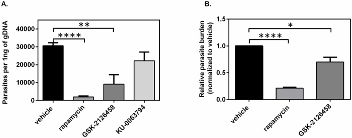
Leishmaniasis is a serious global health problem affecting many people worldwide. While patients with leishmaniasis can be treated with several agents, drug toxicicty and the emergence of resistant strains render available treatments ineffective in the long run. Inhibitors of the mammalian target of rapamycin (mTOR) have been demonstrated to exert anti-pathogen properties. In this study, we tested the therapeutic efficacy of several mTOR inhibitors in controlling infection with Leishmania major. Rapamycin, GSK-2126458 and KU-0063794 were administered to BALB/c mice, which had received an intrafootpad injection of the parasite. Footpad swelling and parasite burden were assessed, and cytokine production by mouse splenocytes and phenotypic changes in draining lymph node cells were evaluated. Treatment with a clinically relevant dose of rapamycin or with GSK-2126458, but not with KU-0063794, dramatically lowered both the footpad swelling and the parasite load in the draining lymph node. Importantly, the employed dose of rapamycin did not kill the promastigotes in vitro as judged by 3-(4,5-dimethylthiazol-2-yl)-2,5-diphenyltetrazolium bromide (MTT) assays and electron microscopy. Moreover, the IL-4 production capacity of splenocytes harvested from infected mice that were treated with rapamycin was significantly reduced. Consequently, the IFN-γ:IL-4 production ratio was elevated, suggesting a T helper-type 1 (Th1)-skewed cytokine profile. Finally, the expression level of CD69, an early activation marker, on splenic and lymph node CD4+ and CD8+ T cells was enhanced in rapamycin-treated mice. Taken together, our findings suggest that select mTOR inhibitors may be used in therapeutic settings for the management of leishmaniasis. We propose that the beneficial effects of such inhibitors stem from their immunomodulatory properties. Therefore, the adjuvanticity of mTOR inhibitors may also be considered in vaccination strategies against Leishmania species.


Conflict of interest statement

The authors have declared that no competing interests exist.

Figures

Fig 1
Fig 1. The effect of treatment with mTOR inhibitors on footpad swelling of L. major-infected mice.
BALB/c mice were inoculated in their footpad with L. major. After 21 days, mice were treated i.p. with vehicle (n = 9), rapamycin (10.2 μg/dose/day, n = 9), GSK-2126458 (10.2 μg/dose/day, n = 9), or KU-0063794 (10.2 μg/dose/day, n = 9). The timeline for infection and treatment with indicated mTOR inhibitors is schematically depicted (A). Control and infected footpads are shown at the experimental end point (day 31 post-infection) before (shown as black) and after treatment with mTOR inhibitors (shown as red) or vehicle (shown as blue) (B). The observed changes in footpad swelling and lesions over the course of the disease/treatment are graphed form one representative experiment (C). Data are pooled from 3 experiments, each of which typically consisted of at least 3 mice/group. Error bars represent standard error of the mean (SEM). The difference in footpad swelling was analyzed using multiple comparisons, two-way ANOVA at each time point. **: p< 0.01***: P<0.001 **** P<0.0001.
Fig 2
Fig 2. Enumeration of Leishmania parasite burden in BALB/c mice treated with vehicle or with daily high doses of mTOR inhibitors.
Following the treatment schedule highlighted in Fig 1A, parasite burden in the draining popliteal lymph nodes (pLN) of the infected footpad was determined by qPCR of bulk genomic DNA. Absolute copy numbers of Leishmania RV1-RV2 in the pLN of mice treated with vehicle or 10.2 μg/dose of rapamycin, GSK-2126458 or KU-0063794 were calculated via a standard curve and values are presented as number of parasites per ng of gDNA (A). In parallel, gDNA for the mammalian GAPDH housekeeping gene was quantified and normalized parasite numbers in mTOR inhibitor-treated mice were quantified relative to that of vehicle-treated mice using the ΔΔCt method (B). The error bars represent the standard error of the mean (SEM), data were pooled from three different experiments, n = 9. Statistical differences in parasite burden between groups were determined using one-way ANOVA, Student t-test was done between untreated group (vehicle) and treated group. *: p< 0.05; **: p< 0.01; ****: p< 0.0001.
Fig 3
Fig 3. In vivo concentrations of rapamycin do not exhibit direct toxic effect on Leishmania promastigotes in vitro.
In vivo serum concentration of rapamycin was determined after 10 days of daily i.p. treatment with 10.2 μg/dose (n = 4) (A). Twenty million stationary phase parasites/mL were treated with different doses of rapamycin (0–50 μg/mL) and IC50 was determined by MTT assay (B). Based on determined IC50, stationary phase parasites treated for 48 h with low, medium and high doses of rapamycin (5, 10 and 20 μg/mL), and then subjected to SEM electron microscopy, the scale was 7μm. (C). Parasites treated with 10 μg/mL of rapamycin were subjected to TEM electron microscopy (D) The scale was 500 nm. n: nucleus; fp: flagella pocket; mi: mitochondrion; g: golgi apparatus; k: kinetoplast; iv: intracellular vacuole; c: carbohydrate droplet; ld: lipid droplet; ed: electron dense structure. Fig 3E: Effect of Rapamycin on the THP-1 cells infected with stationary-phase L. major promastigotes. The growth of the surviving parasites was determined by parasite rescue assay. EC50 of rapamycin for infected THP-1 was 12 μg/mL. Fig 3F: A) Transmission ultra-micrograph of L. major amastigotes in the cytoplasm within THP-1 cells. Numerous intracellular parasites were detected in a single THP-1 cell. Pseudopodia of the cell are visible on the surface.1: intracellular parasite, 2: intracellular vacuole, 3: pseudopodia formation (shown as arrows) B) A THP-1 cell infected with L. major, treated with the EC50 dose of rapamycin (12 μg/mL). The cells were infected with the parasite and treated with rapamycin and incubated for 24 h as previously described. Three injured amastigotes within the infected cell. 1–2: intracellular parasite (shown as arrows) C) THP-1 cell infected with L. major and treated with rapamycin. The cells were treated with EC50 dose of rapamycin and incubated for 48 h and fixed for TEM. No trace of Leishmania amastigote, pseudopodia formation on the surface, or vacuole formation was observed. The intracytoplasmic organelles of the cell remained intact. 1: nucleus (shown as arrow) The scale was 1μm. Fig 3G: Ultra micrograph of THP-1 cell after treatment with 12 μg/mL of rapamycin with no signs of toxicity and normal THP-1 cells. A) THP-1 cells treated with 12 μg/mL of rapamycin 1: nucleus 2: nucleolus (shown as arrows) B) Normal THP-1 cell 1: nucleus (shown as arrow) All The experiment was repeated two times. The scale was 1μm.
Fig 4
Fig 4. Splenocytes from Leishmania-infected and mTOR inhibitor-treated mice demonstrate Th1-polarized cytokine bias.
Splenocytes from Leishmania-infected mice were left untreated or exposed to 10 μg/mL lysate preparation or a 1:20 dilution of culture supernatant obtained from an anti-CD3 mAb-producing hybridoma. Culture supernatants were collected 24 and 72 h after stimulation with lysate or anti-CD3 mAb for the measurement of IL-4 (A) and IFN-γ (B). The IFN-γ/IL-4 ratio was also determined at indicated time points (C). Error bars represent standard error of the mean (SEM) and the data were collected from three different experiments (n = 9). Statistical differences in cytokine production between groups were evaluated using one-way ANOVA, comparison of vehicle control and the other treatments groups at the end of time point were determined, using a Student’s t-test. *: p<0.05; **: p<0.001; ****: p<0.0001.
Fig 5
Fig 5. Treatment with mTOR inhibitors does not impair T cell activation in the lymph nodes and spleen of Leishmania-infected mice.
Following 10 days of daily i.p. treatment with mTOR inhibitors or vehicle, the activation status of CD4+ and CD8+ T cells was determined. Representative FACS plots (left) and pooled data (right) depict the frequency of CD69+ cells as well as the MFI of CD69 expression among TCRβ+CD4+ or TCRβ+CD8+ cells in the spleen (A), in the popliteal lymph nodes of the uninfected footpad (B) and the draining popliteal lymph nodes of the infected footpad (C). Error bars represent SEM from two experiments (n = 8). Statistical differences in were determined by Student’s t-test *: p<0.05; **: p<0.001; ****: p<0.0001.

Similar articles

Cited by

References

    1. Barrett MP, Croft SL (2012) Management of trypanosomiasis and leishmaniasis. British medical bulletin 104: 175–196. 10.1093/bmb/lds031 - DOI - PMC - PubMed
    1. Alvar J, Vélez ID, Bern C, Herrero M, Desjeux P, et al. (2012) Leishmaniasis worldwide and global estimates of its incidence. PloS one 7: e35671 10.1371/journal.pone.0035671 - DOI - PMC - PubMed
    1. Kamhawi S (2006) Phlebotomine sand flies and Leishmania parasites: friends or foes? Trends in parasitology 22: 439–445. 10.1016/j.pt.2006.06.012 - DOI - PubMed
    1. Badirzadeh A, Taheri T, Taslimi Y, Abdossamadi Z, Heidari-Kharaji M, et al. (2017) Arginase activity in pathogenic and non-pathogenic species of Leishmania parasites. PLoS neglected tropical diseases 11: e0005774 10.1371/journal.pntd.0005774 - DOI - PMC - PubMed
    1. Vendrame CM, Souza LD, Carvalho MD, Salgado K, Carvalho EM, et al. (2010) Insulin-like growth factor-I induced and constitutive arginase activity differs among isolates of Leishmania derived from patients with diverse clinical forms of Leishmania braziliensis infection. Transactions of the Royal Society of Tropical Medicine and Hygiene 104: 566–568. 10.1016/j.trstmh.2010.03.007 - DOI - PubMed

Publication types

MeSH terms