. 2018 Aug 22;14(8):e1007266.
doi: 10.1371/journal.ppat.1007266. eCollection 2018 Aug.

NLRC3 negatively regulates CD4+ T cells and impacts protective immunity during Mycobacterium tuberculosis infection

Affiliations

NLRC3 negatively regulates CD4+ T cells and impacts protective immunity during Mycobacterium tuberculosis infection

Shengfeng Hu et al. PLoS Pathog. .

Abstract

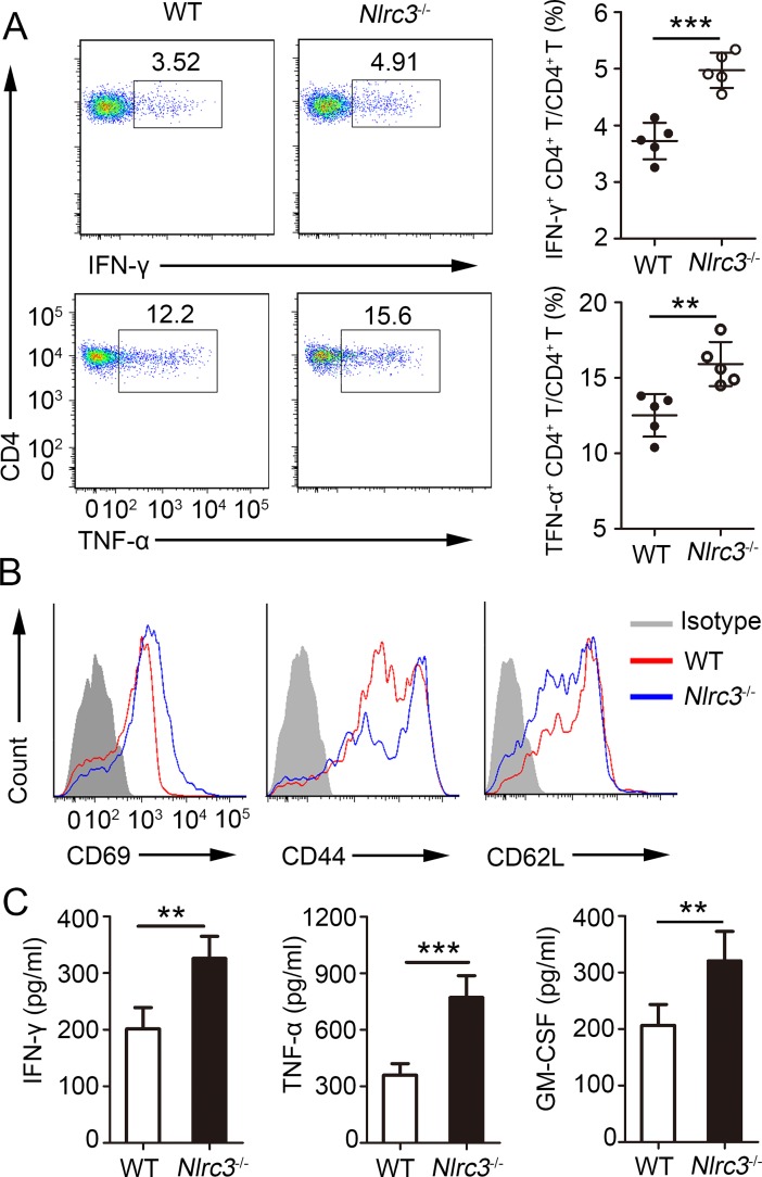
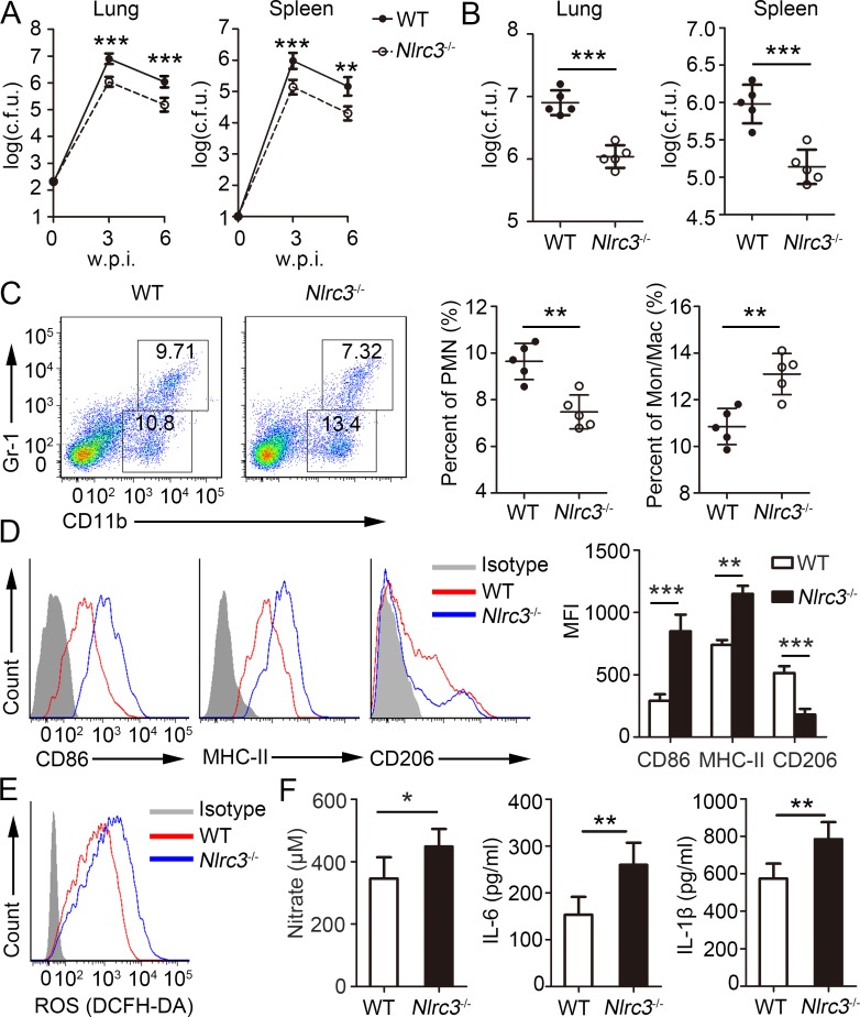
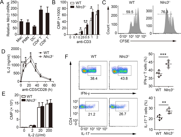
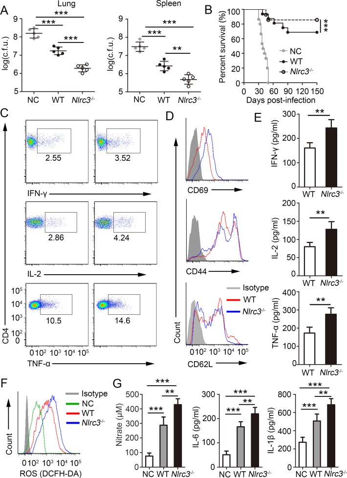
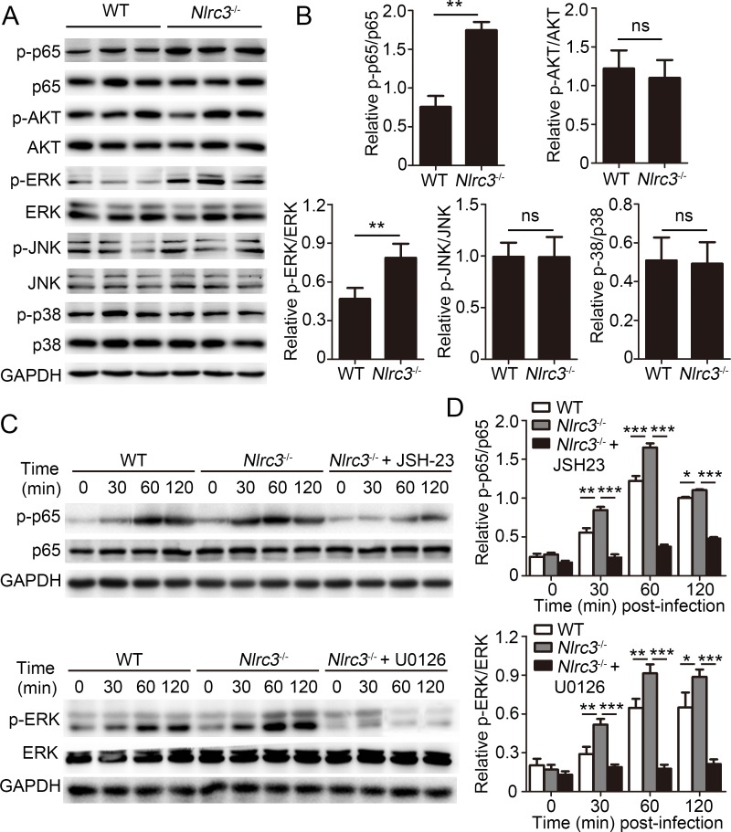
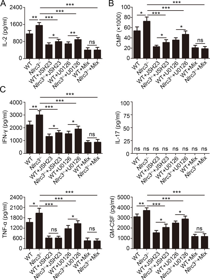
NLRC3, a member of the NLR family, has been reported as a negative regulator of inflammatory signaling pathways in innate immune cells. However, the direct role of NLRC3 in modulation of CD4+ T-cell responses in infectious diseases has not been studied. In the present study, we showed that NLRC3 plays an intrinsic role by suppressing the CD4+ T cell phenotype in lung and spleen, including differentiation, activation, and proliferation. NLRC3 deficiency in CD4+ T cells enhanced the protective immune response against Mycobacterium tuberculosis infection. Finally, we demonstrated that NLRC3 deficiency promoted the activation, proliferation, and cytokine production of CD4+ T cells via negatively regulating the NF-κB and MEK-ERK signaling pathways. This study reveals a critical role of NLRC3 as a direct regulator of the adaptive immune response and its protective effects on immunity during M. tuberculosis infection. Our findings also suggested that NLRC3 serves as a potential target for therapeutic intervention against tuberculosis.


Conflict of interest statement

The authors have declared that no competing interests exist.

Figures

Fig 1
Fig 1. NLRC3 deficiency promotes activation of CD4+ T in M. tuberculosis infection.
WT and Nlrc3-/- mice were infected with M. tuberculosis and mice were harvested at 3 weeks post-infection (w.p.i.). (A) Lung cells were restimulated with M. tuberculosis lysate directly ex vivo and the intracellular production of IFN-γ and TNF-α by CD4+ T cells was determined. Pooled data are presented in the right panel. (B) Expression of activation markers by lung CD4+ T cells. (C) Concentration of IFN-γ, TNF-α and GM-CSF in lungs (homogenized in 2 ml PBS and 0.05% Tween 80) as detected by ELISA. Data shown in (A, C) are the mean ±SD. **P < 0.01 and ***P < 0.001. Data are representative of three independent experiments with similar results.
Fig 2
Fig 2. Nlrc3-/- mice are protected from M. tuberculosis infection.
WT and Nlrc3-/- mice infected with approximately 200 colony-forming units (c.f.u.) of M. tuberculosis were monitored. (A) Bacterial burdens were determined after infection at 3 and 6 w.p.i.. (B) Bacterial burdens were determined after infection at 3 w.p.i.. (C) Frequencies of lung-infiltrating cells that are neutrophils (CD11b+ Gr-1+) or monocyte-macrophages (CD11b+ Gr-1-) at 3 w.p.i.. (D) Expressions of CD86, MHC-II and CD206 were detected on monocyte-macrophages (CD11b+ Gr-1-) via flow cytometry at 3 w.p.i.. (E) ROS production by monocyte-macrophages (CD11b+ Gr-1-) were detected assessed as mean fluorescence intensity (MFI) of intracellular CFDA. (F) Concentrations of nitrate were measured by nitrate reductase assay and concentrations of IL-6 and IL-1β in lungs (homogenized in 2 ml PBS and 0.05% Tween 80) were detected by ELISA at 3 w.p.i.. Data shown in are the mean ±SD. *P < 0.05, **P < 0.01 and ***P < 0.001. Data are representative of three independent experiments with similar results.
Fig 3
Fig 3. T cell activation in vitro requires NLRC3.
(A) Relative expression of Nlrc3 in purified macrophages (CD11b+ Gr-1-), dendritic cells (CD11c+ MHCIIhi), polymorphonuclear leukocytes (PMNs) (CD11b+ Gr-1+), CD4+ T cells (CD3+ CD4+) and CD8+ T cells (CD3+ CD8+). (B) Purified naïve T cells isolated from WT and Nlrc3-/- mice were stimulated with plate bound anti-CD3 (increasing concentrations) and anti-CD28 (1 μg/ml) for 48 hr and the incorporation of thymidine was measured during the final 8 hr. (C) Purified WT and Nlrc3-/- naïve CD4+ T cells were labeled with CFSE and stimulated with anti-CD3 (1 μg/ml) and anti-CD28 (1 μg/ml) for 3 d. (D) Concentrations of IL-2 in supernatants of purified WT and Nlrc3-/- naïve CD4+ T cells stimulated for 0–80 h with anti-CD3 (1 μg/ml) and anti-CD28 (1 μg/ml) were detected by ELISA. (E) Thymidine incorporation in purified WT and Nlrc3-/- naive CD4+ T cells first primed with anti-CD3 and CD28 and then cultured with various concentrations of IL-2. (F) Purified WT and Nlrc3-/- naïve CD4+ T cells were polarized in Th1 or Th17 culture conditions for 4 days. Data shown in (B, C, E, F) are the mean ±SD. *P < 0.05 and **P < 0.01. Data are representative of three independent experiments with similar results.
Fig 4
Fig 4. NLRC3 deficiency promotes differentiation of CD4+ T and IL-2 production in vivo.
Naïve CD4+ T cells from CD45.1+ WT and CD45.2+ Nlrc3-/- mice were mixed at a 1:1 ratio and competitively transferred via tail vein injection into Rag2-/- recipient mice. Then recipient mice were infected with M. tuberculosis and were harvested at 3w.p.i.. (A) Lung cells were restimulated with M. tuberculosis lysate directly ex vivo and the intracellular production of IFN-γ by CD4+ T cells was determined. Representative FACs plots depicting gating of CD4+ T cells are shown. Gating strategies to evaluate cytokine production by WT and Nlrc3-/- CD4+ T cells are provided. Numbers in the quadrants indicate the percent cells in each. Pooled data are presented in the right panel. (B) Expression of CD69 by lung CD4+ T cells. (C) Expression of IL-2 by lung CD4+ T cells. Pooled data are presented in the right panel. Data shown in (A, C) are the mean ±SD. *P < 0.05 and **P < 0.01. Data are representative of three independent experiments with similar results.
Fig 5
Fig 5. NLRC3 deficiency promotes the antibacterial ability in vivo via regulating CD4+ T cells.
Purified WT or Nlrc3-/- naïve CD4+ T cells were adoptively transferred into Rag2-/- mice. Then recipient mice were infected with M. tuberculosis and parts of mice were harvested at 3w.p.i.. (A) Bacterial burdens were determined in lungs and spleen at 3w.p.i.. (B) Survival of mice every other day from 0–150 day post-infection (d.p.i.). (C) Lung cells were restimulated with M. tuberculosis lysate directly ex vivo and the intracellular production of IFN-γ, IL-2, and TNF-α by CD4+ T cells was determined. (D) Expression of activation markers by lung CD4+ T cells. (E) Concentration of IFN-γ, IL-2 and TNF-α in lungs (homogenized in 2 ml PBS and 0.05% Tween 80) as detected by ELISA. (F) ROS production by monocyte-macrophages (CD11b+ Gr-1-) were detected assessed as mean fluorescence intensity (MFI) of intracellular CFDA. (G) Concentrations of nitrate were measured by nitrate reductase assay and concentrations of IL-6 and IL-1β in lungs (homogenized in 2 ml PBS and 0.05% Tween 80) were detected by ELISA. Data shown in (A, E, G) are the mean ±SD. *P < 0.05, **P < 0.01 and ***P < 0.001. Data are representative of three independent experiments with similar results.
Fig 6
Fig 6. NLRC3 negatively regulates NF-κB and ERK Signaling in CD4+ T cells.
(A-B) Purified WT or Nlrc3-/- naïve CD4+ T cells were adoptively transferred into Rag2-/- mice. Then recipient mice were infected with M. tuberculosis and were harvested at 3w.p.i.. Lungs were collected. (A) Immunoblot analysis of lung lysates. Each lane represents an individual mouse. (B) Densitometry quantification of band intensity for A. (C-D) Purified WT and Nlrc3-/- CD4+ T cells were stimulated with anti-CD3 (1 μg/ml) plus anti-CD28 (1 μg/ml) in the presence or absence of the NF-κB inhibitor JSH-23 (20 μM) or MEK1/2-inhibitor U0126 (40 μM). (C) Lysates were probed for total and phosphorylated p65 (p-p65), p65, p-ERK, ERK and GAPDH. (D) Densitometry quantification of band intensity for C. Data shown in (B and D) are the mean ±SD. *P < 0.05, **P < 0.01 and ***P < 0.001. Data are representative of three independent experiments with similar results.
Fig 7
Fig 7. NLRC3 suppresses activation of CD4+ T cells via negatively regulating NF-κB and ERK Signaling.
Purified WT and Nlrc3-/- CD4+ T cells were stimulated for 48 hr with anti-CD3 (1 μg/ml) plus anti-CD28 (1 μg/ml) in the presence or absence of the NF-κB inhibitor JSH-23 (20 μM), MEK1/2-inhibitor U0126 (40 μM) or the mix of the two. (A) Concentrations of IL-2 in supernatants were detected by ELISA. (B) The incorporation of thymidine was measured during the final 8 hr. (C) Concentrations of IFN-γ, IL-17, TNF-α and GM-CSF in supernatants were detected by ELISA. Data shown are the mean ±SD. *P < 0.05, **P < 0.01 and ***P < 0.001. Data are representative of three independent experiments with similar results.

Similar articles

Cited by

References

    1. Yeretssian G (2012) Effector functions of NLRs in the intestine: innate sensing, cell death, and disease. Immunol Res 54: 25–36. 10.1007/s12026-012-8317-3 - DOI - PubMed
    1. Kufer TA, Sansonetti PJ (2011) NLR functions beyond pathogen recognition. Nat Immunol 12: 121–128. 10.1038/ni.1985 - DOI - PubMed
    1. Van Gorp H, Kuchmiy A, Van Hauwermeiren F, Lamkanfi M (2014) NOD-like receptors interfacing the immune and reproductive systems. FEBS J 281: 4568–4582. 10.1111/febs.13014 - DOI - PubMed
    1. Kanneganti TD, Lamkanfi M, Nunez G (2007) Intracellular NOD-like receptors in host defense and disease. Immunity 27: 549–559. 10.1016/j.immuni.2007.10.002 - DOI - PubMed
    1. Rathinam VA, Vanaja SK, Fitzgerald KA (2012) Regulation of inflammasome signaling. Nat Immunol 13: 333–342. 10.1038/ni.2237 - DOI - PMC - PubMed

Publication types

Substances