

Effect of cavity preparation design and ceramic type on the stress distribution, strain and fracture resistance of CAD/CAM onlays in molars
- PMID: 30133672
- PMCID: PMC6110459
- DOI: 10.1590/1678-7757-2018-0004
Effect of cavity preparation design and ceramic type on the stress distribution, strain and fracture resistance of CAD/CAM onlays in molars
Abstract
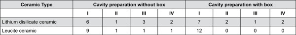
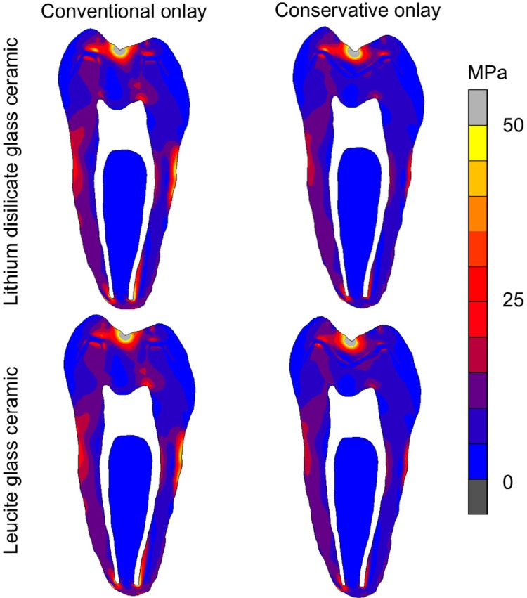
Objective This study aimed to evaluate the effect of the cavity preparation and ceramic type on the stress distribution, tooth strain, fracture resistance and fracture mode of human molar teeth restored with onlays. Material and Methods Forty-eight molars were divided into four groups (n=12) with assorted combinations of two study factors: BL- conventional onlay preparation with boxes made from leucite ceramic (IPS-Empress CAD, Ivoclar Vivadent); NBL- conservative onlay preparation without boxes made from leucite ceramic; BD- conventional onlay preparation with boxes made from lithium disilicate glass ceramic (IPS e.max CAD, Ivoclar Vivadent); NBL- conservative onlay preparation with boxes made from lithium disilicate glass ceramic cuspal deformation (µS) was measured at 100 N and at maximum fracture load using strain gauge. Fracture resistance (N) was measured using a compression test, and the fracture mode was recorded. Finite element analysis was used to evaluate the stress distribution by modified von Mises stress criteria. The tooth strain and fracture resistance data were analyzed using the Tukey test and two-way ANOVA, and the fracture mode was analyzed by the chi-square test (α=0.05). Results The leucite ceramic resulted in higher tooth deformation at 100 N and lower tooth deformation at the maximum fracture load than the lithium disilicate ceramic (P<0.001). The lithium disilicate ceramic exhibited higher fracture resistance than the leucite ceramic (P<0.001). The conservative onlay resulted in higher fracture strength for lithium disilicate ceramic. Finite element analysis results showed the conventional cavity preparation resulted in higher stress concentration in the ceramic restoration and remaining tooth than the conservative onlay preparation. The conservative onlays exhibited increased fracture resistance, reduced stress concentration and more favorable fracture modes. Conclusion Molars restored with lithium disilicate CAD-CAM ceramic onlays exhibited higher fracture resistance than molars restored with leucite CAD-CAM ceramic onlays.
Figures
References
-
- Raigrodski AJ, Chiche GJ. The safety and efficacy of anterior ceramic fixed partial dentures: a review of the literature. J Prosthet Dent. 2001;86(5):520–525. - PubMed
-
- D'Arcangelo C, De Angelis F, Vadini M, D'Amario M. Clinical evaluation on porcelain laminate veneers bonded with light-cured composite: results up to 7 years. Clin Oral Inv. 2012;16(4):1071–1079. - PubMed
-
- Molin MK, Karlsson SL. A randomized 5-year clinical evaluation of 3 ceramic inlay systems. International. J Prosthodont. 2000;13(3):194–200. - PubMed
-
- Morin D, DeLong R, Douglas WH. Cusp reinforcement by the acid-etch technique. J Dent Res. 1984;639(8):1075–1078. - PubMed
Publication types
MeSH terms
Substances
LinkOut - more resources
Full Text Sources
Other Literature Sources
Miscellaneous
