. 2019 Jan;60(1):84-95.
doi: 10.1165/rcmb.2018-0053OC.

IL-13-regulated Macrophage Polarization during Granuloma Formation in an In Vitro Human Sarcoidosis Model

Affiliations

IL-13-regulated Macrophage Polarization during Granuloma Formation in an In Vitro Human Sarcoidosis Model

Landon W Locke et al. Am J Respir Cell Mol Biol. 2019 Jan.

Abstract

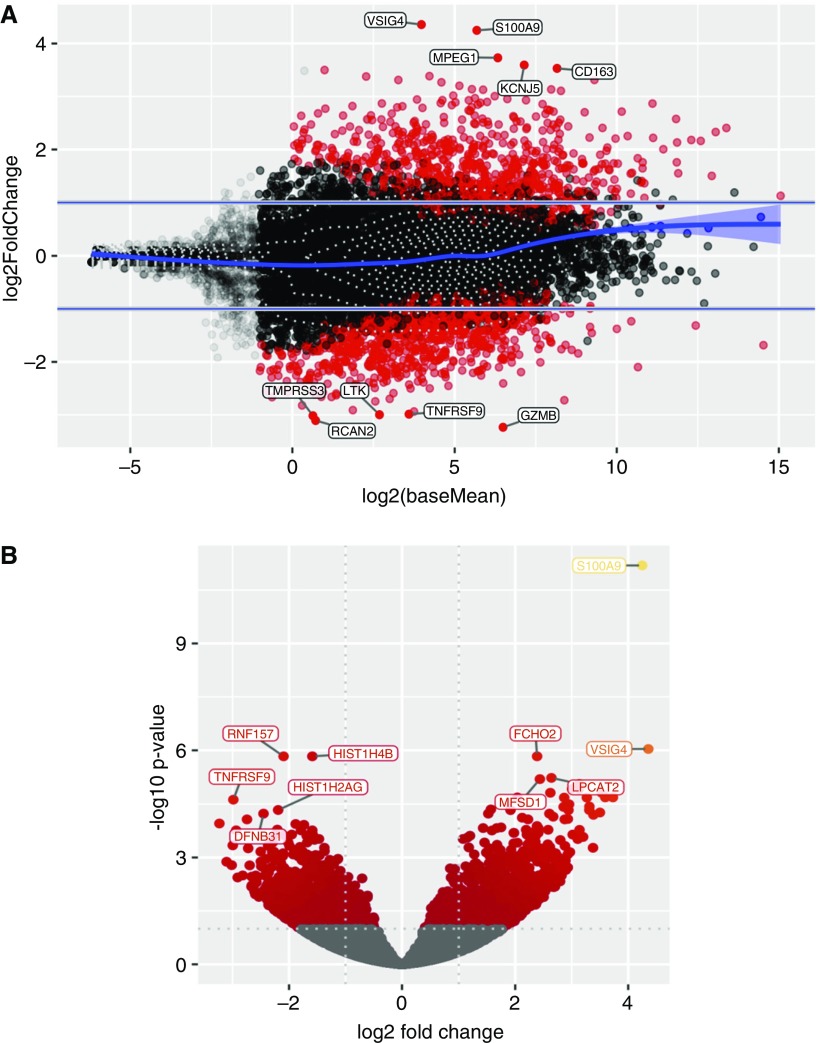
The mechanisms underlying abnormal granuloma formation in patients with sarcoidosis are complex and remain poorly understood. A novel in vitro human granuloma model was used to determine the molecular mechanisms of granuloma genesis in patients with sarcoidosis in response to putative disease-causing mycobacterial antigens. Peripheral blood mononuclear cells (PBMCs) from patients with active sarcoidosis and from normal, disease-free control subjects were incubated for 7 days with purified protein derivative-coated polystyrene beads. Molecular responses, as reflected by differential expression of genes, extracellular cytokine patterns, and cell surface receptor expression, were analyzed. Unbiased systems biology approaches were used to identify signaling pathways engaged during granuloma formation. Model findings were compared with human lung and mediastinal lymph node gene expression profiles. Compared with identically treated PBMCs of control subjects (n = 5), purified protein derivative-treated sarcoidosis PBMCs (n = 6) were distinguished by the formation of cellular aggregates resembling granulomas. Ingenuity Pathway Analysis of differential expression gene patterns identified molecular pathways that are primarily regulated by IL-13, which promotes alternatively activated (M2) macrophage polarization. M2 polarization was further demonstrated by immunohistochemistry performed on the in vitro sarcoidosis granuloma-like structures. IL-13-regulated gene pathways were confirmed in human sarcoidosis lung and mediastinal lymph node tissues. The in vitro human sarcoidosis granuloma model provides novel insights into early granuloma formation, particularly IL-13 regulation of molecular networks that regulate M2 macrophage polarization. M2 macrophages are predisposed to aggregation and multinucleated giant cell formation, which are characteristic features of sarcoidosis granulomas. Clinical trial registered with www.clinicaltrials.gov (NCT01857401).

Keywords: AmpliSeq; macrophage; peripheral blood mononuclear cells; purified protein derivative.


Figures

Figure 1.
Figure 1.
In vitro granuloma-like cell aggregation of sarcoidosis peripheral blood mononuclear cells in response to treatment with purified protein derivative (PPD)-coated beads. Representative light photomicrographs (10×) obtained 7 days after sarcoidosis or control peripheral blood mononuclear cells were incubated with either PPD-coated or uncoated beads. (A) PPD-coated beads induced prominent cellular aggregation in the sarcoidosis group (black arrows) that was comparatively minimal in the (C) similarly treated controls. Treatment with uncoated beads elicited no cellular aggregation in either (B) the sarcoidosis or (D) control group.
Figure 2.
Figure 2.
Differential gene expression after PPD-coated bead treatment of peripheral blood mononuclear cells (PBMCs) derived from patients with sarcoidosis compared with similarly treated PBMCs from normal healthy control subjects. (A) The output type “MvA” plots of normalized gene expression data wherein each point represents a gene, with the y-axis representing a log(base 2) fold change in expression after 7-day treatment with PPD-coated beads of PBMCs derived from patients with sarcoidosis compared with identically treated PBMCs from normal, healthy control subjects. The x-axis is the log average of the gene expression level. All genes with an adjusted P value of 0.05 (representing a 5% false discovery rate) and at least a twofold change (highlighted by the two horizontal gray lines) in the magnitude of gene expression between patients with sarcoidosis and control subjects are shaded red. The blue line represents smoothed local mean expression (fitted using a generalized additive model) with the surrounding 95% confidence interval shaded light blue. The dashed gray contour lines represent a two-dimensional density estimation. Genes that are most differentially expressed in response to PPD-coated beads in patients with sarcoidosis compared with control subjects are highlighted in the top part of the MvA plot, whereas those genes most differentially expressed in control subjects compared with patients with sarcoidosis are at the bottom. (The top five most differentially expressed genes in each case are labeled.) (B) Volcano plots of the same data wherein the y-axis corresponds to transcripts with high statistical significance (−log10 of P value) and the x-axis corresponds to fold change of gene expression (log base 2) were also generated from the same data set. Labeled transcripts on the upper left side of the plot have strong statistical significance with relatively low expression after treatment of PBMCs with PPD-coated beads of sarcoidosis versus controls. Transcripts on the upper right are more highly expressed with strong statistical significance between sarcoidosis and controls after PPD treatment. The dotted line corresponds to a P value cutoff of 0.05, and genes failing to meet this cutoff are shaded gray. Data were generated from all 11 experiments (six sarcoidosis and five controls).
Figure 3.
Figure 3.
Pathway analysis predicts IL-13 as an important upstream regulator of the observed gene expression changes. Predictions are based on two statistical measures reflecting data set genes known to be regulated by each specific cytokine or transcription factor (overlap P value) and activation (z-score) based on the known direction by which the upstream regulator is known to influence the gene target (inhibition vs. activation). Details of the statistical methods are provided by Ingenuity Systems (8). Applying this statistical approach to the differential gene expression pattern of PBMCs of patients with sarcoidosis compared with healthy control subjects determined 7 days after treatment with PPD-coated beads yielded the above IL-13–regulated network. Data were generated from all 11 experiments (six sarcoidosis and five controls).
Figure 4.
Figure 4.
Pathway analysis of gene expression identifies IL-13 to be a mediator of the sarcoidosis granulomatous response. (A) The gene network most highly overrepresented (see the Methods section regarding AmpliSeq analyses) in PBMCs from patients with sarcoidosis (n = 6) relative to control subjects (n = 5) 7 days after PPD treatment was predicted to be strongly regulated by IL-13 and IL-1β. Genes and gene products are represented by their common abbreviated names according to established direct (solid lines) or indirect (dashed lines) interactions and the specific nature of the interaction. These include binding (straight line), activation (arrow), inhibition (truncated line), or either activation or inhibition (truncated line with arrow). Genes that are significantly overexpressed in the sarcoidosis group relative to controls are highlighted in shades of red, wherein darker shades conform to higher differential expression values. The shape of each gene “node” denotes the function of the gene product, as indicated in the key. (B) Pathway analysis of gene expression results (from Affymetrix GeneChip arrays) obtained from human sarcoidosis lymph nodes (n = 5) compared with disease-free control lymph nodes (n = 8) using a cutoff of greater than or equal to twofold change (P < 0.05) identified IL-13 and IL-2 as putative mediators of the granulomatous response, including induction of STAT1 (signal transducer and activator of transcription 1)-mediated inflammatory signaling. (C) Pathway analysis of gene expression results (from Affymetrix GeneChip arrays) obtained from human sarcoidosis lung tissues (n = 6) compared with disease-free control lung tissues (n = 6) using a cutoff of greater than or equal to twofold change (P < 0.05) identified IL-13 as a putative mediator of the granulomatous response, including regulation of metalloproteases MMP-12 and ADAMDEC1, the expression of which correlated significantly with pulmonary sarcoidosis disease severity (3).
Figure 5.
Figure 5.
Immunofluorescence imaging of granuloma-like structures demonstrates persistent macrophage CD163 expression on sarcoidosis PBMCs after PPD stimulation. Representative photomicrographs of immunostained PBMCs in granuloma-like structures 7 days after bead treatment using confocal microscopy. DAPI and CD163 staining are shown in blue and red, respectively (see data supplement for methods and antibodies used). (A) PBS-washed (uncoated) and (B) PPD-coated, bead-stimulated PBMCs obtained from a PPD-naive, healthy control subject showing high CD163 expression on macrophages that was markedly decreased after PPD stimulation. In contrast, images of granuloma-associated macrophages obtained from a patient with sarcoidosis demonstrated high CD163 expression after (C) uncoated bead stimulation that persisted after (D) stimulation with PPD-coated beads. (E) Staining of PPD-stimulated PBMCs from a donor with sarcoidosis using a concentration-matched isotype control (to CD163) antibody. In each panel, a matching differential interference contrast image is shown next to each composite image. All images are representative of the results from four independent experiments. Scale bars: 10 μm.
Figure 6.
Figure 6.
Leflunomide (Lef) pretreatment suppresses PPD-induced granuloma formation in sarcoidosis PBMCs. (A) Representative light photomicrograph of granuloma-like structures 7 days after sarcoidosis PBMCs were treated with PPD-coated beads. (B) The same image shown in A wherein granulomas were identified and color-coded on the basis of size, employing the EVOS FL Cell Imaging System (Thermo Fisher Scientific) and MIPAR analysis software as described in the data supplement. (C) Representative light photomicrograph of granuloma formation in PBMCs from the same patient with sarcoidosis 7 days after PPD stimulation after pretreatment with leflunomide for 30 minutes. (D) The same image shown in C after analysis with the EVOS FL Cell Imaging System and MIPAR analysis software. (E) Significant reduction in the area comprised by granulomas as a percentage of the field of view in sarcoidosis PBMCs 7 days after treatment with PPD-coated beads after initial pretreatment with leflunomide. All images are representative of the results from five independent experiments. Scale bars: 200 μm.

Comment in

References

    1. Grunewald J, Spagnolo P, Wahlström J, Eklund A. Immunogenetics of disease-causing inflammation in sarcoidosis. Clin Rev Allergy Immunol. 2015;49:19–35. - PubMed
    1. Prokop S, Heppner FL, Goebel HH, Stenzel W. M2 polarized macrophages and giant cells contribute to myofibrosis in neuromuscular sarcoidosis. Am J Pathol. 2011;178:1279–1286. - PMC - PubMed
    1. Crouser ED, Culver DA, Knox KS, Julian MW, Shao G, Abraham S, et al. Gene expression profiling identifies MMP-12 and ADAMDEC1 as potential pathogenic mediators of pulmonary sarcoidosis. Am J Respir Crit Care Med. 2009;179:929–938. - PMC - PubMed
    1. Helming L, Gordon S. Macrophage fusion induced by IL-4 alternative activation is a multistage process involving multiple target molecules. Eur J Immunol. 2007;37:33–42. - PubMed
    1. Crouser ED, White P, Caceres EG, Julian MW, Papp AC, Locke LW, et al. A novel in vitro human granuloma model of sarcoidosis and latent tuberculosis infection. Am J Respir Cell Mol Biol. 2017;57:487–498. - PMC - PubMed

Publication types

MeSH terms

Associated data