. 2018 Aug 21;24(8):1939-1948.e4.
doi: 10.1016/j.celrep.2018.07.072.

Living Neurons with Tau Filaments Aberrantly Expose Phosphatidylserine and Are Phagocytosed by Microglia

Affiliations

Living Neurons with Tau Filaments Aberrantly Expose Phosphatidylserine and Are Phagocytosed by Microglia

Jack Brelstaff et al. Cell Rep. .

Abstract

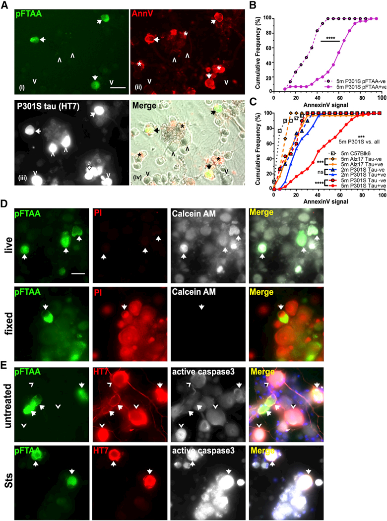
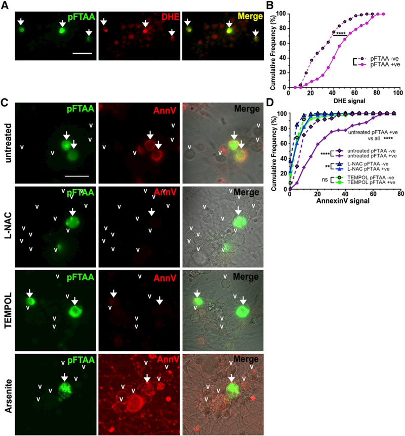
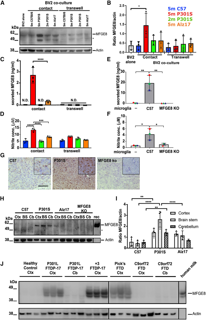
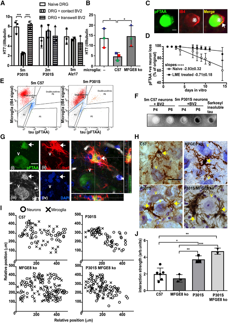
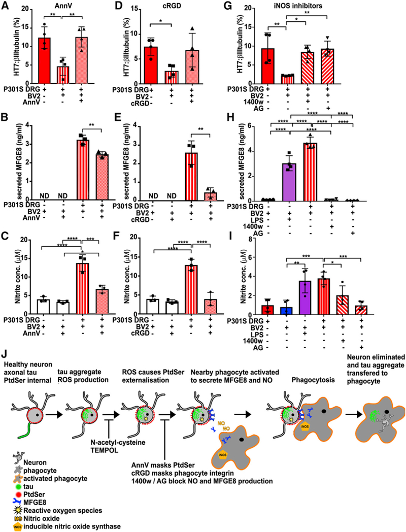
Tau protein forms insoluble filamentous inclusions that are closely associated with nerve cell death in many neurodegenerative diseases. How neurons die in these tauopathies is unclear. We report that living neurons with tau inclusions from P301S-tau mice expose abnormally high amounts of phosphatidylserine because of the production of reactive oxygen species (ROS). Consequently, co-cultured phagocytes (BV2 cells or primary microglia) identify and phagocytose the living neurons, thereby engulfing insoluble tau inclusions. To facilitate engulfment, neurons induce contacting microglia to secrete the opsonin milk-fat-globule EGF-factor-8 (MFGE8) and nitric oxide (NO), whereas neurons with tau inclusions are rescued when MFGE8 or NO production is prevented. MFGE8 expression is elevated in transgenic P301S-tau mouse brains with tau inclusions and in tau inclusion-rich brain regions of several human tauopathies, indicating shared mechanisms of disease. Preventing phagocytosis of living neurons will preserve them for treatments that inhibit tau aggregation and toxicity.

Keywords: MFGE8; cell death; microglia; neurodegeneration; neuroinflammation; nitric oxide; phagocytosis; phosphatidylserine; tau; tauopathies.


Conflict of interest statement

DECLARATION OF INTERESTS

The authors declare no competing interests.

Figures

Figure 1.
Figure 1.. Living Neurons with pFTAA+Tau Filaments Aberrantly Expose PS by a Reversible ROS-Dependent Mechanism
(A) Living DRG neurons from 5-month-old P301S mice with filamentous tau aggregates stained with (i) pFTAA (green) and (ii) AnnV-647 (red) (arrows) (asterisk denotes dead cell debris); (iii) same neurons fixed and stained for human tau (HT7 antibody). pFTAA-/HT7+ neurons do not stain with AnnV-647 (arrowheads). (iv) Nontransgenic (HT7-) neurons are AnnV-; images (i) and (ii) merged with phase contrast. Scale bar, 25 μm. (B) Highersignal intensities ofAnnV-647 binding to pFTAA+ versus pFTAA- neurons in live cultures from 5-month-old P301S mice (****p < 0.0001). Cumulativefrequencyplot, 30 neurons perculture, n = 3 independent experiments. Kolmogorov- Smirnov test. (C) Cumulative frequency plot comparing AnnV- 647 binding intensity values of HT7+ and HT7- neurons in the same cultures from 5-month-old P301S mice (***p < 0.001), 2-month-old P301S- mice (not significant [n.s.]), 5-month-old Alz17 mice (***p < 0.001), and HT7- 5-month-old wild- type C57 mice. AnnV staining of HT7+ neurons from 5-month-old P301S mice is significantly more intense than all others (***p < 0.001). Thirty neurons, n = 3 independent experiments. Kol- mogorov-Smirnov test with Bonferroni correction. (D) pFTAA+ neurons from 5-month-old P301S mice are living cells. Top row: calcein-AM (white) and PI (red) staining showing exclusion of PI and retention of fluorescent calcein. Bottom row: after fixation, PI stains all cells, and there is no calcein retention. Data representative of n = 3 independent experiments. Scale bar, 30 mm. (E) pFTAA+ neurons do not display PS, because of activation of caspase-3. Top row: pFTAA+ (green, arrows) and HT7+ (red)/pFTAA- neurons (arrowheads) showing basal intensities of active caspase-3 staining (white). Bottom row: staur- osporine (Sts) induces high levels of active cas- pase-3 in all the cells.
Figure 2.
Figure 2.. Tau Aggregates Cause PS Externalization through a ROS-Dependent Mechanism
(A and B) Representative image (A) of DRG neurons from 5-month-old P301S mice showing higher intensity of nuclear oxidized DHE staining (red) in pFTAA+ (arrows) versus pFTAA- neurons. Scale bar, 25 μm. (B) Cumulative frequency plot quantifying DHE fluorescence intensities for pFTAA+ and pFTAA- DRG neurons; ≥30 neurons per one culture from n = 3 independent experiments. Kolmogorov-Smirnov test. ****p < 0.0001. (C) Loss of AnnV binding to pFTAA+ DRG neurons (arrows) from 5-month-old P301S mice treated with 5 mM NAC or 5 mM TEMPOL for 2 days before washing and staining live with AnnV-647. Arrowheads indicate pFTAA- neurons. Pro-oxidant arsenite (0.5 mM, 15 min) causes intense AnnV- 647 staining in all neurons without any cell death (PI-). Scale bar, 30 μm. (D) Cumulative frequency plots quantifying AnnV- 647 fluorescence intensity values for pFTAA+ and pFTAA- DRG neurons treated as in (C); >30 neurons from n = 3 independent experiments. Kolmogorov-Smirnov test, Bonferroni corrected. Comparison of pFTAA+ values with all others, ****p < 0.0001. Values comparing pFTAA+ versus pFTAA-: untreated, ****p < 0.0001; NAC, **p < 0.01; TEMPOL, n.s.
Figure 3.
Figure 3.. MFGE8 and NO Are Produced and Secreted by Co-cultured BV2 Microglia Only When Co-cultured in Contact with pFTAA+ Neurons
(A and B) Representative blot (A) ofMFGE8 in BV2 cells lysates cultured alone or co-cultured in direct contact, or via a transwell, with DRG neurons from 5-month-old P301S-tau, 5-month-old C57, 5-month-old Alz17 mice, or 2-month-old P301S mice. Asterisk: either another isoform ofMFGE8 or a breakdown product. (B) Densitometry of MFGE8 expression normalized to β-actin; significantly more MFGE8 is produced only in contact co-cultures containing pFTAA+ neurons. Mean ± SD, n = 3 independent experiments (*p < 0.05), twoway ANOVA, Dunnett’s correction. (C and D) Elevated MFGE8 (C) or NO (D) in medium conditioned by BV2 cells co-cultured in contact with DRG neurons from 5-month-old P301S mice compared with 5-month-old Alz17, 5-month- old C57, or 2-month-old P301S mice (MFGE8: ****p < 0.0001 versus all; NO: ****p < 0.0001 versus 5-month-old C57 or 2-month-old P301S mice; ***p < 0.001 versus 5-month-old Alz17 mice). N.D., none detected. Mean ± SD, n = 3 independent experiments, two-way ANOVA, Bonferroni corrected. (E and F) Elevated MFGE8 (E) or NO (F) in medium conditioned by primary microglia from C57 mice co-cultured in contact with DRG neurons from 5-month-old P301S mice; microglia from MFGE8 KO mice are negative controls. Mean ± SD, n = 3 independent microglial preparations, one-way ANOVA, **p < 0.01 (MFGE8), *p < 0.05 (NO). (G) Increased MFGE8 immunostaining intensity in frontal motor cortex of 5-month-old P301S mice compared with 5-month-old C57 mice; MFGE8 KO mouse is negative control; 25 μm section at inter- aural 5.12 mm, bregma 1.32 mm; brown, DAB; blue, cresyl violet. Scale bar, 130 mm. Inset, 65 μm. (H and I) Elevated MFGE8 (H) in 5-month-old P301S-tau brains. Lysatesfrom cortex(Ctx), brain stem (BS), and cerebellum (Cb) of 5-month-old C57, 5-month-old P301S-tau, and 5-month-old Alz17 mice probed with anti-MFGE8; MFGE8 KO brain lysate is negative control. rec, recombinant mouse MFGE8. (I) Densitometry of MFGE8 expression normalized to β-actin. Significantly higher MFGE8 expression in P301S versus C57BU6 Ctx (**p < 0.01), BS (****p < 0.0001), Cb (**p < 0.01), and P301SversusALz17BS (****p < 0.0001). Mean ± SD, n = 3 independent preparations, two-way ANOVA, Bonferroni corrected. (J) Elevated MFGE8 expression in brain extracts from cortex (Ctx) of FTDP-17T patients with two different MAPT mutations (P301L, +3) or sporadic Pick’s disease but not in extracts from patients with C9orf72 hexanucleotide expansions and TDP-43 aggregates. No expression is found in the cerebellum, where tau pathology is absent in all cases. Human milk MFGE8 is a positive control.
Figure 4.
Figure 4.. pFTAA/HT7+ DRG Neurons Are Preferentially Removed by Phagocytosis
(A) pFTAA+ DRG neuronsfrom 5-month-old P301S mice are depleted after contact co-culture with BV2 cells for 4 days (***p < 0.001), but not if BV2 cells are in transwells. No loss of HT7+ neurons when BV2 cells are contact co-cultured with neurons from 2-month-old P301S mice or 5-month-old Alz17 mice. Mean ± SD, n = 3 independent ex- periments,two-wayANOVA, Bonferroni corrected. (B) pFTAA+ DRG neuronsfrom 5-month-old P301S mice are removed from cultures in contact with primary microglia from C57 mice for 4 days but not those from MFGE8 KO mice. Mean ± SD, n = 3 independent experiments, *p < 0.05, one-way ANOVA, Bonferroni corrected. (C) Endogenous phagocytes mediate the slow removal of cultured pFTAA+ neurons from 5-month-old P301S mice. Representative image of a pFTAA+ neuron (green) surrounded by an IB4+ phagocyte (red). Nuclei (DAPI) in white. Arrow marks pFTAA+ inclusions inside the phagocyte. (D) Removal of pFTAA+ neurons over 14 days is halted by elimination of phagocytes using LME (50 mM). x axis, days in vitro beginning 1 day after phagocyte elimination. Each point shows mean ± SD, n = 3 independent experiments. Lines denote linear regressions depicting rates of neuronal loss; slopes, ****p < 0.0001. (E) Insoluble pFTAA+ tau is transferred from pFTAA+ DRG neurons to BV2 cells in co-contact cultures. FACS sorting of IB4–594 pre-labeled BV2 cells collected 4 days after co-culture with pFTAA+ neurons from 5-month-old P301S-tau or C57 mice. Note double-positive population (4.9%, P6) of BV2 cells present only in cultures from P301S mice. (F) Insoluble tau in BV2 cells. Single IB4-labeled (P4) and double-labeled (P6) populations of BV2 cells shown in (E), extracted in 5% SDSand filtered through cellulose nitrate membrane, which traps insoluble tau. Sarkosyl-insoluble tau fibrils from 5-month-old P301S-tau brains are the positive control. (G) Microglia in the facial nucleus of 5-month-old P301S-mice engulf pFTAA+ neurons with tau. Total fluorescent staining of (i) pFTAA+ neurons, (ii) Iba+ microglia (red), (iii) human (HT7) P301S-tau (white), (iv) nuclei (DAPI, blue). Enlarged image: confocal z section shows the entire pFTAA+ neuron encased inside a microglial cell. The neuronal nucleus (arrow) is also engulfed. Scale bar, 5 μm. (H) Iba-1 staining of microglia in the FN from 5-month-old C57, 5-month-old P301S-tau, 5-month-old P301S-tau/MFGE8 KO with tau pathology, or MFGE8 KO mice. FN neurons are closely apposed by globoid-like microglia. Brown, Iba-1/DAB; blue, cresyl violet. Scale bar, 10 mm. (I) Relative positions of motor neurons and microglia in representative examples of the FN. Microglia and neurons are more closesly distributed in mice with P301S-tau pathology. (J) Quantification of proximity; significant increases in interaction strength between P301S-tau+ neurons and microglia in P301S-tau and P301S-tau/MFGE8 KO mice compared with C57 or MFGE8 KO controls. *p < 0.05, **p < 0.01, and ***p < 0.001, one-way ANOVA, Bonferroni corrected.
Figure 5.
Figure 5.. Blocking Phagocytosis Prevents the Loss of HT7+ Neurons by BV2 Cells
(A-C) Excess AnnV (100 nM) prevents loss of HT7+ neurons (A) (**p < 0.01) and significantly reduces the amount ofsecreted MFGE8 (B) (**p < 0.01) and NO (C) (***p < 0.001). Mean ± SD, n = 4 independent experiments, one-way ANOVA, Bonferroni corrected. (D-F) cRGD (5 μM) partially prevents the loss of HT7+ neurons (p = 0.09) (D) and significantly reduces the amount of secreted MFGE8 (E) (**p < 0.01)and NO (F) (****p < 0.0001). Mean ± SD, n = 3 independent experiments, one-way ANOVA, Bonferroni corrected. (G-I) The iNOS inhibitors 1400W and amino- guanidine (AG) inhibit the loss of HT7+ neurons (G) (1400W, *p < 0.05; AG, **p < 0.01) and the production of MFGE8 (H) (1400W or AG, ****p < 0.0001) and NO (I) (1400W, *p < 0.05; AG, ***p < 0.001). LPS added as a positive control. Mean ± SD, n = 4 independent experiments, oneway ANOVA, Bonferroni corrected. (J) Scheme summarizing the mechanism of death of tau aggregate-bearing neurons by phagocytosis: tau aggregate formation causing ROS- dependent PS exposure, activation of nearby phagocytes to produce NO and MFGE8, leading to engulfment of neurons and tau transfer into phagocytes. Blunt arrows indicate points of mechanism-related drug interception.

References

    1. Aguzzi A, Barres BA, and Bennett ML (2013). Microglia: scapegoat, saboteur, or something else? Science 330, 156–161. - PMC - PubMed
    1. Allen B, Ingram E, Takao M, Smith MJ, Jakes R, Virdee K, Yoshida H, Holzer M, Craxton M, Emson PC, et al. (2002). Abundant tau filaments and nonapoptotic neurodegeneration in transgenic mice expressing human P301S tau protein. J. Neurosci. 22, 9340–9351. - PMC - PubMed
    1. Asano K, Miwa M, Miwa K, Hanayama R, Nagase H, Nagata S, and Tanaka M (2004). Masking of phosphatidylserine inhibits apoptotic cell engulf- ment and induces autoantibody production in mice. J. Exp. Med. 200, 459–467. - PMC - PubMed
    1. Bellucci A, Westwood AJ, Ingram E, Casamenti F, Goedert M, and Spill- antini MG (2004). Induction of inflammatory mediators and microglial activation in mice transgenic for mutant human P301S tau protein. Am. J. Pathol. 165,>1643–1652. - PMC - PubMed
    1. Bellucci A, Bugiani O, Ghetti B, and Spillantini MG (2011). Presence of reactive microglia and neuroinflammatory mediators in a case of frontotemporal dementia with P301S mutation. Neurodegener. Dis. 8,> 221–229. - PMC - PubMed

Publication types

LinkOut - more resources