. 2018 Aug 21;24(8):1958-1966.e5.
doi: 10.1016/j.celrep.2018.07.080.

Site-Specific Glycosylation of Virion-Derived HIV-1 Env Is Mimicked by a Soluble Trimeric Immunogen

Affiliations

Site-Specific Glycosylation of Virion-Derived HIV-1 Env Is Mimicked by a Soluble Trimeric Immunogen

Weston B Struwe et al. Cell Rep. .

Abstract

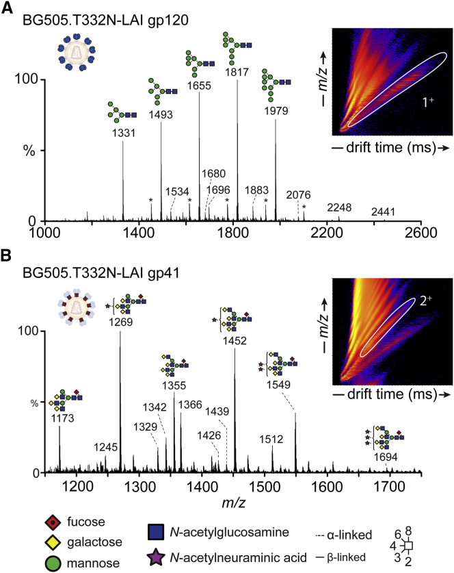
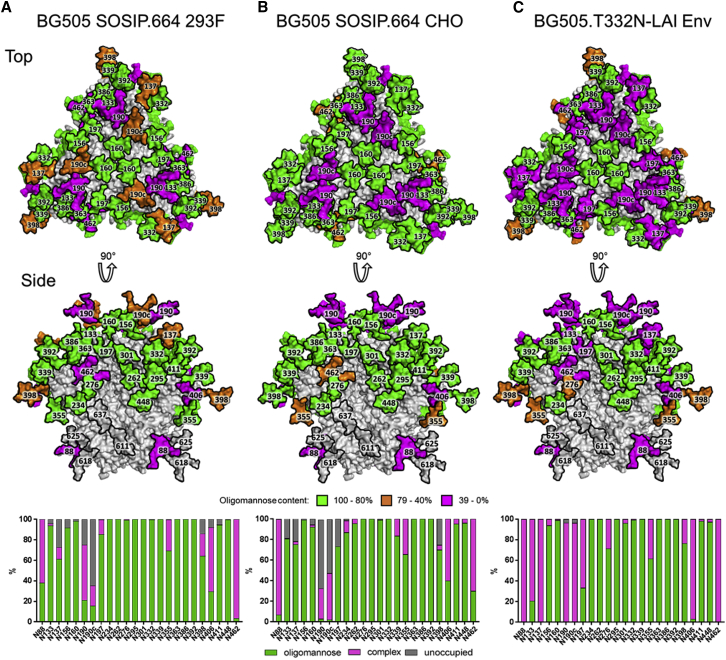
Many broadly neutralizing antibodies (bnAbs) against HIV-1 recognize and/or penetrate the glycan shield on native, virion-associated envelope glycoprotein (Env) spikes. The same bnAbs also bind to recombinant, soluble trimeric immunogens based on the SOSIP design. While SOSIP trimers are close structural and antigenic mimics of virion Env, the extent to which their glycan structures resemble ones on infectious viruses is undefined. Here, we compare the overall glycosylation of gp120 and gp41 subunits from BG505 (clade A) virions produced in a lymphoid cell line with those from recombinant BG505 SOSIP trimers, including CHO-derived clinical grade material. We also performed detailed site-specific analyses of gp120. Glycans relevant to key bnAb epitopes are generally similar on the recombinant SOSIP and virion-derived Env proteins, although the latter do contain hotspots of elevated glycan processing. Knowledge of native versus recombinant Env glycosylation will guide vaccine design and manufacturing programs.

Keywords: HIV; envelope glycoprotein; glycosylation; mass spectrometry.


Figures

None
Graphical abstract
Figure 1
Figure 1
UPLC Analysis of N-Glycans from gp120 and gp41 (A) UPLC chromatogram of N-glycans from BG505.T332N-LAI gp120 gel bands. Peaks sensitive to endoglycosidase H (endoH) digestion (green) represent oligomannose-type glycans. Peaks resistant to endoH (magenta) represent complex-type glycans. Pie charts depict the quantification of oligomannose and complex-type glycans in each sample. Peaks corresponding to oligomannose-type glycans are annotated with the number of mannose residues (Man5–9). (B) UPLC chromatogram of N-glycans released from gp120 from BG505 SOSIP.664 trimers produced in 293F cells. (C) Reproduction of the UPLC analysis of N-glycans released from BG505 SOSIP.664 trimers produced in CHO cells (Dey et al., 2018). (D–F) (D), (E), and (F) show data for gp41 and correspond to gp120 (A), (B), and (C), respectively. See also Figure S1 and Table S2.
Figure 2
Figure 2
Ion-Mobility-Mass Spectrometry Analysis of BG505.T332N-LAI Virion-Derived gp120 and gp41 N-Glycans (A) Mobility-extracted singly charged negative ions from BG505.T332N-LAI gp120. The corresponding singly charged ions ([M+H2PO4]) are encircled in white in the ion mobility drift plot (inset). The series of carbohydrate ions marked with an asterisk is formed by the additional adduction of sodium phosphate to the main oligomannose ions. (B) Mobility-extracted doubly charged negative ions from BG505.T332N-LAI gp41. The corresponding doubly charged ions (2+) are encircled in white in the ion mobility drift plot. See also Figures S2–S4.
Figure 3
Figure 3
Determination of the Predominant Glycoforms Presented at Each Site on Soluble BG505 SOSIP.664 gp120 Expressed in Different Cells and Virus (A–C) A model of the predominant glycoform from 293F-derived BG505 SOSIP.664 using the cryo-EM model of BG505 SOSIP.664 (Behrens et al., 2016) is colored according to the predominant glycan species determined by glycosidase-treated glycopeptides for 293F cells (A), CHO cells (B), and BG505.T332N-LAI virus (C). The bar graphs represent the relative amounts of digested glycopeptides possessing the footprints for oligomannose glycans (green), complex-type glycans (magenta), and unoccupied PNGs (gray) at each gp120 site, listed from N to C terminus. See also Figure S1.
Figure 4
Figure 4
Compositional Site-Specific Analysis of Intact Glycopeptides from BG505 SOSIP.664 Trimers Produced in 293F or CHO Cells and from BG505.T332N-LAI Viruses (A) Model of the N-glycan compositions of BG505.T332N-LAI Env highlighting three regions that are important for bnAb binding (reproduced from Figure 3C). (B–E) Site-specific compositions of N-linked glycans located on (B) the trimer apex, (C) the mannose patch, (D) proximal to the CD4 binding site (CD4bs), and (E) at remaining sites on the trimer. (F) Neutralization by bnAbs targeting the apex (red), mannose patch (blue), and CD4bs (orange). Data corresponding to CHO-cell derived BG505 SOSIP.664 trimers are reproduced from Dey et al. (2018) and included here to assist in comparisons with other datasets. To simplify data presentation, complex glycans are grouped according to the presence or absence of fucose and the number of terminal antenna (e.g., FA2). Data are represented as means ± SEMs. See also Figure S1.

References

    1. Andrabi R., Su C.Y., Liang C.H., Shivatare S.S., Briney B., Voss J.E., Nawazi S.K., Wu C.Y., Wong C.H., Burton D.R. Glycans function as anchors for antibodies and help drive HIV broadly neutralizing antibody development. Immunity. 2017;47:524–537.e3. - PMC - PubMed
    1. Behrens A.J., Crispin M. Structural principles controlling HIV envelope glycosylation. Curr. Opin. Struct. Biol. 2017;44:125–133. - PMC - PubMed
    1. Behrens A.J., Vasiljevic S., Pritchard L.K., Harvey D.J., Andev R.S., Krumm S.A., Struwe W.B., Cupo A., Kumar A., Zitzmann N. Composition and antigenic effects of individual glycan sites of a trimeric HIV-1 envelope glycoprotein. Cell Rep. 2016;14:2695–2706. - PMC - PubMed
    1. Behrens A.J., Harvey D.J., Milne E., Cupo A., Kumar A., Zitzmann N., Struwe W.B., Moore J.P., Crispin M. Molecular architecture of the cleavage-dependent mannose patch on a soluble HIV-1 envelope glycoprotein trimer. J. Virol. 2017;91 e01894-16. - PMC - PubMed
    1. Binley J.M., Sanders R.W., Clas B., Schuelke N., Master A., Guo Y., Kajumo F., Anselma D.J., Maddon P.J., Olson W.C., Moore J.P. A recombinant human immunodeficiency virus type 1 envelope glycoprotein complex stabilized by an intermolecular disulfide bond between the gp120 and gp41 subunits is an antigenic mimic of the trimeric virion-associated structure. J. Virol. 2000;74:627–643. - PMC - PubMed

Publication types

LinkOut - more resources