. 2018 Aug 21;10(9):1130.
doi: 10.3390/nu10091130.

A Natural Dietary Supplement with a Combination of Nutrients Prevents Neurodegeneration Induced by a High Fat Diet in Mice

Affiliations

A Natural Dietary Supplement with a Combination of Nutrients Prevents Neurodegeneration Induced by a High Fat Diet in Mice

Domenico Nuzzo et al. Nutrients. .

Abstract

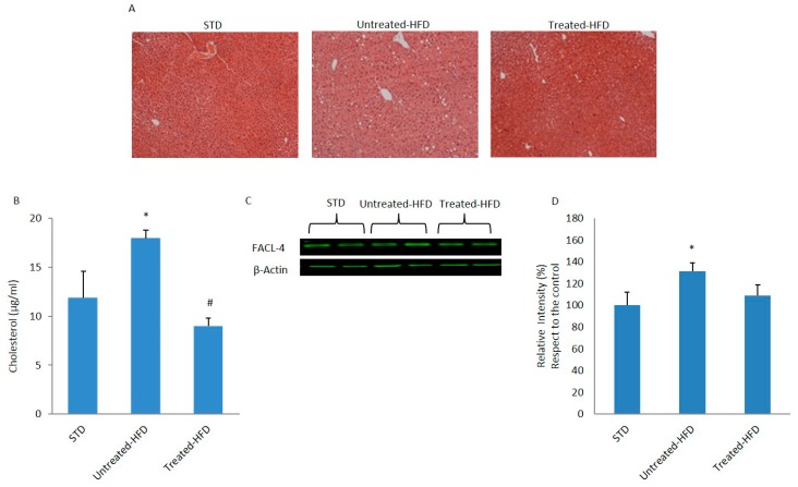
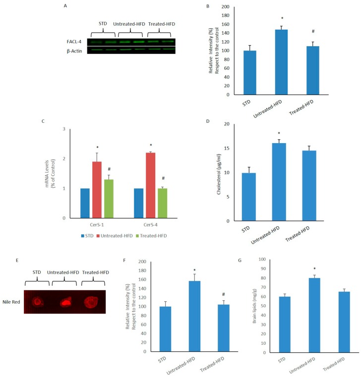
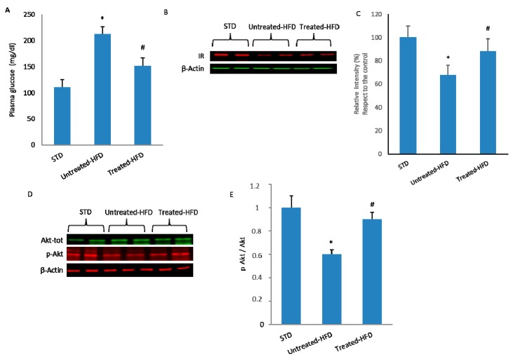
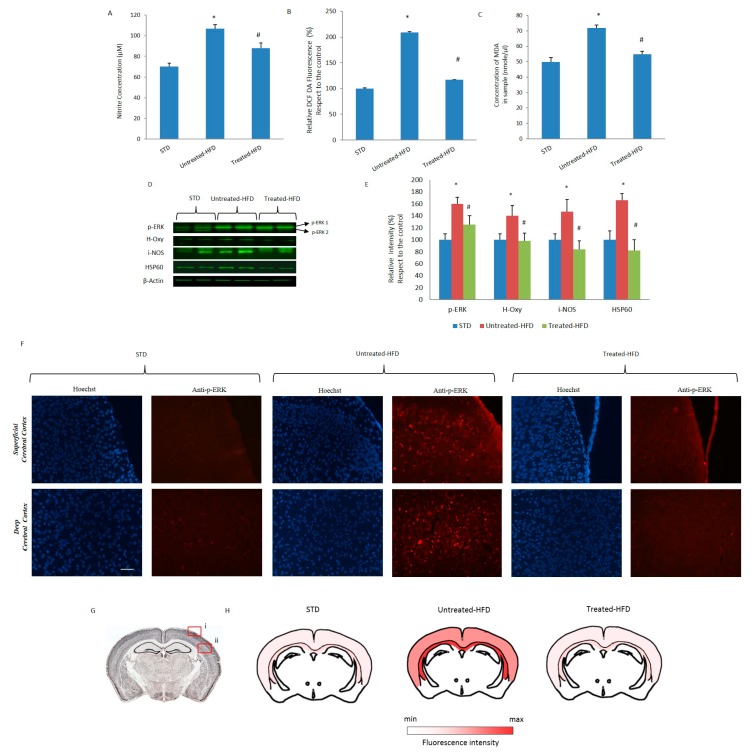
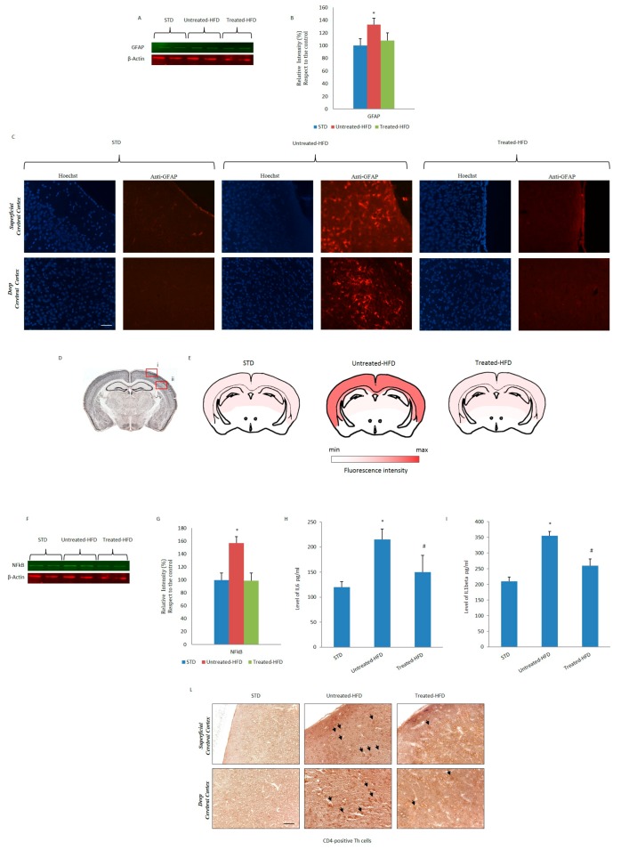
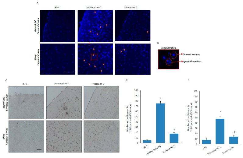
Obesity and metabolic disorders can be risk factors for the onset and development of neurodegenerative diseases. The aim of the present study was to investigate the protective effects of a natural dietary supplement (NDS), containing Curcuma longa, silymarin, guggul, chlorogenic acid and inulin, on dysmetabolism and neurodegeneration in the brains of high fat diet (HFD)-fed mice. Decrease in the expression of FACL-4, CerS-1, CerS-4, cholesterol concentration and increase in the insulin receptor expression and insulin signaling activation, were found in brains of NDS-treated HFD brains in comparison with HFD untreated-mice, suggesting that NDS is able to prevent brain lipid accumulation and central insulin resistance. In the brains of NDS-treated HFD mice, the levels of RNS, ROS and lipid peroxidation, the expression of p-ERK, H-Oxy, i-NOS, HSP60, NF-kB, GFAP, IL-1β, IL-6 and CD4 positive cell infiltration were lower than in untreated HFD mice, suggesting antioxidant and anti-inflammatory effects of NDS. The decreased expression of p-ERK and GFAP in NDS-treated HFD mice was confirmed by immunofluorescence. Lastly, a lower number of apoptotic nuclei was found in cortical sections of NDS-treated HFD mice. The present data indicate that NDS exerts neuroprotective effects in HFD mice by reducing brain fat accumulation, oxidative stress and inflammation and improving brain insulin resistance.

Keywords: HFD mice; insulin resistance; natural antioxidants; neurodegeneration; obesity.


Conflict of interest statement

The authors declare no conflict of interest.

Figures

Figure 1
Figure 1
Effects of NDS on the liver. (A) Liver sections of control (STD), HFD (untreated-HFD) and NDS-treated (treated-HFD) mice; (B) Intrahepatic cholesterol concentration; (C) Western blot of proteins extracted from livers of STD, untreated-HFD and NDS treated-HFD mice and incubated with anti-fatty acid-CoA ligase-4 (FACL-4) and anti-β-Actin (loading control); (D) Quantification of immunoreactivity was performed using densitometric analysis. Data are the mean values ± SEM (n = 8/group). * p ≤ 0.05 vs. STD; # p ≤ 0.05 vs. untreated-HFD.
Figure 2
Figure 2
Lipid accumulation in the brain. (A) Western blot of proteins extracted from brains of STD, untreated-HFD and NDS treated-HFD mice and incubated with anti-FACL-4 and anti-β-Actin (loading control); (B) Quantification of immunoreactivity was performed using densitometric analysis; (C) CerS-1 and CerS-4 transcript levels determined by quantitative Real-Time PCR; (D) Cholesterol concentration in the brain tissue; (E) Nile Red staining of brain homogenates; (F) Quantification of E fluorescence intensity; (G) Total brain lipid levels. Data are the mean values ± SD (n = 8/group). * p ≤ 0.05 vs. STD. # p ≤ 0.05 vs. untreated-HFD.
Figure 3
Figure 3
NDS decreases the HFD-induced insulin signaling alterations in the brain. (A) Fasting plasma glucose concentration in STD, untreated-HFD and treated-HFD mice; (B) Western blot of proteins extracted from brains of STD, untreated-HFD and NDS treated-HFD mice and incubated with anti-Insulin Receptor (IR) and anti-β-Actin (loading control); (C) Quantification of immunoreactivity was performed using densitometric analysis; (D) Western blot of proteins extracted from brains of STD, untreated-HFD and NDS treated-HFD mice and incubated with anti-Akt, anti-phospho-Akt (p-Akt) and anti-β-Actin (loading control); (E) Quantification of Akt/p-Akt ratio. Data are the mean values ± SD (n = 8/group). * p ≤ 0.05 vs. STD. # p ≤ 0.05 vs. untreated-HFD.
Figure 4
Figure 4
Nitric and oxidative stress and lipid peroxidation in the brains of HFD-mice were prevented by NDS treatment. (A) Nitrite concentration in STD, untreated-HFD and treated-HFD brains; (B) Levels of ROS in STD, untreated-HFD and treated-HFD brains (C) Lipid peroxidation levels in STD, untreated-HFD and treated-HFD brains; (D) Western blot of proteins extracted from brains of STD, untreated-HFD and treated-HFD mice and incubated with anti-p-ERK, anti-H-Oxy, anti-i-NOS, anti-HSP60 and anti-β-Actin (loading control); (E) Quantification of immunoreactivity was performed using densitometric analysis; (F) Immunofluorescence of superficial and deep cerebral cortex sections of STD, untreated-HFD and treated-HFD mice incubated with anti-phospho-ERK; (G) Schematic representation of superficial (i) and deep (ii) cerebral cortex positive areas; (H) Brain map indicating the levels of positive p-ERK staining. Representative images from 3 animals per group are shown. Bar 20 μm. Data are the mean values ± SD (n = 8/group). * p ≤ 0.05 vs. STD. # p ≤ 0.05 vs. untreated-HFD.
Figure 5
Figure 5
NDS prevents HFD-induced inflammation and immunological response in the brain. (A) Western blot of proteins extracted from brains of STD, untreated-HFD and treated-HFD mice and incubated with anti-GFAP and anti-β-Actin (loading control); (B) Quantification of immunoreactivity was performed using densitometric analysis; (C) Immunofluorescence of superficial and deep cerebral cortex sections incubated with anti-GFAP; (D) Schematic representation of superficial (i) and deep (ii) cerebral cortex positive areas; (E) Brain map indicating the levels of positive staining of GFAP; (F) Western blot of proteins extracted from brains of STD, untreated-HFD and treated-HFD mice and incubated with anti-NFkB and anti-β-Actin (loading control); (G) Quantification of immunoreactivity was performed using densitometric analysis; (H,I) Levels of IL-6 and IL-1β in brains of STD, untreated-HFD and treated-HFD mice, quantified by ELISA assay. (L) Brain sections from STD, untreated-HFD and treated-HFD mice incubated with anti-CD4. Representative images from 3 animals per group are shown. Bar 20 μm. Data are the mean values ± SD (n = 8/group). * p ≤ 0.05 vs. STD. # p ≤ 0.05 vs. untreated-HFD.
Figure 6
Figure 6
NDS inhibits neurodegeneration induced by HFD. (A) Brain sections from STD, untreated-HFD and treated-HFD mice incubated with Hoechst 33342. Fragmented apoptotic nuclei are indicated by the arrows; (B) The outlined area is enlarged in the squares; (C) TUNEL assay on superficial and deep cortical regions from STD, untreated-HFD and treated-HFD mice; (D) Number of apoptotic nuclei in the superficial cerebral cortex positive area; (E) Number of apoptotic nuclei in the deep cerebral cortex positive area. Representative images from 3 animals per group are shown. Bar 20 μm. Data are the mean values ± SD (n = 8/group). * p ≤ 0.05 vs. STD. # p ≤ 0.05 vs. untreated-HFD.

References

    1. Castelli V., Grassi D., Bocale R., D’Angelo M., Antonosante A., Cimini A., Ferri C., Desideri G. Diet and brain health: Which role for polyphenols? Curr. Pharm. Des. 2017;24:227–238. doi: 10.2174/1381612824666171213100449. - DOI - PubMed
    1. S Roriz-Filho J., Sá-Roriz T.M., Rosset I., Camozzato A.L., Santos A.C., Chaves M.L., Moriguti J.C., Roriz-Cruz M. (Pre)Diabetes, Brain, Aging and Cognition. Biochim. Biophys. Acta. 2009;1792:432–443. doi: 10.1016/j.bbadis.2008.12.003. - DOI - PubMed
    1. Jayarama A., Pike C.J. Alzheimer’s Disease and Type 2 Diabetes: Multiple Mechanisms Contribute to Interactions. Curr. Diab. Rep. 2014;14:476. doi: 10.1007/s11892-014-0476-2. - DOI - PMC - PubMed
    1. Lee E.B. Obesity, leptin, and Alzheimer’s disease. Ann. N. Y. Acad. Sci. 2011;1243:15–29. doi: 10.1111/j.1749-6632.2011.06274.x. - DOI - PMC - PubMed
    1. Ghareeb D.A., Hafez H.S., Hussien H.M., Kabapy N.F. Non-alcoholic fatty liver induces insulin resistance and metabolic disorders with development of brain damage and dysfunction. Metab. Brain Dis. 2011;26:253–267. doi: 10.1007/s11011-011-9261-y. - DOI - PubMed

MeSH terms