. 2018 Mar 19;24(1):1.
doi: 10.1186/s10020-018-0009-6.

Analysis of menstrual effluent: diagnostic potential for endometriosis

Affiliations

Analysis of menstrual effluent: diagnostic potential for endometriosis

Laura A Warren et al. Mol Med. .

Abstract

Background: Endometriosis is a chronic and underdiagnosed disease which affects 5-10% of women of childbearing age and is characterized by growth of endometrial tissue outside of the uterus, most often in the peritoneal cavity. Delay in diagnosis is a major problem for management of this disorder, and treatment is often not initiated until the disease has progressed for many years. Although the exact etiology of endometriosis remains unknown, retrograde menstruation is recognized as a common underlying factor leading to the deposit of menstrual effluent (ME) into the peritoneal cavity. Differences in the cellular biology and genetics of the cells within ME are therefore likely to explain why endometriosis develops in only a subset of women.

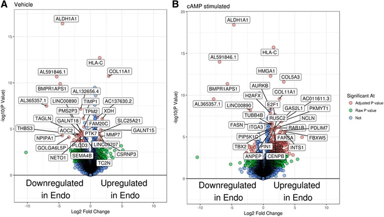
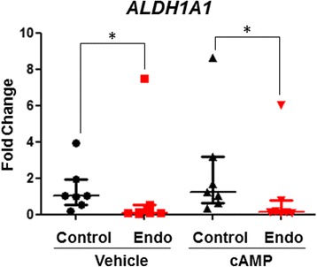
Methods: Patients with and without endometriosis were consented to provide ME. ME was analyzed by flow cytometry for CD45- and CD45+ cell populations or used to isolate stromal fibroblast cells. ME-derived stromal fibroblast cells were assessed using decidualization assays following the addition of cAMP and IGFBP-1 concentrations in the culture supernatants were measured by ELISA. In addition, RNA was collected and analyzed by RNA-Seq and qPCR for markers of decidualization and to identify differentially expressed genes in ME-derived stromal fibroblast cells obtained from controls and subjects with endometriosis (±cAMP).

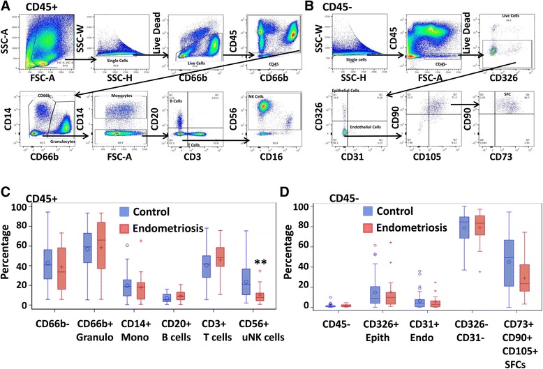
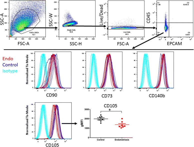
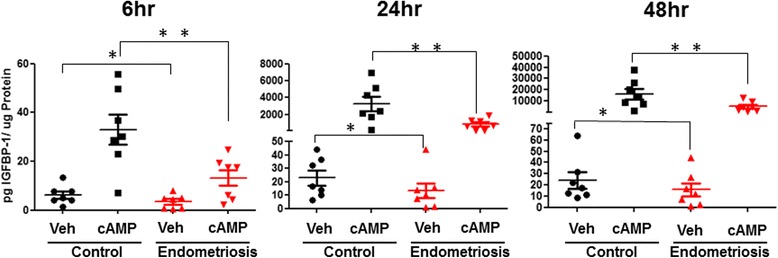
Results: Flow cytometry analysis of cell subsets within the CD45+ fraction of ME revealed a significant decrease in the number of uterine NK cells in endometriosis patients compared with controls (p < 0.01). No other significant differences within either the CD45+ or CD45- cell populations were observed. Most strikingly, ME-derived stromal fibroblast cells cultured from endometriosis subjects showed impaired decidualization potential compared with controls. Highly significant differences in decidualization response were detected by measuring IGFBP-1 production at multiple time points after cAMP stimulation (p = 0.0025 at 6 h; p = 0.0045 at 24 h; p = 0.0125 at 48 h). RNA-Seq and qPCR analyses were used to identify genes differentially expressed by ME-derived stromal fibroblast cells obtained from endometriosis and control subjects.

Conclusions: Menstrual effluent can be useful for investigating the pathobiology of endometriosis and for developing a non-invasive diagnostic for endometriosis which may lead to earlier and more effective treatments for this common disorder.

Keywords: Biomarkers; Decidualization; Menstruation; Stromal fibroblast cells.


Conflict of interest statement

Ethics approval and consent to participate

Prior to recruiting, enrolling and consenting of participants with and without endometriosis, the human subjects research protocols (IRB#13-376A and IRB#13-627A, respectively) were reviewed and approved by the Institutional Review Board (IRB) of Northwell Health.;

Consent for publication

All subjects consent to the publication of research results without personal identifiers (i.e. in a de-identified manner).

Competing interests

The authors declare that they have no financial, professional, or other conflicts related to this manuscript.

Publisher’s Note

Springer Nature remains neutral with regard to jurisdictional claims in published maps and institutional affiliations.

Figures

Fig. 1
Fig. 1
Flow Cytometric analysis of ME from endometriosis and control subjects CD45+ (n = 14 control, n = 8 endometriosis) (a) and CD45- (n = 14 control, n = 6 endometriosis) (b) flow cytometry staining scheme for ME. Box plots showing the cellular composition CD45+ subsets (CD66b + granulocytes [Granulo], CD14+ monocytes [Mono], CD20+ B cells, CD3+ T cells, and CD56+ uterine natural killer (uNK) cells) (c) and CD45- subsets (CD45-, CD326+ epithelial cells [Epith], CD31+ endothelial cells [Endo], CD326-/CD31- cells, and CD73+/CD90+/CD105+ [SFCs] (d) found in menstrual effluent from women with and without endometriosis. The CD66b + and CD66b- populations were normalized to the CD45+ population cell counts and the CD14+ Mono, CD20+ B Cells, CD3+ T cells, and CD56+ uNK cell populations were normalized to CD66b-population cell counts. The Epith, Endo, CD326-/CD31- cells, and SFC populations were normalized to the CD45- population cell counts. Data are shown as box plots depicting the median and interquartile ranges for each cell subset; significance for uNK cells **p = 0.01 (c)
Fig. 2
Fig. 2
Cultured ME-derived SFCs express CD73, CD90, CD105, and CD140b but not CD45 or CD326. Flow cytometry gating of cultured ME-derived SFCs to show CD45-/CD326- population (upper panel). The CD45- population of ME derived SFCs from endometriosis (n = 7) and control (n = 7) subjects at passage 1 were further analyzed by flow cytometry for CD90, CD73, CD140b, and CD105 expression (lower panel). CD105 expression is lower on endometriosis-SFCs when compared to control subject-SFCs (lower right panel). Data are shown as geometric mean fluorescence intensity (gMFI), for each subject’s SFC culture and the horizontal lines are the group means, *p = 0.03
Fig. 3
Fig. 3
ME-derived SFCs obtained from endometriosis subjects exhibit reduced decidualization capacity. Time course of IGFBP-1 secretion by vehicle (Veh) and cAMP treated (0.5 mM) ME-derived SFCs isolated from endometriosis and control subjects (n = 7 control, n = 7 endometriosis). Data are shown as IGFBP-1 values for each subject’s SFC culture and the horizontal lines represent group means. ** posterior probabilities (Pr) < 0.01 *Pr < 0.05. Statistics were performed on log transformed data, as described in the methods, see Additional file 1: Table S3)
Fig. 4
Fig. 4
Numerous genes are differentially expressed when comparing vehicle-treated and cAMP-treated SFCs obtained from subjects with endometriosis vs. controls. Volcano plots of genes differentially regulated between endometriosis and control subjects in the vehicle-treated group (a) and the cAMP-treated group (b) (n = 3 control, n = 3 endometriosis for A and B)
Fig. 5
Fig. 5
SFCs from endometriosis subjects exhibit reduced ALDH1A1 expression. ALDH1A1 expression by vehicle and cAMP-treated SFC obtained from subjects with and without endometriosis (n = 7 subjects per group) was confirmed by qPCR. Data are shown as relative gene expression (fold-change) for each subject’s SFC culture; horizontal lines represent group medians and vertical lines represent interquartile ranges, *p < 0.05, calculated using Mann Whitney Testᅟ

References

    1. Acimovic M, Vidakovic S, Milic N, Jeremic K, Markovic M, Milosevic-Djeric A, et al. Survivin and VEGF as novel biomarkers in diagnosis of endometriosis. J med. Biochemist. 2016;35(1):63–68. - PMC - PubMed
    1. Aghajanova L, Tatsumi K, Horcajadas JA, Zamah AM, Esteban FJ, Herndon CN, et al. Unique transcriptome, pathways, and networks in the human endometrial fibroblast response to progesterone in endometriosis. Biol Reprod. 2011;84(4):801–815. doi: 10.1095/biolreprod.110.086181. - DOI - PMC - PubMed
    1. Arruda MS, Petta CA, Abrão MS, Benetti-Pinto CL. Time elapsed from onset of symptoms to diagnosis of endometriosis in a cohort study of Brazilian women. Hum Reprod. 2003;18(4):756–759. doi: 10.1093/humrep/deg136. - DOI - PubMed
    1. Ballard K, Lowton K, Wright J. What's the delay? A qualitative study of women's experiences of reaching a diagnosis of endometriosis. Fertil Steril. 2006;86(5):1296–1301. doi: 10.1016/j.fertnstert.2006.04.054. - DOI - PubMed
    1. Barragan F, Irwin JC, Balayan S, Erikson DW, Chen JC, Houshdaran S, et al. Human endometrial fibroblasts derived from mesenchymal progenitors inherit progesterone resistance and acquire an inflammatory phenotype in the endometrial niche in endometriosis. Biol Reprod. 2016. 94(5):118, 1–20-, 1–20. - PMC - PubMed

Publication types