. 2018 May 29;24(1):26.
doi: 10.1186/s10020-018-0029-2.

B-1a cells protect mice from sepsis-induced acute lung injury

Affiliations

B-1a cells protect mice from sepsis-induced acute lung injury

Monowar Aziz et al. Mol Med. .

Abstract

Background: Sepsis morbidity and mortality are aggravated by acute lung injury (ALI) or acute respiratory distress syndrome (ARDS). Mouse B-1a cells are a phenotypically and functionally unique sub-population of B cells, providing immediate protection against infection by releasing natural antibodies and immunomodulatory molecules. We hypothesize that B-1a cells ameliorate sepsis-induced ALI.

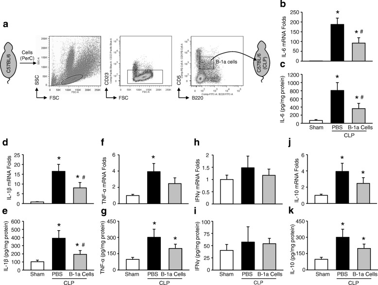
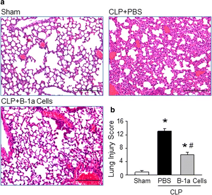
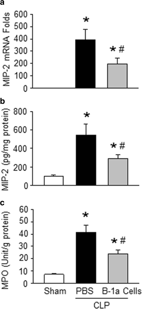
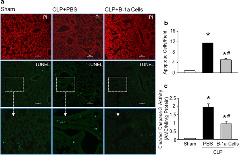
Methods: Sepsis was induced in C57BL/6 mice by cecal ligation and puncture (CLP). PBS or B-1a cells were adoptively transferred into the septic mice intraperitoneally. After 20 h of CLP, lungs were harvested and assessed by PCR and ELISA for pro-inflammatory cytokines (IL-6, IL-1β) and chemokine (MIP-2) expression, by histology for injury, by TUNEL and cleaved caspase-3 for apoptosis, and by myeloperoxidase (MPO) assay for neutrophil infiltration.

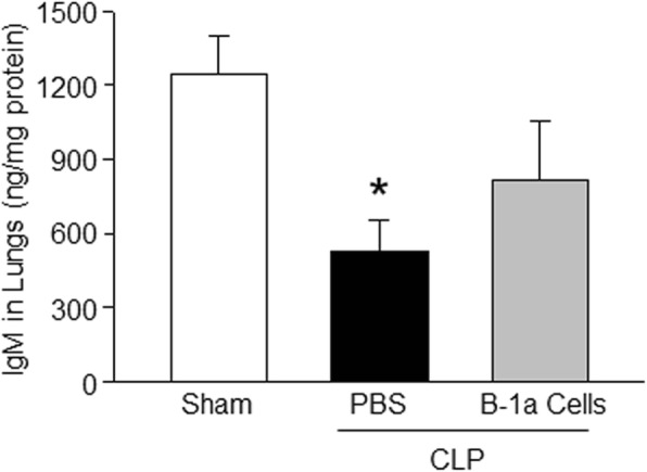
Results: We found that septic mice adoptively transferred with B-1a cells significantly decreased the mRNA and protein levels of IL-6, IL-1β and MIP-2 in the lungs compared to PBS-treated mice. Mice treated with B-1a cells showed dramatic improvement in lung injury compared to PBS-treated mice after sepsis. We found apoptosis in the lungs was significantly inhibited in B-1a cell injected mice compared to PBS-treated mice after sepsis. B-1a cell treatment significantly down-regulated MPO levels in the lungs compared to PBS-treated mice in sepsis. The protective outcomes of B-1a cells in ALI was further confirmed by using B-1a cell deficient CD19-/- mice, which showed significant increase in the lung injury scores following sepsis as compared to WT mice.

Conclusions: Our results demonstrate a novel therapeutic potential of B-1a cells to treat sepsis-induced ALI.

Keywords: Acute lung injury; B-1a cells; IL-10; Inflammation; Neutrophils; Sepsis.


Conflict of interest statement

Ethics approval

All animal protocols were approved by our Institutional Animal Care and Use Committee of the Feinstein Institute for Medical Research.

Consent for publication

All authors have contributed to, read and approved the final version of this manuscript for submission and publication in the journal Molecular Medicine.

Competing interests

The authors declare that they have no competing interests.

Publisher’s Note

Springer Nature remains neutral with regard to jurisdictional claims in published maps and institutional affiliations.

Figures

Fig. 1
Fig. 1
Adoptive transfer of B-1a cells attenuates lung inflammation. a Peritoneal washout cells isolated from healthy mice were stained with anti-mouse Pacific Blue-CD23, FITC-B220 and PE-Cy5 Abs and subjected to sort purification by using a flow cytometry-based cell sorting system. A total of 5 × 105 B-1a cells suspended in 150 μl of PBS were delivered into the peritoneal cavity of CLP mice. After 20 h, lung tissue was harvested and mRNA and protein expression of b, c IL-6, d, e IL-1β, f, g TNF-α, h, i IFNγ and j, k IL-10 were assessed, respectively. Data are expressed as means ± SE (n = 9 mice/group) and compared by one-way ANOVA and SNK method (*p < 0.05 vs. sham mice; #p < 0.05 vs. PBS-treated CLP mice). CLP, cecal ligation and puncture; IL, interleukin
Fig. 2
Fig. 2
Treatment with B-1a cells improves the histopathological score of lung tissue damage in sepsis. a Lung tissue was collected after 20 h from sham-operated, and either PBS- or B-1a cell-treated CLP mice and stained with H&E. Each slide was observed under light microscopy at × 100 original magnification in a blinded fashion. Representative images for each group are shown. Scale bar, 100 μm. b Histological injury scores of the lungs in different groups were quantified as described in Materials and Methods. Data from three independent experiments are expressed as means ± SE (n = 6 mice/group) and compared by one-way ANOVA and SNK method (*p < 0.05 vs. shams; #p < 0.05 vs. PBS-treated CLP mice). CLP, cecal ligation and puncture; H&E, hematoxylin and eosin
Fig. 3
Fig. 3
B-1a cells attenuate MIP-2 and MPO levels in lungs after sepsis. a, b At the time of CLP, mice were treated with either PBS as vehicle or 5 × 105 PerC B-1a cells in 150 μl of PBS by i.p. injection. After 20 h, lung tissue was harvested and mRNA and protein expression of MIP-2 were assessed, respectively. c MPO activity in lungs of sham-operated, and PBS or B-1a cell-treated CLP mice was determined. Data are expressed as means ± SE (n = 9 mice/group from 3 independent experiments) and compared by one-way ANOVA and SNK method (*p < 0.05 vs. shams; #p < 0.05 vs. PBS-treated CLP mice). CLP, cecal ligation and puncture; MIP-2, macrophage-inflammatory protein-2; MPO, myeloperoxidase
Fig. 4
Fig. 4
Treatment with B-1a cells attenuates apoptosis in lungs after sepsis. After 20 h of CLP, lung tissues were collected from PBS or B-1a cell treated mice. a Lung tissue sections were prepared for TUNEL staining shown in green, and for nuclear staining using PI shown in red. Representative images at × 100 original magnification are shown. Scale bar, 100 μm. b TUNEL positive apoptotic cells were counted at 18 random fields in a blinded fashion, and the average numbers of cells per field are shown. c Cleaved Caspase-3 activity in total lung tissues of sham-operated, and PBS or B-1a cell-treated CLP mice was determined. Data are expressed as means ± SE (n = 6 mice/group) and compared by one-way ANOVA and SNK method (*p < 0.05 vs. shams; #p < 0.05 vs. PBS-treated CLP mice). CLP, cecal ligation and puncture; TUNEL, terminal deoxynucleotidyl transferase dUTP nick end labeling; PI, propidium iodide
Fig. 5
Fig. 5
Treatment with B-1a cells increases IgM levels in the lungs following sepsis. A total of 5 × 105 sorted B-1a cells were delivered into the peritoneal cavity of CLP mice. After 20 h, lung tissue was harvested from sham, PBS-, and B-1a cell-treated mice and assessed IgM levels in total extracted proteins by ELISA. Data are expressed as means ± SE (n = 9 mice/group) and compared by one-way ANOVA and SNK method (*p < 0.05 vs. sham mice). CLP, cecal ligation and puncture; ELISA, enzyme-linked immunosorbent assay
Fig. 6
Fig. 6
Deficiency of B-1a cells exaggerates lung injury during sepsis. a After 20 h of CLP induced in WT and CD19−/− mice, lung tissues were harvested and stained with H&E. The slides were observed under light microscopy at × 100 original magnification in a blinded fashion. Representative images for each group are shown. Scale bar, 100 μm. b Histological injury scores of the lungs in WT and CD19−/− mice were quantified as described in Materials and Methods. Data obtained from three independent experiments are expressed as means ± SE (n = 6 mice/group) and compared by one-way ANOVA and SNK method (*p < 0.05 vs. shams; #p < 0.05 vs. WT CLP mice). CLP, cecal ligation and puncture; H&E, hematoxylin and eosin

Similar articles

Cited by

References

    1. Abraham E. Neutrophils and acute lung injury. Crit Care Med. 2003;31:S195–S199. doi: 10.1097/01.CCM.0000057843.47705.E8. - DOI - PubMed
    1. Aziz M, Holodick NE, Rothstein TL, Wang P. The role of B-1 cells in inflammation. Immunol Res. 2015;63:153–166. doi: 10.1007/s12026-015-8708-3. - DOI - PMC - PubMed
    1. Aziz M, Holodick NE, Rothstein TL, Wang P. B-1a cells protect mice from sepsis: critical role of CREB. J Immunol. 2017;199:750–760. doi: 10.4049/jimmunol.1602056. - DOI - PMC - PubMed
    1. Aziz M, Jacob A, Wang P. Revisiting caspases in sepsis. Cell Death Dis. 2014;5:e1526. doi: 10.1038/cddis.2014.488. - DOI - PMC - PubMed
    1. Aziz M, Jacob A, Yang WL, Matsuda A, Wang P. Current trends in inflammatory and immunomodulatory mediators in sepsis. J Leukoc Biol. 2013;93:329–342. doi: 10.1189/jlb.0912437. - DOI - PMC - PubMed

Publication types