

Changes in expression profiles of internal jugular vein wall and plasma protein levels in multiple sclerosis
- PMID: 30134823
- PMCID: PMC6085618
- DOI: 10.1186/s10020-018-0043-4
Changes in expression profiles of internal jugular vein wall and plasma protein levels in multiple sclerosis
Abstract
Background: Multiple sclerosis (MS) is an inflammatory, demyelinating and degenerative disorder of the central nervous system (CNS). Several observations support interactions between vascular and neurodegenerative mechanisms in multiple sclerosis (MS). To investigate the contribution of the extracranial venous compartment, we analysed expression profiles of internal jugular vein (IJV), which drains blood from CNS, and related plasma protein levels.
Methods: We studied a group of MS patients (n = 19), screened by echo-color Doppler and magnetic resonance venography, who underwent surgical reconstruction of IJV for chronic cerebrospinal venous insufficiency (CCSVI). Microarray-based transcriptome analysis was conducted on specimens of IJV wall from MS patients and from subjects undergoing carotid endarterectomy, as controls. Protein levels were determined by multiplex assay in: i) jugular and peripheral plasma from 17 MS/CCSVI patients; ii) peripheral plasma from 60 progressive MS patients, after repeated sampling and iii) healthy individuals.
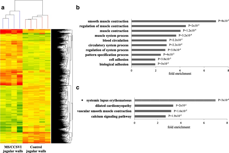
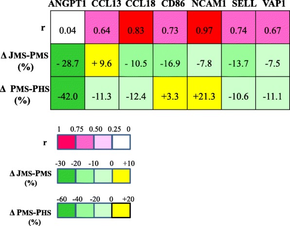
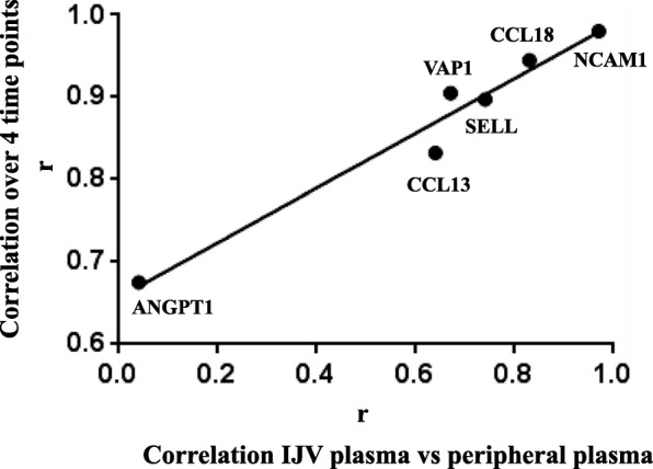
Results: Of the differentially expressed genes (≥ 2 fold-change, multiple testing correction, P < 0.05), the immune-related CD86 (8.5 fold-change, P = 0.002) emerged among the up regulated genes (N = 409). Several genes encoding HOX transcription factors and histones potentially regulated by blood flow, were overexpressed. Smooth muscle contraction and cell adhesion processes emerged among down regulated genes (N = 515), including the neuronal cell adhesion L1CAM as top scorer (5 fold-change, P = 5 × 10- 4). Repeated measurements in jugular/peripheral plasma and overtime in peripheral plasma showed conserved individual plasma patterns for immune-inflammatory (CCL13, CCL18) and adhesion (NCAM1, VAP1, SELL) proteins, despite significant variations overtime (SELL P < 0.0001). Both age and MS disease phenotypes were determinants of VAP1 plasma levels. Data supported cerebral related-mechanisms regulating ANGPT1 levels, which were remarkably lower in jugular plasma and correlated in repeated assays but not between jugular/peripheral compartments.
Conclusions: This study provides for the first time expression patterns of the IJV wall, suggesting signatures of altered vascular mRNA profiles in MS disease also independently from CCSVI. The combined transcriptome-protein analysis provides intriguing links between IJV wall transcript alteration and plasma protein expression, thus highlighting proteins of interest for MS pathophysiology.
Keywords: Adhesion molecules; Chemokines; Chronic cerebrospinal venous insufficiency; Gene expression; Jugular plasma protein levels; Jugular vein wall; Multiple sclerosis; Multiplex protein assay; Venous abnormalities.
Conflict of interest statement
The study was approved by the Ethical Committee of the S. Anna University-Hospital of Ferrara. Written informed consent was obtained from all participants enrolled in the study.
Not applicable.
The authors declare that they have no competing interests.
Springer Nature remains neutral with regard to jurisdictional claims in published maps and institutional affiliations.
Figures
References
Publication types
MeSH terms
Substances
LinkOut - more resources
Full Text Sources
Other Literature Sources
Medical
Research Materials
Miscellaneous
