. 2018 Aug 22;19(1):622.
doi: 10.1186/s12864-018-4982-5.

Deciphering the oviductal extracellular vesicles content across the estrous cycle: implications for the gametes-oviduct interactions and the environment of the potential embryo

Affiliations

Deciphering the oviductal extracellular vesicles content across the estrous cycle: implications for the gametes-oviduct interactions and the environment of the potential embryo

C Almiñana et al. BMC Genomics. .

Abstract

Background: The success of early reproductive events depends on an appropriate communication between gametes/embryos and the oviduct. Extracellular vesicles (EVs) contained in oviductal secretions have been suggested as new players in mediating this crucial cross-talk by transferring their cargo (proteins, mRNA and small ncRNA) from cell to cell. However, little is known about the oviductal EVs (oEVS) composition and their implications in the reproductive success. The aim of the study was to determine the oEVs content at protein, mRNA and small RNA level and to examine whether the oEVs content is under the hormonal influence of the estrous cycle.

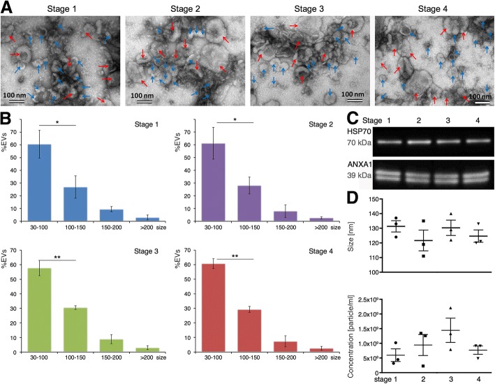
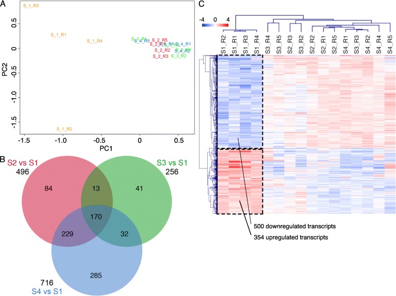
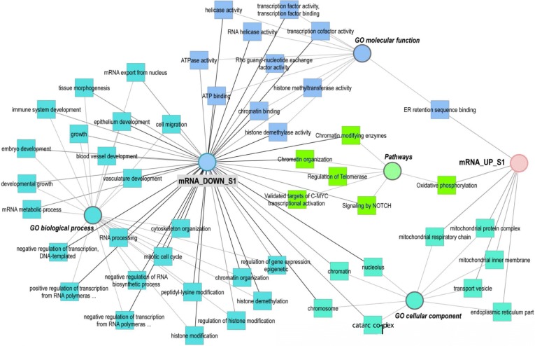
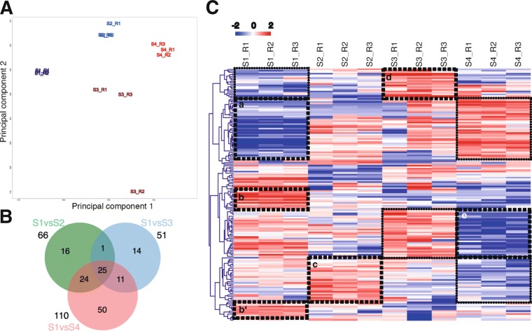
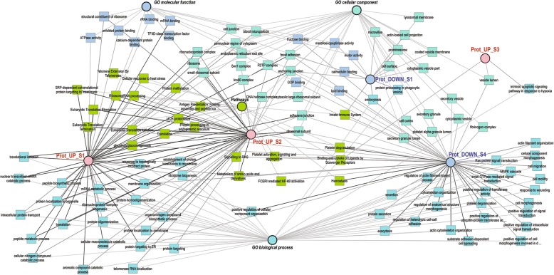
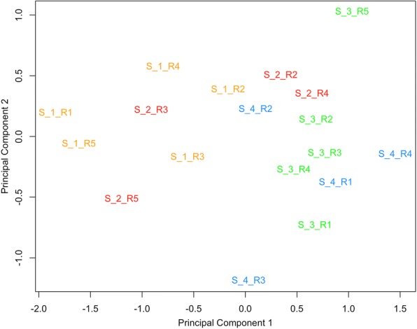
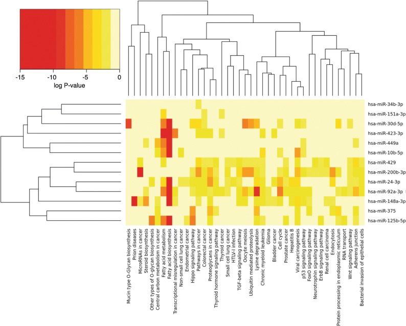
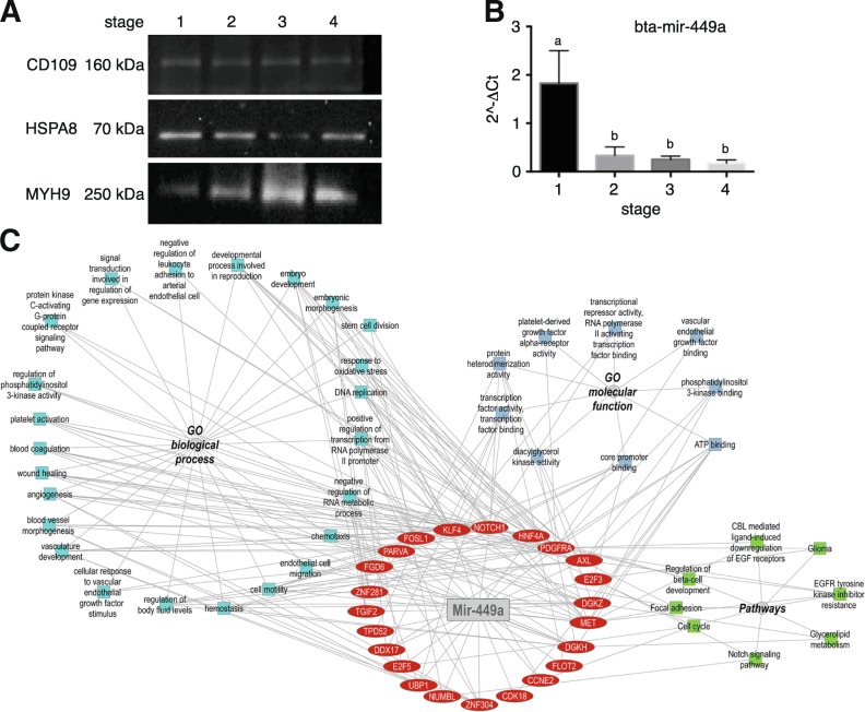
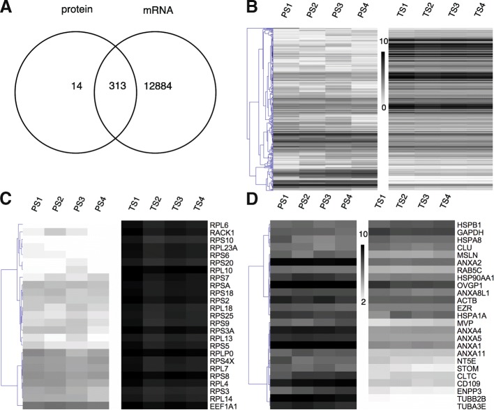
Results: We identified the presence of oEVs, exosomes and microvesicles, in the bovine oviductal fluid at different stages of the estrous cycle (postovulatory-stage, early luteal phase, late luteal phase and pre-ovulatory stage) and demonstrated that their composition is under hormonal regulation. RNA-sequencing identified 903 differentially expressed transcripts (FDR < 0.001) in oEVs across the estrous cycle. Moreover, small RNA-Seq identified the presence of different types of ncRNAs (miRNAs, rRNA fragments, tRNA fragments, snRNA, snoRNA, and other ncRNAs), which were partially also under hormonal influence. Major differences were found between post-ovulatory and the rest of the stages analyzed for mRNAs. Interesting miRNAs identified in oEVs and showing differential abundance among stages, miR-34c and miR-449a, have been associated with defective cilia in the oviduct and infertility. Furthermore, functional annotation of the differentially abundant mRNAs identified functions related to exosome/vesicles, cilia expression, embryo development and many transcripts encoding ribosomal proteins. Moreover, the analysis of oEVs protein content also revealed changes across the estrous cycle. Mass spectrometry identified 336 clusters of proteins in oEVs, of which 170 were differentially abundant across the estrous cycle (p-value< 0.05, ratio < 0.5 or ratio > 2). Our data revealed proteins related to early embryo development and gamete-oviduct interactions as well as numerous ribosomal proteins.

Conclusions: Our study provides with the first molecular signature of oEVs across the bovine estrous cycle, revealing marked differences between post- and pre-ovulatory stages. Our findings contribute to a better understanding of the potential role of oEVs as modulators of gamete/embryo-maternal interactions and their implications for the reproductive success.

Keywords: Exosomes; Gamete/embryo-maternal interaction; Hormones; Microvesicles; Oviduct; Proteomics; RNA-sequencing.


Conflict of interest statement

Ethics approval and consent to participate

Not applicable.

Consent for publication

Not applicable.

Competing interests

The authors declare that they have no competing interests.

Publisher’s Note

Springer Nature remains neutral with regard to jurisdictional claims in published maps and institutional affiliations.

Figures

Fig. 1
Fig. 1
Oviduct EVs characterization and quantification across the bovine estrous cycle. a Representative images of exosomes (blue arrows, 30–100 nm size) and microvesicles (red arrows > 100 nm size) across the different stages of the bovine estrous cycle (Stage 1, Stage 2, Stage 3 and Stage 4) by TEM. b Histograms showing the size distribution of oEVs across the estrous cycle. A batch of 12 samples containing 3 replicates/stage were analyzed using TEM and ImageJ software. Comparison between populations of exosomes and microvesicles for each stage were performed (*P < 0.05; **P < 0.01, T-test. c Western blotting characterization of bovine oEVs at all 4 stages analyzed for known exosomal protein markers. A pool of samples containing 3 biological replicates at the 4 different stages analyzed was used, showing oEVs from all 4 stages were positive for HSP70 and ANAX1.). d Bovine oEVs size average and concentration was measured by Nanosight analysis from a different batch of 12 samples (3 replicates /stage)
Fig. 2
Fig. 2
Comparative analysis of the oviduct EVs mRNA content across the bovine estrous cycle. a Principal Component analysis (PCA) on mRNA oviduct EVs content at 4 different stages of the estrous cycle (S1 in yellow; S2 in red, S3 in green and S4 in blue). b Venn Diagram demonstrating relations among differential transcripts (DT) for selected comparisons during the estrous cycle. Data were analyzed with a cut-off FDR < 0.001. c Dendrogram representing results of unsupervised hierarchical clustering (HCL) created with Pearson correlation coefficient by MeV. Rows indicate single differential transcripts (DT), while columns represent individual samples collected at different stages of the estrous cycle. Mean-centered expression values (log2 counts per million of sample – mean of log2 counts per million of all samples) for the samples of the 4 stages are shown. Color scale is from −4 (blue, lower than mean) to 4 (red, higher than mean)
Fig. 3
Fig. 3
Network of enriched GO terms associated to up and downregulated transcripts during stage 1 (S1). Comparative enrichment analysis of GO terms associated to transcripts up or downregulated in oviduct EVs at stage 1 of the estrous cycle compared to the rest of the stages (S2, S3, S4). The network shows enriched GO terms (biological process, a molecular function and cellular component) and pathways specifically overrepresented for the up and downregulated transcripts
Fig. 4
Fig. 4
Comparative analysis of the oviduct EVs protein content across the bovine estrous cycle. A) Principal Component analysis (PCA) on protein oviduct EVs content at 4 different stages of the estrus cycle (S1 in dark blue; S2 in light blue; S3 in brown and S4 in red) based on normalized weighted spectra data. B) Venn Diagram demonstrating relations among differential proteins (DP) for selected comparisons during the estrous cycle. Data were analyzed with cut-off FDR < 0.001. C) Dendrogram representing results of unsupervised hierarchical clustering (HCL) by MeV. Mean-centered expression values (log2 expression value of sample – mean of log2 expression values of all samples) for the samples of the 4 stages are shown. Rows indicate single DP, while columns represent different stages of the estrous cycle. Clusters of proteins strongly differentially regulated among stages are pointed with doted squares in HCL figure. These clusters of proteins were used for functional analysis and GO enriched terms are shown in proteins network in Fig. 5
Fig. 5
Fig. 5
Network of enriched GO terms associated to up and downregulated proteins during stage 1–4. Comparative enrichment analysis of GO terms associated to clusters of differential proteins up or downregulated in oviduct EVs at 1–4 stages of the estrus cycle. The network shows the enriched GO terms (biological process, a molecular function and cellular component) and pathways specifically overrepresented for the different protein cluster lists showing differential expression across the estrous cycle. The 5 clusters of differential proteins used for building the network are represented in Fig. 4C
Fig. 6
Fig. 6
RNA type distribution of the oviduct EVs small ncRNA content across the bovine estrous cycle. a Pie graph shows the percentage of sequences attributed to each RNA type among all ncRNAs identified by small RNA sequencing in oviduct EVs across the cycle (miRNAs, rRNA fragments, tRNA fragments, snRNA, snoRNA, and other ncRNAs). b Bar graph shows the percent of reads attributed to each RNA type per stage of cycle. c Bar graph shows the percent of reads attributed to each RNA type for each sample
Fig. 7
Fig. 7
Comparative analysis of the snRNA expression across the bovine estrous cycle. Principal Component analysis (PCA) on snRNA oviduct EVs content at 4 different stages of the estrous cycle (Stage 1: S1 in yellow; Stage 2: S2 in red, Stage 3: S3 in green and Stage 4: S4 in blue) for all snRNA identified. Five different replicates for each stage were analysed (represented by R1, R2, R3, R4 and R5) in the plot. PCA showed a partial separation between stage 1 and 2 (S1, S2) and stages 3 and 4 (S3, S4)
Fig. 8
Fig. 8
Selected miRNA and their associated pathways. Selected miRNAs based on differential expression and high abundance in EVs in Stage 1 and Stage 4 were used for clustering analysis based on the presence of interactions of target genes and pathways using DIANA-miRPath v3.0. Heatmap clustering of 13 miRNAs versus their associated pathways based on significance levels. Color scale from red (highest significance) to yellow (not significant)
Fig. 9
Fig. 9
Validation of Mass Spectrometry and miRNA analyses of bovine oEVs by Western immunoblotting and qPCR. a Western blot analysis confirmed MS results for 3 proteins associated with reproductive roles and showing differential abundance or not across the bovine estrus cycle: CD109, HSPA8 and MYH9. A pool of samples containing 3 biological replicates at the 4 different stages analyzed was used. b qPCR analysis confirmed miRNA-seq results for Mir-449a, which dysregulation has been associated with defective cilia in the oviduct and infertility. Mir-449a was more abundant in S1 compared to the rest of the stages (p < 0.05, T-test). A new batch of oEVs isolated from oviducts classified in the four stages of the estrous cycle (n = 12) was used. c Network of Mir-449a target genes and overrepresented functional categories
Fig. 10
Fig. 10
Overlap of all transcripts and proteins identified in oviduct EVs across the bovine estrus cycle. a Venn Diagram demonstrating the overlap among all transcripts and proteins identified in EVs; b Heatmap representing results of unsupervised hierarchical clustering (HCL) by MeV. Rows indicate single proteins and transcripts, while columns represent different stages of the estrous cycle for transcripts (TS1-TS4) and proteins (PS1-PS4) analyses. Expression values for proteins and transcripts were log2-transformed and scaled to have values in a similar numerical range. c Heatmap representing the 25 top most abundant proteins which were attributed to known exosomal proteins (Annexins, Heat Shock proteins) and proteins associated to reproductive roles (OVGP, HSPA8). d Heatmap showing the 25 top most abundant transcripts representing mainly mRNAs encoding different ribosomal proteins (RPS, RPL)

Similar articles

Cited by

References

    1. Valadi H, Ekstrom K, Bossios A, Sjostrand M, Lee JJ, Lotvall JO. Exosome-mediated transfer of mRNAs and microRNAs is a novel mechanism of genetic exchange between cells. Nat Cell Biol. 2007;9(6):654–659. doi: 10.1038/ncb1596. - DOI - PubMed
    1. Camussi G, Deregibus MC, Bruno S, Grange C, Fonsato V, Tetta C. Exosome/microvesicle-mediated epigenetic reprogramming of cells. Am J Cancer Res. 2011;1(1):98–110. - PMC - PubMed
    1. Yanez-Mo M, Siljander PR, Andreu Z, Zavec AB, Borras FE, Buzas EI, Buzas K, Casal E, Cappello F, Carvalho J, et al. Biological properties of extracellular vesicles and their physiological functions. J Extracell Vesicles. 2015;4:27066. doi: 10.3402/jev.v4.27066. - DOI - PMC - PubMed
    1. Sullivan R, Saez F. Epididymosomes, prostasomes, and liposomes: their roles in mammalian male reproductive physiology. Reproduction. 2013;146(1):R21–R35. doi: 10.1530/REP-13-0058. - DOI - PubMed
    1. da Silveira JC, Veeramachaneni DN, Winger QA, Carnevale EM, Bouma GJ. Cell-secreted vesicles in equine ovarian follicular fluid contain miRNAs and proteins: a possible new form of cell communication within the ovarian follicle. Biol Reprod. 2012;86(3):71. doi: 10.1095/biolreprod.111.093252. - DOI - PubMed

MeSH terms