

Computer-aided detection in chest radiography based on artificial intelligence: a survey
- PMID: 30134902
- PMCID: PMC6103992
- DOI: 10.1186/s12938-018-0544-y
Computer-aided detection in chest radiography based on artificial intelligence: a survey
Abstract
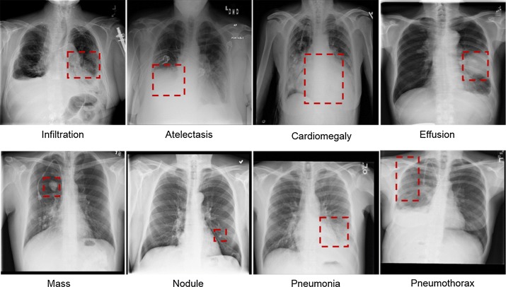
As the most common examination tool in medical practice, chest radiography has important clinical value in the diagnosis of disease. Thus, the automatic detection of chest disease based on chest radiography has become one of the hot topics in medical imaging research. Based on the clinical applications, the study conducts a comprehensive survey on computer-aided detection (CAD) systems, and especially focuses on the artificial intelligence technology applied in chest radiography. The paper presents several common chest X-ray datasets and briefly introduces general image preprocessing procedures, such as contrast enhancement and segmentation, and bone suppression techniques that are applied to chest radiography. Then, the CAD system in the detection of specific disease (pulmonary nodules, tuberculosis, and interstitial lung diseases) and multiple diseases is described, focusing on the basic principles of the algorithm, the data used in the study, the evaluation measures, and the results. Finally, the paper summarizes the CAD system in chest radiography based on artificial intelligence and discusses the existing problems and trends.
Keywords: Artificial intelligence; Chest radiography; Computer-aided detection; Disease classification.
Figures
Similar articles
-
Computer-aided diagnosis and artificial intelligence in clinical imaging.Semin Nucl Med. 2011 Nov;41(6):449-62. doi: 10.1053/j.semnuclmed.2011.06.004. Semin Nucl Med. 2011. PMID: 21978447 Review.
-
Computer-aided diagnostic scheme for the detection of lung nodules on chest radiographs: localized search method based on anatomical classification.Med Phys. 2006 Jul;33(7):2642-53. doi: 10.1118/1.2208739. Med Phys. 2006. PMID: 16898468
-
Computer-aided diagnosis in chest radiography: beyond nodules.Eur J Radiol. 2009 Nov;72(2):226-30. doi: 10.1016/j.ejrad.2009.05.061. Epub 2009 Jul 14. Eur J Radiol. 2009. PMID: 19604661 Review.
-
Image-processing technique for suppressing ribs in chest radiographs by means of massive training artificial neural network (MTANN).IEEE Trans Med Imaging. 2006 Apr;25(4):406-16. doi: 10.1109/TMI.2006.871549. IEEE Trans Med Imaging. 2006. PMID: 16608057
-
Gradient vector flow based active shape model for lung field segmentation in chest radiographs.Annu Int Conf IEEE Eng Med Biol Soc. 2009;2009:3561-4. doi: 10.1109/IEMBS.2009.5334886. Annu Int Conf IEEE Eng Med Biol Soc. 2009. PMID: 19964999
Cited by
-
Artificial Intelligence and Echocardiography.J Cardiovasc Imaging. 2021 Jul;29(3):193-204. doi: 10.4250/jcvi.2021.0039. Epub 2021 May 3. J Cardiovasc Imaging. 2021. PMID: 34080347 Free PMC article. Review.
-
Using Transfer Learning of Convolutional Neural Network on Neck Radiographs to Identify Acute Epiglottitis.J Digit Imaging. 2023 Jun;36(3):893-901. doi: 10.1007/s10278-023-00774-4. Epub 2023 Jan 19. J Digit Imaging. 2023. PMID: 36658377 Free PMC article.
-
Analysis of Tuberculosis in Chest Radiographs for Computerized Diagnosis using Bag of Keypoint Features.J Med Syst. 2019 Feb 28;43(4):87. doi: 10.1007/s10916-019-1222-8. J Med Syst. 2019. PMID: 30820678
-
Detection and visualization of abnormality in chest radiographs using modality-specific convolutional neural network ensembles.PeerJ. 2020 Mar 17;8:e8693. doi: 10.7717/peerj.8693. eCollection 2020. PeerJ. 2020. PMID: 32211231 Free PMC article.
-
Deep Learning-Based Classification and Semantic Segmentation of Lung Tuberculosis Lesions in Chest X-ray Images.Diagnostics (Basel). 2024 Apr 30;14(9):952. doi: 10.3390/diagnostics14090952. Diagnostics (Basel). 2024. PMID: 38732366 Free PMC article.
References
-
- X-ray (radiography)—chest. https://www.radiologyinfo.org/en/info.cfm?pg=chestrad. Accessed 10 June 2018.
-
- Zakirov AN, Kuleev RF, Timoshenko AS, Vladimirov AV. Advanced approaches to computer-aided detection of thoracic diseases on chest X-rays. Appl Math Sci. 2015;9(88):4361–4369.
-
- Krizhevsky A, Sutskever I, Hinton GE. ImageNet classification with deep convolutional neural networks. Commun ACM. 2017;60(6):84–90. doi: 10.1145/3065386. - DOI
Publication types
MeSH terms
LinkOut - more resources
Full Text Sources
Other Literature Sources
Research Materials
Miscellaneous
