. 2018 Oct 12;92(21):e00704-18.
doi: 10.1128/JVI.00704-18. Print 2018 Nov 1.

Cottontail Rabbit Papillomavirus E1 and E2 Proteins Mutually Influence Their Subcellular Localizations

Affiliations

Cottontail Rabbit Papillomavirus E1 and E2 Proteins Mutually Influence Their Subcellular Localizations

Markus Schneider et al. J Virol. .

Abstract

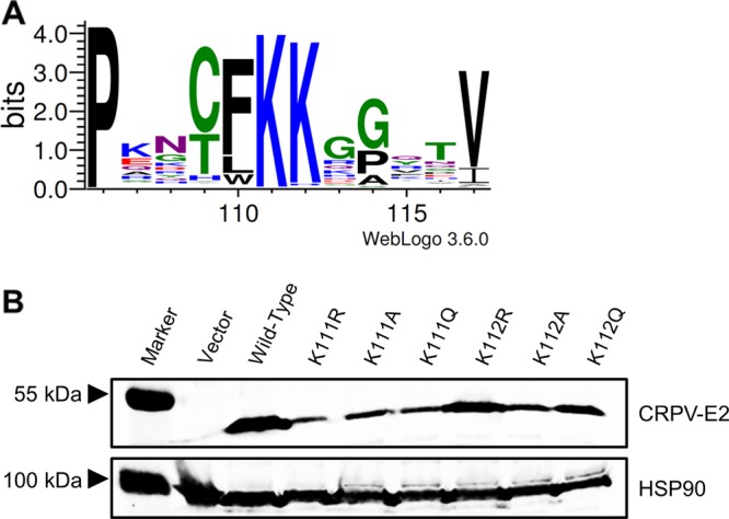
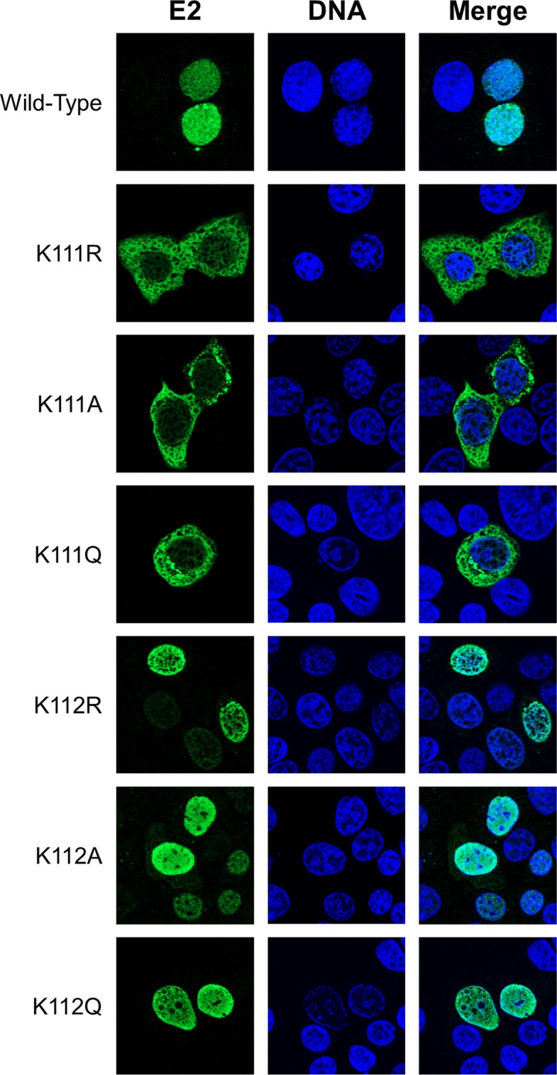
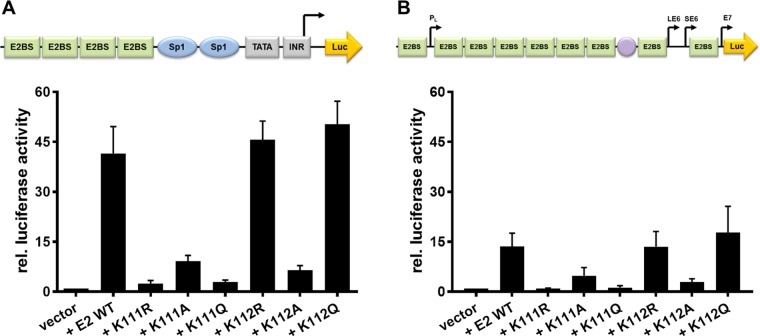
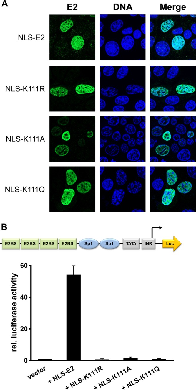
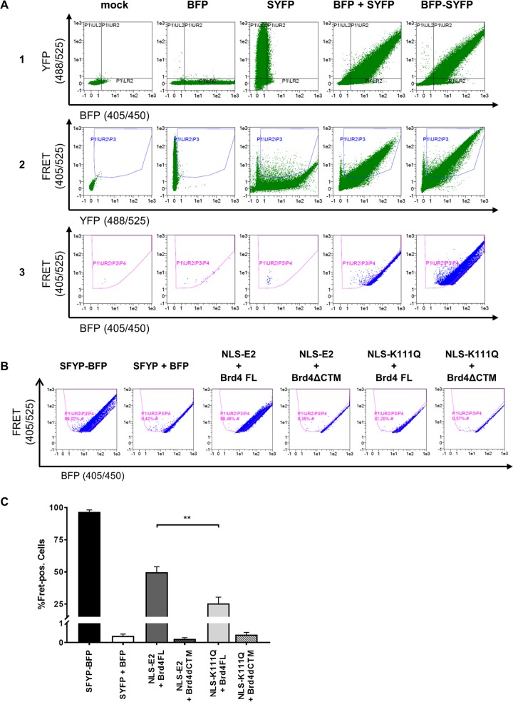
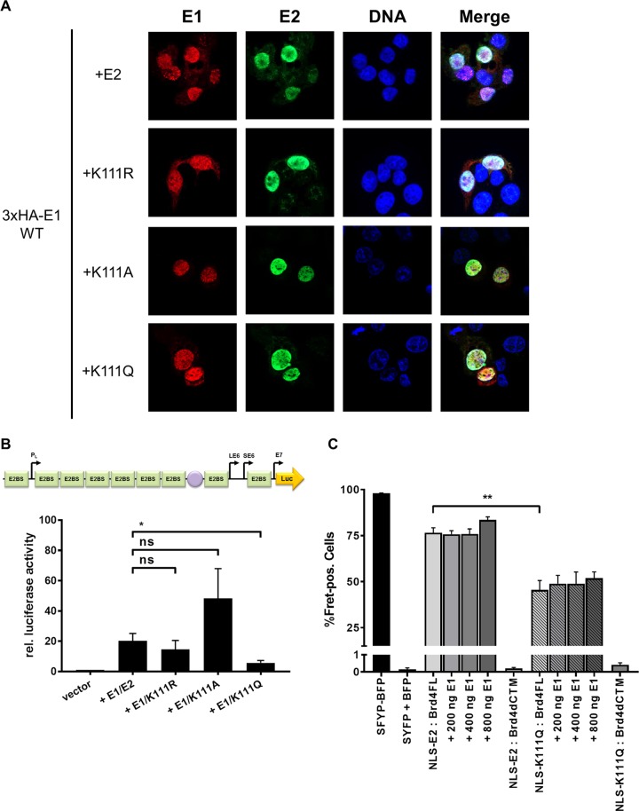
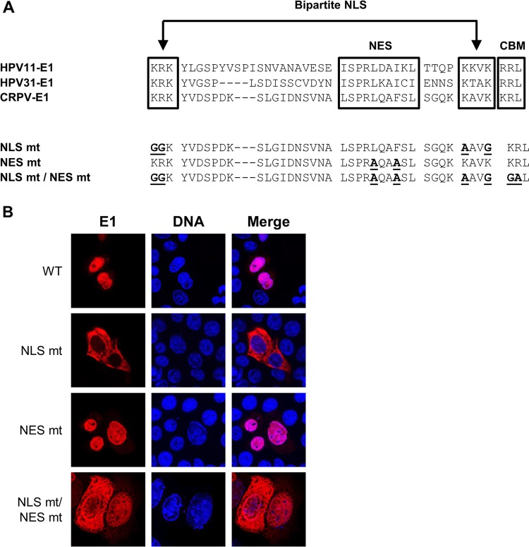
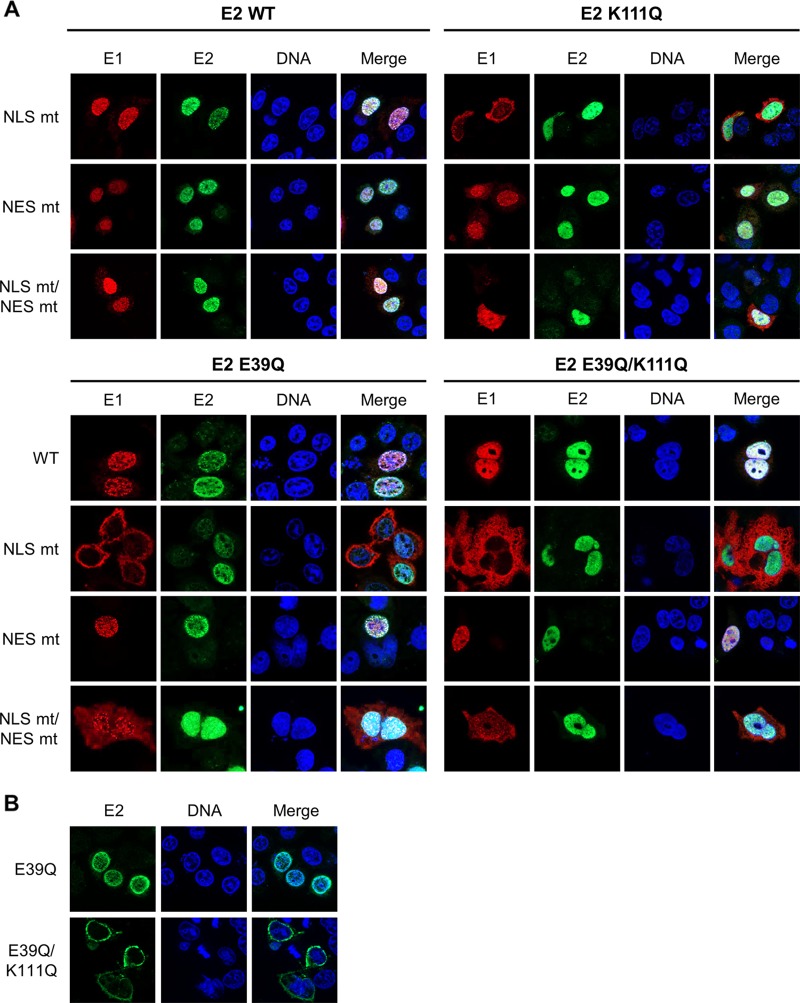
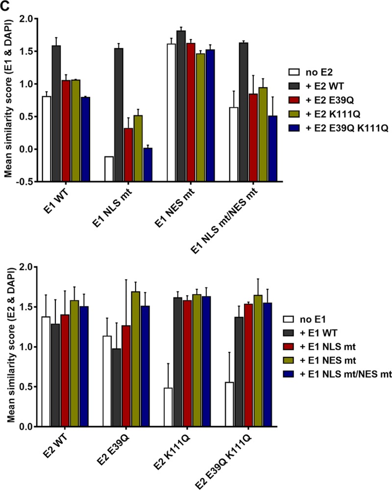
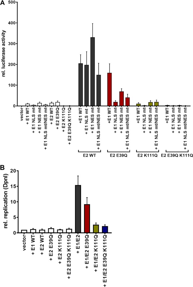
The papillomavirus (PV) E2 protein is a nuclear, sequence-specific DNA-binding protein that regulates transcription and nuclear retention of viral genomes. E2 also interacts with the viral E1 protein to replicate the viral genome. E2 residue K111 is highly conserved among PV and has been implicated in contributing to nuclear transport, transcription, and replication. Cottontail rabbit (Sylvilagus floridanus) PV (CRPV or SfPV1) E2 K111R, A, or Q mutations are transcription deficient and localized to the cytoplasm, comparable to other PV types. The addition of a nuclear localization signal (NLS) resulted in nuclear E2 K111 mutant proteins but did not restore transcriptional activation, and this is most likely due to an impaired binding to the cellular Brd4 protein. Surprisingly, coexpression of E1 with E2 K111 mutations resulted in their nuclear localization and, for K111A and R mutations, the activation of an E1/E2-dependent reporter construct. Interestingly, the nuclear localization of E2 K111Q mutant protein was independent from the presence of the conserved bipartite NLS in E1 and the direct interaction between E1 and E2. On the other hand, the cytoplasmic E1 NLS mutation could be targeted to the nucleus by wild-type E2, and this was dependent upon an interaction between E1 and E2. In summary, our studies have uncovered that E1 and E2 control each other's subcellular localization: direct binding of E2 to E1 can direct E1 to the nucleus independently from the E1 NLS, and E1 can direct E2 to the nucleus without an intact NLS or direct binding to E2.IMPORTANCE Papillomaviruses encode the DNA-binding E1 and E2 proteins, which form a complex and are essential for genome replication. Both proteins are targeted to the nucleus via nuclear localization signals. Our studies have uncovered that cytoplasmic mutant E1 or E2 proteins can be localized to the nucleus when E1 or E2 is also present. An interaction between E1 and E2 is necessary to target cytoplasmic E1 mutant proteins to the nucleus, but cytoplasmic E2 mutant proteins can be targeted to the nucleus without a direct interaction, which points to a novel function of E1.

Keywords: E1; E2; nuclear localization; papillomavirus; replication.


Figures

FIG 1
FIG 1
Expression levels of CRPV E2 K111 and K112 mutant proteins. (A) Partial sequence alignment of the region between amino acids 106 and 117 of all 353 annotated E2 proteins. The sequences were obtained from https://pave.niaid.nih.gov/ and adjusted to each other. A sequence logo was generated using WebLogo 3 (http://weblogo.threeplusone.com/) (33). (B) C33a cells were transiently transfected with expression vectors for E2 wt or E2 mt proteins. An empty expression vector (pSG5) was used as a control. At 48 h after transfection, whole-cell extracts were analyzed in immunoblot analysis using the indicated antibodies.
FIG 2
FIG 2
Mutation of K111 affects subcellular localization of the E2 protein. C33a cells were transfected with 500 ng expression vector for each E2 protein (as indicated on the left side). Forty-eight hours after transfection, cells were stained with an anti-CRPV E2 antibody to detect E2 proteins (green) and analyzed by immunofluorescence microscopy. DNA was stained with DAPI (blue).
FIG 3
FIG 3
K111 mutations do not enable transcriptional activity. C33a cells were either transiently transfected with 50 ng pC18-Sp1-luc (A) or 50 ng pGL CRPV (PL-PE7) firefly reporter construct (B), 10 ng of the empty expression vector (pSG5), or the expression vectors for the different CRPV E2 proteins. A schematic presentation of the reporter constructs is shown above the respective graphs. The values represent the relative luciferase activities of the E2 proteins (wild type or mutants) to the basal activity of the used firefly reporter construct. Error bars indicate the standard error of the mean (SEM) from at least six independent experiments.
FIG 4
FIG 4
Relocalization of cytoplasmic K111 mutant proteins does not lead to a gain of transcriptional activity. (A) C33a cells were transfected with 500 ng expression vector, and 48 h after transfection cells were stained with an anti-CRPV E2 antibody to detect E2 proteins (green) and analyzed by immunofluorescence microscopy. DNA was stained with DAPI (blue). (B) C33a cells were transfected with 50 ng pC18-Sp1-luc and 10 ng of the empty expression vector (pSG5) or expression vectors for the different CRPV E2 proteins. The values represent the luciferase activities of the E2 proteins relative to the basal activity of the used firefly reporter construct. Error bars indicate the standard errors of the means (SEM) from at least three independent experiments.
FIG 5
FIG 5
Flow cytometry-FRET analyses indicate a reduced interaction of K111Q with Brd4 in the nucleus. (A) Gating strategy to determine E2 and Brd4 interactions in a flow cytometry-based FRET assay. Untransfected C33a cells were used as a mock control or transfected with 500 ng expression vectors for BFP, SYFP, BFP, and SYFP as well as a BFP-SYFP fusion protein. As a first step, the BFP/SYFP-double positive cells were gated (row 1). Cells giving a false-positive FRET signal resulting from YFP excitation at 405 nm were excluded (row 2). The remaining cells were further analyzed for a positive FRET signal by setting a gate incorporating cells which are cotransfected with the expression vectors for BFP and SYFP, consequently generating no FRET signal (row 3). (B) FACS plots showing FRET-positive cells in living C33a cells cotransfected with the indicated BFP and SYFP fusion proteins. SYFP-BFP fusion protein was used as a positive control, and cotransfected BFP and SYFP expression vectors were negative controls. (C) Mean values of FRET-positive cells from at least three independent experiments that were analyzed as shown in panel B. Error bars indicate the standard errors of the means (SEM). A paired two-tailed t test was used to determine statistical significance (**, P < 0.01).
FIG 6
FIG 6
E2 K111 mutant proteins relocalize to nucleus in the presence of CRPV E1. (A) C33a cells were cotransfected with 500 ng E1 expression vector (pSG 3×HA:CRPV E1) and 500 ng E2 expression vectors (as indicated on the left). Forty-eight hours after transfection, cells were stained with an anti-HA antibody to detect the E1 protein (red) and an anti-CRPV E2 antibody to detect E2 proteins (green) and analyzed by immunofluorescence microscopy. DNA was stained with DAPI (blue). (B) C33a cells were transiently transfected with 50 ng pGL CRPV (PL-PE7) firefly reporter construct, 100 ng E1 expression vector (pSG CRPV E1), and 10 ng of the different E2 expression vectors. Empty expression vector (pSG5) was used to adjust differences in DNA amounts. The values represent the relative luciferase activities of E1/E2 (E2 wild-type or E2 K111 mutant proteins) to the basal activity of the used firefly reporter construct. Error bars indicate the standard errors of the means (SEM) from at least five independent experiments. A paired two-tailed t test was used to determine statistical significance (*, P < 0.05). ns, nonsignificant. (C) C33a cells were cotransfected with expression vectors for BFP and SYFP fusion proteins and, as indicated, increasing DNA amounts of CRPV E1 expression vector (pSG CRPV E1). SYFP-BFP fusion protein was used as a positive control, and cotransfected BFP and SYFP expression vectors were negative controls. Shown are the mean values of FRET-positive cells from at least three independent experiments. Error bars indicate the SEM. A paired two-tailed t test was used to determine statistical significance (**, P < 0.01).
FIG 7
FIG 7
Generation and subcellular localization of CRPV E1 NLS, NES, and NLS/NES mutant proteins. (A) Partial sequence alignment of HPV11, HPV31, and CRPV E1 proteins. Sequences were obtained from https://pave.niaid.nih.gov/. Highlighted are the bipartite nuclear localization signal (NLS), the embedded nuclear export signal (NES), and the cyclin binding motif (CBM). Shown below are the sequences of the generated CRPV E1 mutant proteins. Mutated residues are shown in boldface and are underlined. (B) C33a cells were transfected with 500 ng E1 expression vectors (pSG 3×HA:CRPV E1, pSG 3×HA:CRPV E1 NLS mt, pSG 3×HA:CRPV E1 NES mt, or pSG 3×HA:CRPV E1 NLS mt/NES mt). Forty-eight hours after transfection, cells were stained with an anti-HA antibody to detect E1 proteins (red) and analyzed by immunofluorescence microscopy. DNA was stained with DAPI (blue).
FIG 8
FIG 8
CRPV E1 does not recruit CRPV E2 into the nucleus by direct binding. (A) C33a cells were cotransfected with 500 ng E1 expression vector (pSG 3×HA:CRPV E1, pSG 3×HA:CRPV E1 NLS mt, pSG 3×HA:CRPV E1 NES mt, or pSG 3×HA:CRPV E1 NLS mt/NES mt) and 500 ng E2 expression vector (pSG CRPV E2, pSG CRPV E2 E39Q, pSG CRPV E2 K111Q, or pSG CRPV E2 E39Q/K111Q). Forty-eight hours after transfection, cells were stained with an anti-HA antibody to detect E1 proteins (red) and an anti-CRPV-E2 antibody to detect E2 proteins (green). (B) C33a cells were transfected with 500 ng E2 expression vectors (pSG CRPV E2 E39Q or pSG CRPV E2 E39Q/K111Q). After 48 h, cells were stained with an anti-CRPV-E2 antibody to detect E2 proteins. Cells were analyzed by immunofluorescence microscopy. DNA was stained with DAPI (blue). (C) Subcellular distribution of E1 and E2 wt and mt proteins. C33a cells were transfected with wt and mt E1 and E2 alone (no E1, no E2) or in combination as indicated, and mean similarity scores for E1 and DAPI (upper) and E2 and DAPI (lower) were obtained after staining for DAPI, anti-E1 (HA), and anti-E2. Single-cell images in the focus plane were recorded and analyzed using IDEAS image analysis software. The data shown are from two independent experiments and are derived from at least 247 E2-positive and 637 E1-positive cells per experiment.
FIG 8
FIG 8
CRPV E1 does not recruit CRPV E2 into the nucleus by direct binding. (A) C33a cells were cotransfected with 500 ng E1 expression vector (pSG 3×HA:CRPV E1, pSG 3×HA:CRPV E1 NLS mt, pSG 3×HA:CRPV E1 NES mt, or pSG 3×HA:CRPV E1 NLS mt/NES mt) and 500 ng E2 expression vector (pSG CRPV E2, pSG CRPV E2 E39Q, pSG CRPV E2 K111Q, or pSG CRPV E2 E39Q/K111Q). Forty-eight hours after transfection, cells were stained with an anti-HA antibody to detect E1 proteins (red) and an anti-CRPV-E2 antibody to detect E2 proteins (green). (B) C33a cells were transfected with 500 ng E2 expression vectors (pSG CRPV E2 E39Q or pSG CRPV E2 E39Q/K111Q). After 48 h, cells were stained with an anti-CRPV-E2 antibody to detect E2 proteins. Cells were analyzed by immunofluorescence microscopy. DNA was stained with DAPI (blue). (C) Subcellular distribution of E1 and E2 wt and mt proteins. C33a cells were transfected with wt and mt E1 and E2 alone (no E1, no E2) or in combination as indicated, and mean similarity scores for E1 and DAPI (upper) and E2 and DAPI (lower) were obtained after staining for DAPI, anti-E1 (HA), and anti-E2. Single-cell images in the focus plane were recorded and analyzed using IDEAS image analysis software. The data shown are from two independent experiments and are derived from at least 247 E2-positive and 637 E1-positive cells per experiment.
FIG 9
FIG 9
Functional analysis of E1 and E2 mutant proteins. C33a cells were transiently transfected with 50 ng pGL CRPV(PL-PE7) firefly reporter construct, 100 ng E1 expression vectors, and 10 ng E2 expression vectors (as indicated). Empty expression vector (pSG5) was used to adjust differences in DNA amounts. (A) The values represent the relative luciferase activities of either E1, E2, or E1/E2 to the basal activity of the used firefly reporter construct. Error bars indicate the standard errors of the means (SEM) from at least six independent experiments. (B) Total DNA was extracted and digested with DpnI. The extent of DNA replication of the pGL CRPV (PL-PE7) plasmid was determined by qPCR. Data are shown relative to the extent of replication in the presence of the empty vector (vector). Error bars indicate the SEM.

References

    1. Parkin DM, Bray F. 2006. Chapter 2: the burden of HPV-related cancers. Vaccine 24(Suppl 3):S3/11–S3/25. doi:10.1016/j.vaccine.2005.01.101. - DOI - PubMed
    1. Bergvall M, Melendy T, Archambault J. 2013. The E1 proteins. Virology 445:35–56. doi:10.1016/j.virol.2013.07.020. - DOI - PMC - PubMed
    1. McBride AA. 2013. The papillomavirus E2 proteins. Virology 445:57–79. doi:10.1016/j.virol.2013.06.006. - DOI - PMC - PubMed
    1. McPhillips MG, Oliveira JG, Spindler JE, Mitra R, McBride AA. 2006. Brd4 is required for e2-mediated transcriptional activation but not genome partitioning of all papillomaviruses. J Virol 80:9530–9543. doi:10.1128/JVI.01105-06. - DOI - PMC - PubMed
    1. Schweiger MR, You J, Howley PM. 2006. Bromodomain protein 4 mediates the papillomavirus E2 transcriptional activation function. J Virol 80:4276–4285. doi:10.1128/JVI.80.9.4276-4285.2006. - DOI - PMC - PubMed

Publication types

MeSH terms

Substances

LinkOut - more resources