

The Novel Activity of Carbamazepine as an Activation Modulator Extends from NaV1.7 Mutations to the NaV1.8-S242T Mutant Channel from a Patient with Painful Diabetic Neuropathy
- PMID: 30135145
- PMCID: PMC7501587
- DOI: 10.1124/mol.118.113076
The Novel Activity of Carbamazepine as an Activation Modulator Extends from NaV1.7 Mutations to the NaV1.8-S242T Mutant Channel from a Patient with Painful Diabetic Neuropathy
Abstract
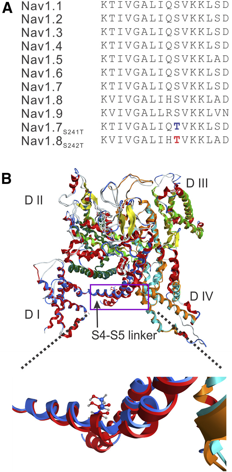
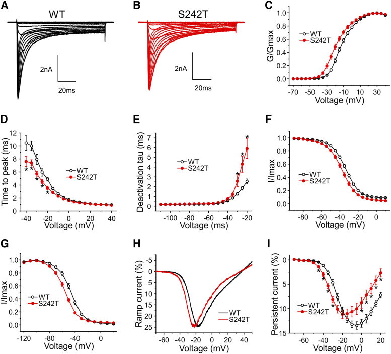
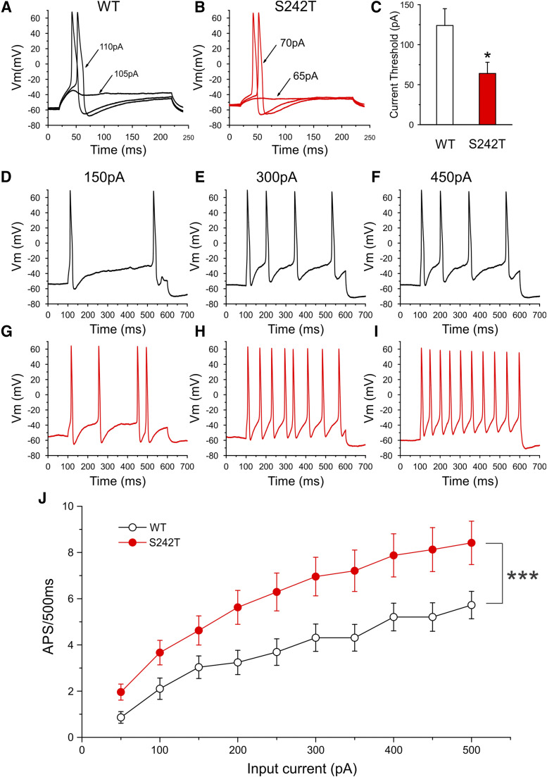
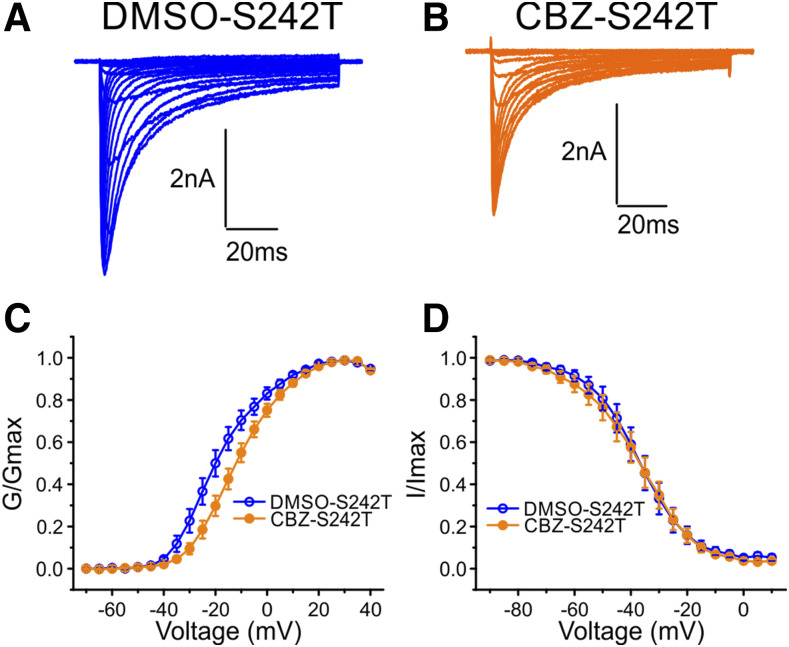
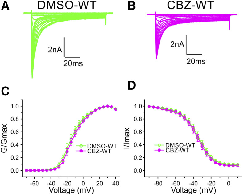
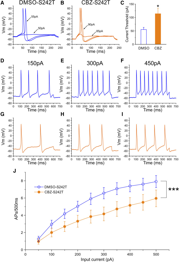
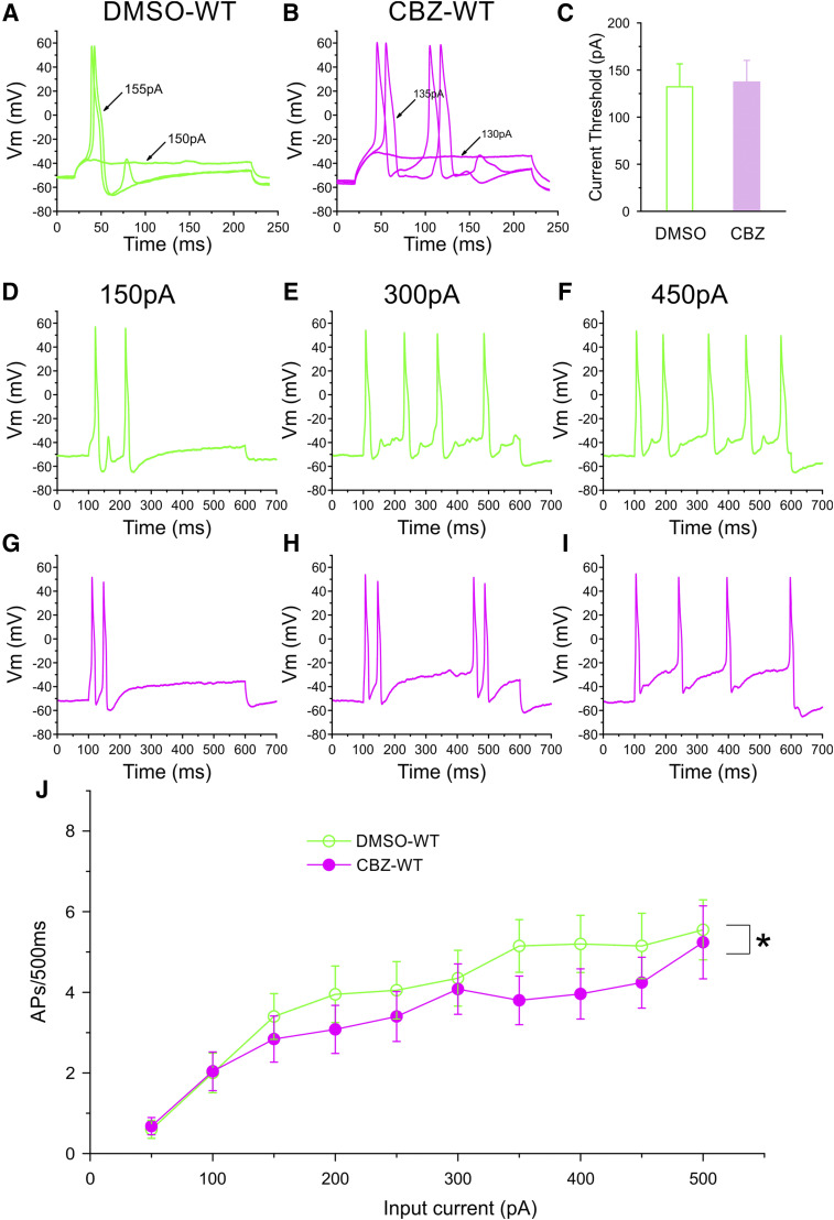
Neuropathic pain in patients carrying sodium channel gain-of-function mutations is generally refractory to pharmacotherapy. However, we have shown that pretreatment of cells with clinically achievable concentration of carbamazepine (CBZ; 30 μM) depolarizes the voltage dependence of activation in some NaV1.7 mutations such as S241T, a novel CBZ mode of action of this drug. CBZ reduces the excitability of dorsal root ganglion (DRG) neurons expressing NaV1.7-S241T mutant channels, and individuals carrying the S241T mutation respond to treatment with CBZ. Whether the novel activation-modulating activity of CBZ is specific to NaV1.7, and whether this pharmacogenomic approach can be extended to other sodium channel subtypes, are not known. We report here the novel NaV1.8-S242T mutation, which corresponds to the NaV1.7-S241T mutation, in a patient with neuropathic pain and diabetic peripheral neuropathy. Voltage-clamp recordings demonstrated hyperpolarized and accelerated activation of NaV1.8-S242T. Current-clamp recordings showed that NaV1.8-S242T channels render DRG neurons hyperexcitable. Structural modeling shows that despite a substantial difference in the primary amino acid sequence of NaV1.7 and NaV1.8, the S242 (NaV1.8) and S241 (NaV1.7) residues have similar position and orientation in the domain I S4-S5 linker of the channel. Pretreatment with a clinically achievable concentration of CBZ corrected the voltage dependence of activation of NaV1.8-S242T channels and reduced DRG neuron excitability as predicted from our pharmacogenomic model. These findings extend the novel activation modulation mode of action of CBZ to a second sodium channel subtype, NaV1.8.
Copyright © 2018 by The American Society for Pharmacology and Experimental Therapeutics.
Figures
Similar articles
-
Nav1.8 and Chronic Pain: From Laboratory Animals to Clinical Patients.Biomolecules. 2025 May 10;15(5):694. doi: 10.3390/biom15050694. Biomolecules. 2025. PMID: 40427587 Free PMC article. Review.
-
A novel gain-of-function Nav1.7 mutation in a carbamazepine-responsive patient with adult-onset painful peripheral neuropathy.Mol Pain. 2018 Jan-Dec;14:1744806918815007. doi: 10.1177/1744806918815007. Epub 2018 Nov 5. Mol Pain. 2018. PMID: 30392441 Free PMC article.
-
Nav1.7-A1632G Mutation from a Family with Inherited Erythromelalgia: Enhanced Firing of Dorsal Root Ganglia Neurons Evoked by Thermal Stimuli.J Neurosci. 2016 Jul 13;36(28):7511-22. doi: 10.1523/JNEUROSCI.0462-16.2016. J Neurosci. 2016. PMID: 27413160 Free PMC article.
-
Pharmacotherapy for Pain in a Family With Inherited Erythromelalgia Guided by Genomic Analysis and Functional Profiling.JAMA Neurol. 2016 Jun 1;73(6):659-67. doi: 10.1001/jamaneurol.2016.0389. JAMA Neurol. 2016. PMID: 27088781
-
Painful and painless mutations of SCN9A and SCN11A voltage-gated sodium channels.Pflugers Arch. 2020 Jul;472(7):865-880. doi: 10.1007/s00424-020-02419-9. Epub 2020 Jun 29. Pflugers Arch. 2020. PMID: 32601768 Free PMC article. Review.
Cited by
-
Differential effect of lacosamide on Nav1.7 variants from responsive and non-responsive patients with small fibre neuropathy.Brain. 2020 Mar 1;143(3):771-782. doi: 10.1093/brain/awaa016. Brain. 2020. PMID: 32011655 Free PMC article. Clinical Trial.
-
Manipulation of a spider peptide toxin alters its affinity for lipid bilayers and potency and selectivity for voltage-gated sodium channel subtype 1.7.J Biol Chem. 2020 Apr 10;295(15):5067-5080. doi: 10.1074/jbc.RA119.012281. Epub 2020 Mar 5. J Biol Chem. 2020. PMID: 32139508 Free PMC article.
-
Pain-causing stinging nettle toxins target TMEM233 to modulate NaV1.7 function.Nat Commun. 2023 Apr 28;14(1):2442. doi: 10.1038/s41467-023-37963-2. Nat Commun. 2023. PMID: 37117223 Free PMC article.
-
Nav1.8 and Chronic Pain: From Laboratory Animals to Clinical Patients.Biomolecules. 2025 May 10;15(5):694. doi: 10.3390/biom15050694. Biomolecules. 2025. PMID: 40427587 Free PMC article. Review.
-
New perspectives in diabetic neuropathy.Neuron. 2023 Sep 6;111(17):2623-2641. doi: 10.1016/j.neuron.2023.05.003. Epub 2023 May 31. Neuron. 2023. PMID: 37263266 Free PMC article. Review.
References
-
- Ahn HS, Dib-Hajj SD, Cox JJ, Tyrrell L, Elmslie FV, Clarke AA, Drenth JP, Woods CG, Waxman SG. (2010) A new Nav1.7 sodium channel mutation I234T in a child with severe pain. Eur J Pain 14:944–950. - PubMed
-
- Akopian AN, Sivilotti L, Wood JN. (1996) A tetrodotoxin-resistant voltage-gated sodium channel expressed by sensory neurons. Nature 379:257–262. - PubMed
-
- Ambrósio AF, Soares-Da-Silva P, Carvalho CM, Carvalho AP. (2002) Mechanisms of action of carbamazepine and its derivatives, oxcarbazepine, BIA 2-093, and BIA 2-024. Neurochem Res 27:121–130. - PubMed
-
- Bennett DL, Woods CG. (2014) Painful and painless channelopathies. Lancet Neurol 13:587–599. - PubMed
-
- Bierhaus A, Fleming T, Stoyanov S, Leffler A, Babes A, Neacsu C, Sauer SK, Eberhardt M, Schnölzer M, Lasitschka F, et al. (2012) Methylglyoxal modification of Nav1.8 facilitates nociceptive neuron firing and causes hyperalgesia in diabetic neuropathy [published correction appears in Nat Med (2012) 18:1445]. Nat Med 18:926–933. - PubMed
Publication types
MeSH terms
Substances
Grants and funding
LinkOut - more resources
Full Text Sources
Other Literature Sources
Medical
