. 2018 Oct 5;293(40):15678-15690.
doi: 10.1074/jbc.RA118.003080. Epub 2018 Aug 22.

Phosphoserine acidic cluster motifs bind distinct basic regions on the μ subunits of clathrin adaptor protein complexes

Affiliations

Phosphoserine acidic cluster motifs bind distinct basic regions on the μ subunits of clathrin adaptor protein complexes

Rajendra Singh et al. J Biol Chem. .

Abstract

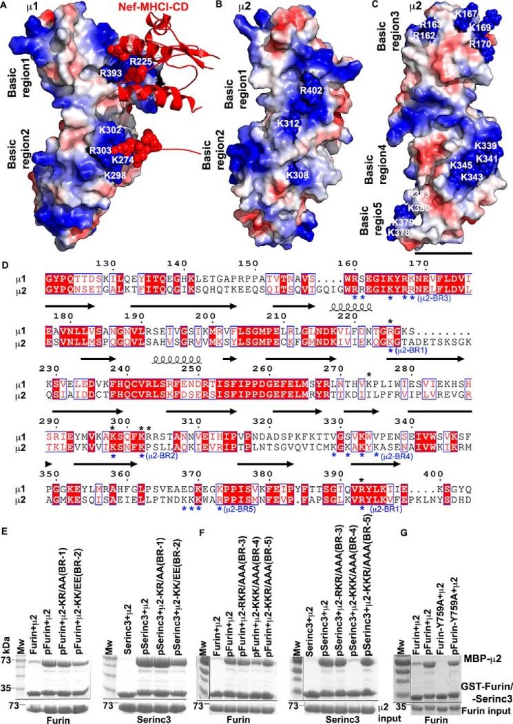
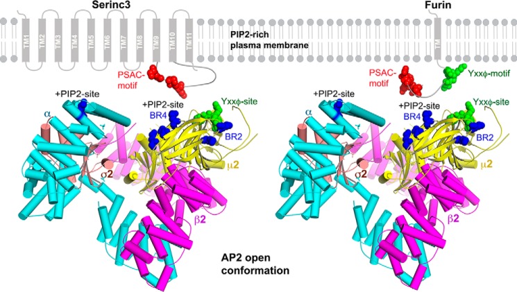
Protein trafficking in the endosomal system involves the recognition of specific signals within the cytoplasmic domains (CDs) of transmembrane proteins by clathrin adaptors. One such signal is the phosphoserine acidic cluster (PSAC), the prototype of which is in the endoprotease furin. How PSACs are recognized by clathrin adaptors has been controversial. We reported previously that HIV-1 Vpu, which modulates cellular immunoreceptors, contains a PSAC that binds to the μ subunits of clathrin adaptor protein (AP) complexes. Here, we show that the CD of furin binds the μ subunits of AP-1 and AP-2 in a phosphorylation-dependent manner. Moreover, we identify a potential PSAC in a cytoplasmic loop of the cellular transmembrane Serinc3, an inhibitor of the infectivity of retroviruses. The two serines within the PSAC of Serinc3 are phosphorylated by casein kinase II and mediate interaction with the μ subunits in vitro The sites of these serines vary among mammals in a manner suggesting host-pathogen conflict, yet the Serinc3 PSAC seems dispensable for anti-HIV activity and for counteraction by HIV-1 Nef. The CDs of Vpu and furin and the PSAC-containing loop of Serinc3 each bind the μ subunit of AP-2 (μ2) with similar affinities, but they appear to utilize different basic regions on μ2. The Serinc3 loop requires a region previously reported to bind the acidic plasma membrane lipid phosphatidylinositol 4,5-bisphosphate. These data suggest that the PSACs within different proteins recognize different basic regions on the μ surface, providing the potential to inhibit the activity of viral proteins without necessarily affecting cellular protein trafficking.

Keywords: HIV-1 Vpu; Serinc3; acidic cluster; adaptor protein; clathrin; furin; host-pathogen interaction; human immunodeficiency virus (HIV); kinetics; medium subunit; membrane trafficking; phosphorylation; phosphoserine; protein chemistry; protein complex.


Conflict of interest statement

The authors declare that they have no conflicts of interest with the contents of this article

Figures

Figure 1.
Figure 1.
Cytoplasmic domains containing PSACs bind directly to the μ subunits of AP-1 and AP-2. A, diagram of the transmembrane proteins HIV-1 Vpu, furin, and Serinc3. Vpu and furin are single-pass, type I transmembrane proteins. Serinc3 is a multipass transmembrane protein. Extracellular/luminal and cytoplasmic regions (loops) of Serinc3 are numbered N-terminally to C-terminally as L1–L12. L10 contains a potential PSAC. B, CDs of Vpu and furin and L10 of Serinc3. TMD, transmembrane domain. Potential clathrin AP complex–binding motifs, including PSACs, are underlined. Serines in red are known or potential sites of phosphorylation by CK-II. C, sites of serine phosphorylation in L10 of Serinc3. GST-Serinc3-L10 was co-expressed with CK-II in E. coli, purified, and analyzed by LC/MS. Phosphopeptides and serine-threonine (ST) phosphorylation are indicated, as are the sites of phosphorylation. Mascot scores for the peptide matches are indicated. D, phosphorylation of the CDs of Vpu and furin and L10 of Serinc3 detected by Phos-tag staining. GST-Vpu CD, GST-furin CD, or GST-Serinc3-L10 was expressed in E. coli either with or without co-expression of CK-II. The proteins were purified by affinity chromatography using GSH-Sepharose, ion-exchange, and size-exclusion chromatography and then stained with either Phos-tag stain or Coomassie Blue. E, Vpu binds μ1 and μ2 in a CK-II–dependent manner. GST or GST-Vpu proteins expressed in E. coli either with or without CK-II were tested for binding to μ1 and μ2 in a pulldown assay; proteins co-expressed with CK-II are denoted by p for presumed phosphorylation. The μ proteins are N-terminally truncated and fused to MBP as a solubility tag. SDS-polyacrylamide gels were stained with Coomassie Blue. F, the CD of furin and L10 of Serinc3 bind μ1 and μ2 in a CK-II–dependent manner. GST-furin CD or GST-Serinc3-L10 was expressed either with or without CK-II (proteins co-expressed with CK-II are denoted by p). The purified proteins were then used to capture either μ1 or μ2. SDS-polyacrylamide gels were stained with Coomassie Blue.
Figure 2.
Figure 2.
Mutational analysis of the PSAC of Serinc3-L10 with respect to μ-binding. A, GST-Serinc3-L10 or the indicated Ser/Asn or Ser/Asp substitution mutants were expressed in E. coli either with (p) or without (no p) co-expression of CK-II; the purified proteins were used to pull down MBP-μ1 or MBP-μ2. B, MBP-μ2-GST was used to pull down phospho-MBP-Serinc3-L10. This “reverse” pulldown relative to A confirmed the key roles of Ser-380 and Ser-383 in the interaction, and it indicated that Ser-356, Ser-359, Ser-367, Ser-369, and Ser-371 are dispensable. C, binding of GST-Serinc3-L10 or related mutants to MBP-μ1 was quantified by band densitometry in duplicate experiments. Values are expressed relative to WT phospho-Serinc3-μ1, which was set at 100%. Individual data points are shown. Error bars, S.D. One-way analysis of variance followed by multiple comparisons using Tukey's post hoc test were done using GraphPad Prism version 7 software. Statistically significant differences between the binding of various Serinc3 mutants to μ1 are indicated: ****, p < 0.0001; **, p < 0.0054. ns, nonsignificant differences. D, binding of GST-Serinc3-L10 or related mutants to μ2 was quantified by band densitometry in triplicate experiments. Values are expressed relative to WT phospho-Serinc3-μ2, which was set at 100%. Individual data points are shown. Error bars, S.D. Statistical analyses were done as above: ****, p < 0.0001; ***, p = 0.0009; **, p < 0.0065. E, binding of MBP-μ2-GST to MBP-Serinc3-L10 was quantified by band densitometry in triplicate experiments. Statistical analyses were done as above. F, binding of WT Serinc3-L10 or a 384DEED/AAAA mutant to μ2; mutation of the acidic residues abrogates the interaction.
Figure 3.
Figure 3.
Binding between the Vpu CD, furin CD, or loop 10 of Serinc3 and μ2 measured by SEC and biolayer interferometry. A, GST-Vpu CD, GST-furin CD, and GST-Serinc3-L10 were fractionated by SEC either alone (P2; blue) or after mixing in equimolar amounts with MBP-μ2 (P1; red). MBP-μ2 was also fractionated alone (P3; green). Top, protein absorbance values versus elution volume for the three independent SEC experiments. Bottom, Coomassie Blue–stained SDS-polyacrylamide gels of the indicated peaks (P1, P2, and P3). P1 contains the protein complexes. B, biolayer interferometry. His-tagged MBP-μ2 was immobilized on a Ni-NTA chip surface; GST-Vpu CD, GST-furin CD, GST-Serinc3-L10, and the GST-Serinc3-L10 S380N/383N mutant were used as analytes at the indicated concentrations. The vertical dotted lines separate the association (left) from the dissociation (right) steps of the experiments. The calculated dissociation constants (KD) and the on- and off-rates for the interactions are shown in Table 1.
Figure 4.
Figure 4.
Identification of basic patches on μ2 and their roles in binding the CD of furin and loop 10 of Serinc3. A, surface representation of the C-terminal two-thirds of μ1 complexed with HIV-1 Nef fused to the CD of the class I MHC α chain (red ribbon; red space-fill for the Nef acidic cluster) (PDB code 4EN2). Basic regions (blue) 1 and 2 (numbered residues) were shown previously to be essential for the interaction. B, surface representation of μ2 (PDB code 1BW8) with basic regions analogous to regions 1 and 2 of μ1. C, surface representation of μ2 rotated relative to B and indicating additional basic regions 3, 4, and 5. Region 4 is a binding site for the acidic membrane lipid PIP2. D, structure-based sequence alignment of μ1 and μ2 was done using ESPript3 (49). Black asterisks, positions of residues constituting basic regions 1 and 2 of μ1. Blue asterisks, basic regions 1–5 of μ2. Secondary structures are indicated by arrows (β-stands) and coils (α-helices) above the sequence. Red boxes indicate strict identity. E, pulldown of MBP-μ2 or the indicated basic region (BR) mutants by GST-furin CD or GST-Serinc3-L10. p, co-expression of the GST fusion proteins with CK-II. Neither basic region 1 nor 2 of μ2 is required for binding. F, pulldown of MBP-μ2 or the indicated basic region mutants by GST-furin CD or GST-Serinc3-L10. Basic region 4 (BR-4) is required for binding Serinc3-L10. G, pulldown of MBP-μ2 by GST-furin CD or a YXXφ mutant of the furin CD (Y759A). Tyr-759 is required for the phosphorylation-independent binding of the furin CD to μ2 but contributes minimally to the binding of the phosphorylated furin CD.
Figure 5.
Figure 5.
Cladogram of placental mammals depicting the coordinated evolution at codon position 380 and 383 of the Serinc3 loop 10. Blue branches indicate amino acid substitutions at site 380, red branches indicate amino acid substitutions at 383, and purple branches indicate amino acid substitutions at both sites. Plus signs indicate a substitution toward serine; minus signs indicate a substitution away from serine. The cladogram is based on a maximum likelihood tree of 133 eutherian serinc3 mRNA sequences. Major mammalian clades are identified.
Figure 6.
Figure 6.
The Serinc3 PSAC is dispensable for antiviral activity and for counteraction by HIV-1 Nef. A, WT or nef-negative (ΔNef) HIV-1 virions were produced in Jurkat TAg (−SERINC3/−SERINC5) cells. The cells were co-transfected to express WT or mutant Serinc3 whose PSAC SDEED sequence was either deleted or partially substituted with alanines, as indicated. Viral infectivity was measured in HeLa-CD4 indicator cells; the infectivity (initially calculated as infectious centers (IC) per ng of p24 capsid antigen) of each virus (WT or nef-negative) in the presence of Serinc3 is expressed relative to its no-Serinc control (set at 100). Error bars, S.D. of triplicate, independent experiments. B, Western blots of a representative experiment showing Serinc3 (HA) in virions (top) and virion-producer cells (bottom). p24 is the virion-capsid antigen; GAPDH is a cellular protein used as a loading control. C, protein band intensities of Serinc3 (HA) and loading controls (p24 or GAPDH) in virions and cells were quantified from three Western blots. Serinc3 protein intensity was normalized to loading controls, and data are expressed relative to the WT HIV-1, WT Serinc3 control (set at 1).
Figure 7.
Figure 7.
Possible mode of interaction of AP-2 with the PSAC-containing CDs of Serinc3 and furin. The AP2 core is shown in an “open” conformation (PDB code 2XA7) (23). A peptide derived from TGN38 and bound to the YXXφ-binding pocket of μ2 is shown by green spheres. Basic regions −2 and −4 are shown by blue spheres. Two PIP2-binding sites on AP-2 are indicated: one on the α subunit and the other on μ2, which is the same as basic region 4. The PIP2 sites and cargo-binding sites are co-planar and face the membrane. Serinc3 and furin are shown schematically, with the TMs of Serinc3 numbered; loop 10, which contains the PSAC, is between TM9 and TM10. PSACs are red; YXXφ motifs are green. The distances from residue Asp-176 of the YXXφ-binding site to the residues of basic region 4 are 26 Å (to Lys-341), 27.3 Å (to Lys-343), and 30.9 Å (to Lys-345); the distances from Asp-176 to the residues of basic region 2 are 31.8 Å (to Lys-308) and 20.3 Å (to Lys-312). These distances are consistent with simultaneous binding of the furin CD to the YXXφ-binding site via its YXXφ motif and to basic region 4 or 2 via its PSAC.

Similar articles

Cited by

References

    1. Bonifacino J. S., and Traub L. M. (2003) Signals for sorting of transmembrane proteins to endosomes and lysosomes. Annu. Rev. Biochem. 72, 395–447 10.1146/annurev.biochem.72.121801.161800 - DOI - PubMed
    1. Robinson M. S. (2004) Adaptable adaptors for coated vesicles. Trends Cell Biol. 14, 167–174 10.1016/j.tcb.2004.02.002 - DOI - PubMed
    1. Owen D. J., Collins B. M., and Evans P. R. (2004) Adaptors for clathrin coats: structure and function. Annu. Rev. Cell Dev. Biol. 20, 153–191 10.1146/annurev.cellbio.20.010403.104543 - DOI - PubMed
    1. Voorhees P., Deignan E., van Donselaar E., Humphrey J., Marks M. S., Peters P. J., and Bonifacino J. S. (1995) An acidic sequence within the cytoplasmic domain of furin functions as a determinant of trans-Golgi network localization and internalization from the cell surface. EMBO J. 14, 4961–4975 - PMC - PubMed
    1. Owen D. J., and Evans P. R. (1998) A structural explanation for the recognition of tyrosine-based endocytotic signals. Science 282, 1327–1332 10.1126/science.282.5392.1327 - DOI - PMC - PubMed

Publication types

MeSH terms

Substances