. 2018 Oct 12;293(41):15991-16005.
doi: 10.1074/jbc.RA118.003832. Epub 2018 Aug 22.

Peptide-MHC (pMHC) binding to a human antiviral T cell receptor induces long-range allosteric communication between pMHC- and CD3-binding sites

Affiliations

Peptide-MHC (pMHC) binding to a human antiviral T cell receptor induces long-range allosteric communication between pMHC- and CD3-binding sites

Sneha Rangarajan et al. J Biol Chem. .

Abstract

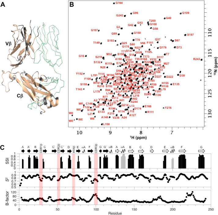
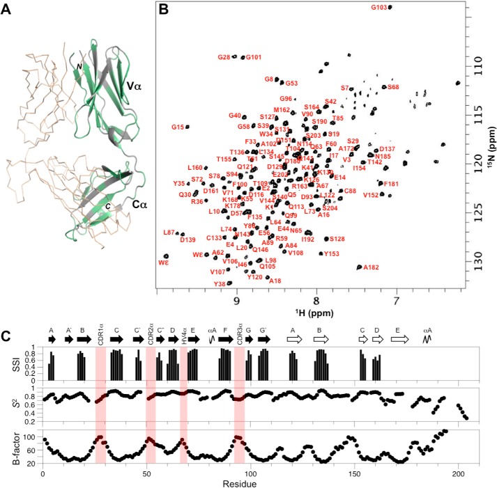
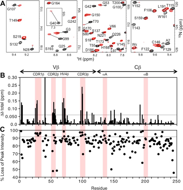
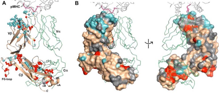
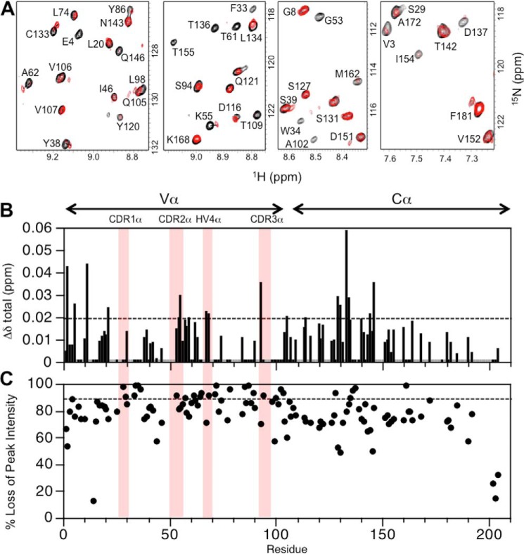
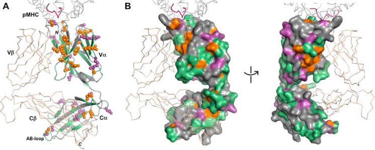
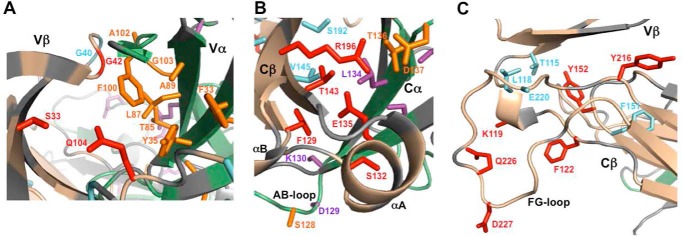
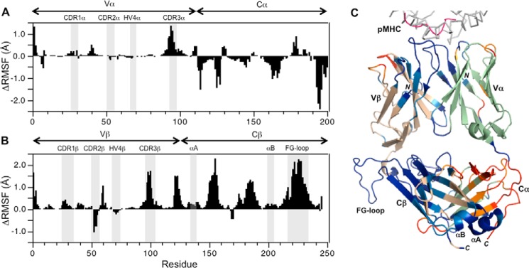
T cells generate adaptive immune responses mediated by the T cell receptor (TCR)-CD3 complex comprising an αβ TCR heterodimer noncovalently associated with three CD3 dimers. In early T cell activation, αβ TCR engagement by peptide-major histocompatibility complex (pMHC) is first communicated to the CD3 signaling apparatus of the TCR-CD3 complex, but the underlying mechanism is incompletely understood. It is possible that pMHC binding induces allosteric changes in TCR conformation or dynamics that are then relayed to CD3. Here, we carried out NMR analysis and molecular dynamics (MD) simulations of both the α and β chains of a human antiviral TCR (A6) that recognizes the Tax antigen from human T cell lymphotropic virus-1 bound to the MHC class I molecule HLA-A2. We observed pMHC-induced NMR signal perturbations in the TCR variable (V) domains that propagated to three distinct sites in the constant (C) domains: 1) the Cβ FG loop projecting from the Vβ/Cβ interface; 2) a cluster of Cβ residues near the Cβ αA helix, a region involved in interactions with CD3; and 3) the Cα AB loop at the membrane-proximal base of the TCR. A biological role for each of these allosteric sites is supported by previous mutational and functional studies of TCR signaling. Moreover, the pattern of long-range, ligand-induced changes in TCR A6 revealed by NMR was broadly similar to that predicted by the MD simulations. We propose that the unique structure of the TCR β chain enables allosteric communication between the TCR-binding sites for pMHC and CD3.

Keywords: T cell activation; T cell receptor (TCR); adaptive immunity; allosteric regulation; conformational change; major histocompatibility complex (MHC); molecular dynamics; nuclear magnetic resonance (NMR); signaling.


Conflict of interest statement

The authors declare that they have no conflicts of interest with the contents of this article

Figures

Figure 1.
Figure 1.
NMR assignment of the TCR ectodomain A6α[β-2H,13C,15N]. A, extent of backbone assignments for the TCR A6 β chain mapped onto the A6 X-ray structure (PDB accession code 3QH3) (34). Assigned regions of the β chain are shown in wheat; unassigned regions including prolines are shown in gray; and the α chain is in green ribbon format. B, two-dimensional 1H-15N TROSY–HSQC spectrum of A6α[β-2H,13C,15N] with backbone amide assignments. C, TCR A6 β chain secondary structure and dynamics from chemical shift data. Hypervariable regions are highlighted (light red). Top panel, secondary structure elements from chemical shift analysis. Confidence levels from TALOS-N (35) are plotted versus residue number for β-strands (black) and α-helices (gray). Center panel, chemical shift derived order parameters (S2) from TALOS-N. Bottom panel, crystallographic temperature (B) factors for the Cα atoms in the TCR A6 β chain, obtained using PDB 3QH3.
Figure 2.
Figure 2.
NMR assignment of the TCR ectodomain A6[α-2H,13C,15N]β. A, extent of backbone assignments for the TCR A6 α chain mapped onto the A6 X-ray structure (3QH3) (34). Assigned regions of the α chain are shown in green; unassigned regions including prolines are shown in gray; and the β chain is in wheat ribbon format. B, two-dimensional 1H-15N TROSY–HSQC spectrum of A6[α-2H,13C,15N]β with backbone amide assignments. C, TCR A6 α chain secondary structure and dynamics from chemical shift data. Hypervariable regions are highlighted (light red). Top panel, secondary structure elements from chemical shift analysis. Confidence levels from TALOS-N (35) are plotted versus residue number for β-strands (black). Center panel, chemical shift derived order parameters (S2) from TALOS-N. Bottom panel, crystallographic temperature (B) factors for Cα atoms in the TCR A6 α chain, obtained using PDB 3QH3.
Figure 3.
Figure 3.
Summary of changes to the NMR spectrum of A6α[β-2H,15N] upon addition of the pMHC ligand, Tax–HLA-A2. A, regions from the overlaid 2D 1H-15N TROSY–HSQC spectra of free A6α[β-2H,15N] (black) and Tax–HLA-A2-bound A6α[β-2H,15N] (red). For visual comparison purposes only, the bound-state spectrum is scaled so that peak intensities of most signals match those of the unbound state, highlighting the differential loss of peak intensity. B, combined 1H and 15N chemical shift perturbations, Δδtotal (ppm), in the TCR A6 β chain as a function of residue number. The dotted line indicates the mean value of Δδtotal plus 1 S.D. Gray histogram bars indicate prolines and unassigned residues. Pink columns indicate residue boundaries of CDR1β, CDR2β, HV4β, CDR3β, αA and αBβ. C, plot of percent loss of peak intensity versus residue number. The dotted line represents the mean percent loss of peak intensity plus 1 S.D. Hypervariable regions and the αA and αB helices in the Cβ domain are highlighted.
Figure 4.
Figure 4.
Effect of Tax–HLA-A2 binding on TCR A6 β chain residues. A, TCR A6 β chain residues with experimentally significant changes (≥ mean plus 1 S.D.) upon addition of Tax–HLA-A2 are highlighted. The A6 color scheme is as follows: β chain (wheat); unassigned β chain residues, including prolines (gray); significant CSPs (cyan); significant peak intensity changes (red); and α chain (green). The portion of the Tax–HLA-A2 at the interface with TCR A6 is shown as a gray ribbon for the MHC and pink for the peptide (PDB code 1QRN) (33). B, surface representation of the TCR A6 β chain in two orientations with the same color scheme as in A.
Figure 5.
Figure 5.
Summary of changes to the NMR spectrum of A6[α-2H,13C,15N]β upon addition of the pMHC ligand, Tax–HLA-A2. A, regions from the overlaid 2D 1H-15N TROSY–HSQC spectra of free A6[α-2H,15N]β (black) and Tax–HLA-A2-bound A6[α-2H,15N]β (red). The bound state spectrum is scaled as in Fig. 3A. B, combined 1H and 15N chemical shift perturbations, Δδtotal (ppm), in the TCR A6 α chain as a function of residue number. The dotted line indicates the mean value of Δδtotal plus 1 S.D. Gray histogram bars indicate prolines and unassigned residues. Pink columns indicate residue boundaries of CDR1β, CDR2β, HV4β, CDR3β, αA and αBβ. C, plot of percent loss of peak intensity versus residue. The dotted line represents the mean percent loss of peak intensity plus 1 S.D. Hypervariable regions are highlighted.
Figure 6.
Figure 6.
Effect of Tax–HLA-A2 binding on TCR A6 α residues. A, TCR A6 α chain residues with experimentally significant changes (≥ mean plus 1 S.D.) upon addition of Tax–HLA-A2 are highlighted. The color scheme is as follows: α chain (green); unassigned α chain residues including prolines (gray); significant CSPs (purple); significant peak intensity changes (orange); and β chain (wheat). The portion of Tax–HLA-A2 at the interface with TCR A6 is shown as a gray ribbon for the MHC and pink for the peptide (PDB code 1QRN) (33). B, surface representation of the A6 α chain in two orientations with the same color scheme as in A.
Figure 7.
Figure 7.
Residues in interfacial regions of TCR A6 with NMR signals perturbed by Tax–HLA-A2 binding. A, Vβ/Vα interface with the following color scheme: β chain (wheat); α chain (green); unassigned/proline residues (gray); experimentally significant CSPs (β chain/cyan and α chain/purple); experimentally significant loss of peak intensities (β chain/red and α chain/orange). B, Cβ/Cα interface. C, Vβ/Cβ interface. Color schemes in B and C are as in A.
Figure 8.
Figure 8.
Molecular dynamics simulations of TCR A6 in unbound and pMHC-bound states. A, ΔRMSF (unbound − bound) values for the TCR A6 α chain. Positive values indicate A6 α chain regions that become more rigid upon binding to Tax–HLA-A2, whereas negative values indicate increased TCR α chain flexibility. Hypervariable regions are highlighted. B, ΔRMSF values for the A6 β chain. C, ΔRMSF values mapped onto the X-ray structure of TCR A6 in complex with Tax–HLA-A2 (1QRN) (33). The individual structural components of the complex are color-coded as follows: HLA-A2 (gray); Tax peptide (magenta); TCR A6 α chain (green); TCR A6 β chain (wheat). The ΔRMSF values are mapped onto the TCR in the following way: ΔRMSF >0.2 Å (blue); 0.1 < ΔRMSF<0.2 Å (light blue); −0.2 < ΔRMSF < −0.1 Å (orange); and ΔRMSF < −0.2 Å (red).
Figure 9.
Figure 9.
Vα/Cα and Vβ/Cβ domain interfaces in human and mouse TCR structures. A, structures shown are 28 TCR α chain X-ray structures (Table S1). B, structures shown are 27 β chain X-ray structures (Table S2). The structures are nonredundant by TRAV or TRBV gene usage and are superposed by Vα or Vβ domains. The orientation between Vα and Cα domains displays greater variability than that between Vβ and Cβ domains. Selected frequently observed V/C interface residues are shown as sticks and labeled by residue number (based on A6 TCR numbering). Highly conserved residues are labeled by predominant or sole amino acid(s) observed at that position in structurally characterized TCRs and human and mouse germline genes.

Similar articles

Cited by

References

    1. Wucherpfennig K. W., Gagnon E., Call M. J., Huseby E. S., and Call M. E. (2010) Structural biology of the T-cell receptor: insights into receptor assembly, ligand recognition, and initiation of signaling. Cold Spring Harb. Perspect. Biol. 2, a005140 - PMC - PubMed
    1. Kuhns M. S., and Badgandi H. B. (2012) Piecing together the family portrait of TCR–CD3 complexes. Immunol. Rev. 250, 120–143 10.1111/imr.12000 - DOI - PubMed
    1. Blum J. S., Wearsch P. A., and Cresswell P. (2013) Pathways of antigen processing. Annu. Rev. Immunol. 31, 443–473 10.1146/annurev-immunol-032712-095910 - DOI - PMC - PubMed
    1. Rudolph M. G., Stanfield R. L., and Wilson I. A. (2006) How TCRs bind MHCs, peptides, and coreceptors. Annu. Rev. Immunol. 24, 419–466 10.1146/annurev.immunol.23.021704.115658 - DOI - PubMed
    1. Marrack P., Scott-Browne J. P., Dai S., Gapin L., and Kappler J. W. (2008) Evolutionarily conserved amino acids that control TCR-MHC interaction. Annu. Rev. Immunol. 26, 171–203 10.1146/annurev.immunol.26.021607.090421 - DOI - PMC - PubMed

Publication types

MeSH terms

Substances

LinkOut - more resources