. 2018 Aug 22;10(455):eaaq0693.
doi: 10.1126/scitranslmed.aaq0693.

An extracellular matrix fragment drives epithelial remodeling and airway hyperresponsiveness

Affiliations

An extracellular matrix fragment drives epithelial remodeling and airway hyperresponsiveness

Dhiren F Patel et al. Sci Transl Med. .

Abstract

It is anticipated that bioactive fragments of the extracellular matrix (matrikines) can influence the development and progression of chronic diseases. The enzyme leukotriene A4 hydrolase (LTA4H) mediates opposing proinflammatory and anti-inflammatory activities, through the generation of leukotriene B4 (LTB4) and degradation of proneutrophilic matrikine Pro-Gly-Pro (PGP), respectively. We show that abrogation of LTB4 signaling ameliorated inflammation and airway hyperresponsiveness (AHR) in a murine asthma model, yet global loss of LTA4H exacerbated AHR, despite the absence of LTB4 This exacerbated AHR was attributable to a neutrophil-independent capacity of PGP to promote pathological airway epithelial remodeling. Thus, we demonstrate a disconnect between airway inflammation and AHR and the ability of a matrikine to promote an epithelial remodeling phenotype that negatively affects lung function. Subsequently, we show that substantial quantities of PGP are detectable in the sputum of moderate-severe asthmatics in two distinct cohorts of patients. These studies have implications for our understanding of remodeling phenotypes in asthma and may rationalize the failure of LTA4H inhibitors in the clinic.


Conflict of interest statement

Competing interests: The authors declare that they have no competing interests.

Figures

Fig. 1.
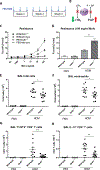
Fig. 1.. Augmented airway resistance in HDM-treated lta4h−/− mice despite reduced airway inflammation.
(A) Wild-type and lta4h−/− mice were administered HDM or phosphate-buffered saline (PBS) intranasally three times per week for 3 weeks. (B) Schematic depicting the opposing proinflammatory (LTB4 generation) and anti-inflammatory (PGP degradation) activities of LTA4H. Abrogation of LTA4H activity (red crosses) will result in reduced LTB4 but increased PGP. (C) Airway resistance (R) to increasing doses of methacholine (Mch) was measured 24 hours after the final HDM/PBS exposure. (D) Airway resistance at 100 mg/ml methacholine. At 24 hours after final HDM/PBS exposure, total cell numbers in the BAL were assessed by trypan blue exclusion (E), and airway eosinophils (F), CD4+ T cells expressing T1/ST2 (G), and CD4+ T cells expressing IL-13 (H) were assessed by flow cytometry. Figures present combined data from two independent experiments with four to six mice per group in each experiment. Results depicted as means ± SEM. *P < 0.05, **P < 0.01, ***P < 0.001, using Mann-Whitney statistical test.
Fig. 2.
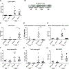
Fig. 2.. HDM-treated lta4h−/− mice exhibit reduced concentrations of TH2 inflammatory mediators relative to their littermate controls.
Wild-type and lta4h−/− mice were administered HDM or PBS intranasally three times per week for 3 weeks, and BALF and serum were collected 24 hours after the final HDM/PBS exposure. Concentrations of BALF IL-4 (A), BALF IL-5 (B), BALF IL-13 (C), BALF eotaxin-2 (D), serum MCPT-1 (E), BALF albumin (F), serum HDM-specific IgE antibodies (G), and serum HDM-specific IgG1 antibodies (H) were assessed by enzyme-linked immunosorbent assay (ELISA). Figures present combined data from two independent experiments with four to six mice per group in each experiment. Results depicted as means ± SEM. **P < 0.01, ***P < 0.001, using Mann-Whitney statistical test.
Fig. 3.
Fig. 3.. HDM-treated lta4h−/− mice exhibit PGP accumulation but comparable neutrophil infiltrate relative to littermate controls.
Wild-type and lta4h−/− mice were administered HDM or PBS intranasally three times per week for 3 weeks, and BALF and lung tissue were collected 24 hours after the final HDM/PBS exposure. Amounts of MMP-9 in BALF were assessed by ELISA (A) and prolylendopeptidase by Western blot (B). The concentration of PGP in BALF was determined by mass spectrometry (C). BALF was incubated with exogenous PGP and degradation assessed after 2 hours by mass spectrometry (D) or release of free proline (E). The number of neutrophils recruited into the lung tissue (F) and airways (G) was determined by flow cytometry. The concentration of MPO (H) in the BALF was determined by ELISA. Figures present combined data from two independent experiments with four to six mice per group in each experiment. Results depicted as means ± SEM. ***P < 0.001, using Mann-Whitney statistical test.
Fig. 4.
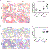
Fig. 4.. HDM-treated lta4h−/− mice exhibit augmented airway epithelial remodeling relative to littermate controls.
Wild-type and lta4h−/− mice were administered HDM or PBS intranasally three times per week for 3 weeks, and 24 hours after the final HDM/PBS exposure, lungs were removed for histological examination. (A) Representative H&E-stained lung sections from wild-type and lta4h−/− mice administered PBS or HDM. (B) Epithelial cell height around medium-sized conducting airways was assessed from H&E-stained lung sections. Data presented are an average per mouse. (C) Representative PAS-stained lung sections from wild-type and lta4h−/− mice administered PBS or HDM. (D) Goblet cells were scored from PAS-stained sections. The sum of airway scores from each lung was divided by the number of airways examined and expressed as mucus cell score in arbitrary units. Figures present combined data from two independent experiments with four to six mice per group in each experiment. Results depicted as means ± SEM. *P < 0.05, ***P < 0.001, using Mann-Whitney statistical test. Scale bars, 20 μm.
Fig. 5.
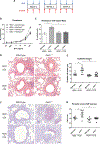
Fig. 5.. PGP neutralization attenuates the heightened airway resistance and epithelial remodeling observed in HDM-exposed lta4h−/− mice.
(A) Wild-type and lta4h−/− mice were administered HDM intranasally three times per week for 3 weeks. Mice were concomitantly administered PGP antagonist, RTR, or control peptide, ASA. (B) Airway resistance to increasing doses of methacholine was measured 24 hours after the final HDM exposure. (C) Airway resistance at 100 mg/ml methacholine. (D) Representative H&E-stained lung sections from wild-type and lta4h−/− mice administered HDM with either RTR or ASA. (E) Epithelial cell height around medium-sized conducting airways was assessed from H&E-stained lung sections. Data presented are an average per mouse. (F) Representative PAS-stained lung sections from wild-type and lta4h−/− mice administered HDM with either RTR or ASA. (G) Goblet cells were scored from PAS-stained sections. The sum of airway scores from each lung was divided by the number of airways examined and expressed as mucus cell score in arbitrary units. Figures present combined data from two independent experiments with five mice per group in each experiment. Results depicted as means ± SEM. *P < 0.05, **P < 0.01, using Mann-Whitney statistical test. Scale bars, 20 μm.
Fig. 6.
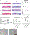
Fig. 6.. AcPGP induces remodeling of human bronchial epithelial cells.
Normal human bronchial epithelial cells were cultured at ALI. Respective wells were treated with media or media supplemented with AcPGP (10 μg/ml) and/or IL-13 (10 ng/ml) for 7 days. Apical supernatants were collected, and cells were fixed for histological analysis. (A) Representative H&E- and PAS-stained sections of ALI culture epithelium after 7 days of treatment. (B) Epithelial cell height was assessed from H&E-stained sections, with data presented as an average per well. (C) Apical supernatant Muc5AC was assessed by ELISA at day 7 after treatment. Undifferentiated normal human bronchial epithelial cells were cultured in media or media supplemented with AcPGP (10 ng/ml) and visualized over a period of 72 hours using a JuLI Stage automated cell imaging system. In some groups, AcPGP-treated cells were preincubated with either anti-CXCR1/2 antibodies or IgG2a isotype control antibody. (D) Intragroup fold change in percent cell confluence of cells at 72 hours after each treatment relative to 0 hours. (E) Fold change in cell confluence over 72-hour period depicted for individual wells in each treatment group. (F) After 72 hours, the cells were fixed and stained for Ki-67, with Ki-67–positive cells expressed as a percentage of 4′,6-diamidino-2-phenylindole (DAPI)–positive cells. (G) Bright-field image of epithelial cells treated with media or AcPGP (10 ng/ml) after 72 hours. Figures (A) to (C) present combined data from two independent experiments with two wells per group in each experiment. Figures (D) to (F) represent data combined from two independent experiments with five wells per group in each experiment. Results depicted as means ± SEM. *P < 0.05, **P < 0.01, ***P < 0.001, using Mann-Whitney statistical test (B and C) or analysis of variance (ANOVA) with Bonferroni correction (E and F). Scale bars, 20 μm.
Fig. 7.
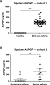
Fig. 7.. The matrikine AcPGP is elevated in the sputum of severe asthmatics.
(A) The concentration of AcPGP peptide in the sputum of patient cohort 1 (UK cohort), consisting of healthy controls (n = 15) and moderate-severe asthmatics (mod-sev asthma; n = 42), as determined by mass spectrometry. (B) The concentration of AcPGP peptide in the sputum of patient cohort 2 (US cohort), consisting of severe (n = 8) and nonsevere asthmatics (n = 9), as determined by mass spectrometry. The horizontal bar depicts the median of each group. Results depicted as means ± SEM. **P < 0.01, ***P < 0.001, using Mann-Whitney statistical test.

Comment in

Similar articles

Cited by

References

    1. Saglani S, Lloyd CM, Novel concepts in airway inflammation and remodelling in asthma. Eur. Respir. J 46, 1796–1804 (2015). - PubMed
    1. Haeggström JZ, Tholander F, Wetterholm A, Structure and catalytic mechanisms of leukotriene A4 hydrolase. Prostaglandins Other Lipid Mediat. 83, 198–202 (2007). - PubMed
    1. Tager AM, Luster AD, BLT1 and BLT2: The leukotriene B4 receptors. Prostaglandins Leukot. Essent. Fatty Acids 69, 123–134 (2003). - PubMed
    1. Di Gennaro A, Haeggström JZ, The leukotrienes: Immune-modulating lipid mediators of disease. Adv. Immunol 116, 51–92 (2012). - PubMed
    1. Di Gennaro A, Haeggström JZ, Targeting leukotriene B4 in inflammation. Expert Opin. Ther. Targets 18, 79–93 (2014). - PubMed

Publication types

MeSH terms