. 2018 Aug 23;3(16):e121882.
doi: 10.1172/jci.insight.121882.

Enterovirus D68 infection induces IL-17-dependent neutrophilic airway inflammation and hyperresponsiveness

Affiliations

Enterovirus D68 infection induces IL-17-dependent neutrophilic airway inflammation and hyperresponsiveness

Charu Rajput et al. JCI Insight. .

Abstract

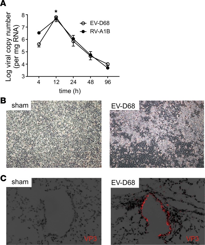
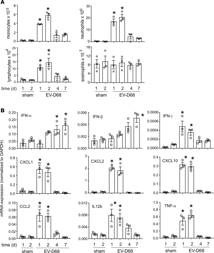
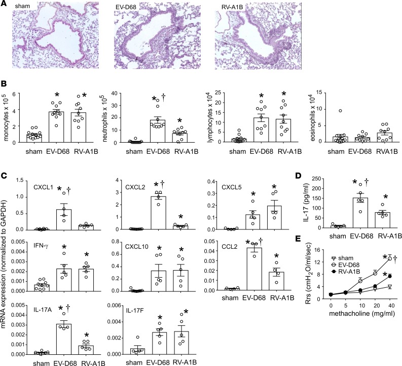
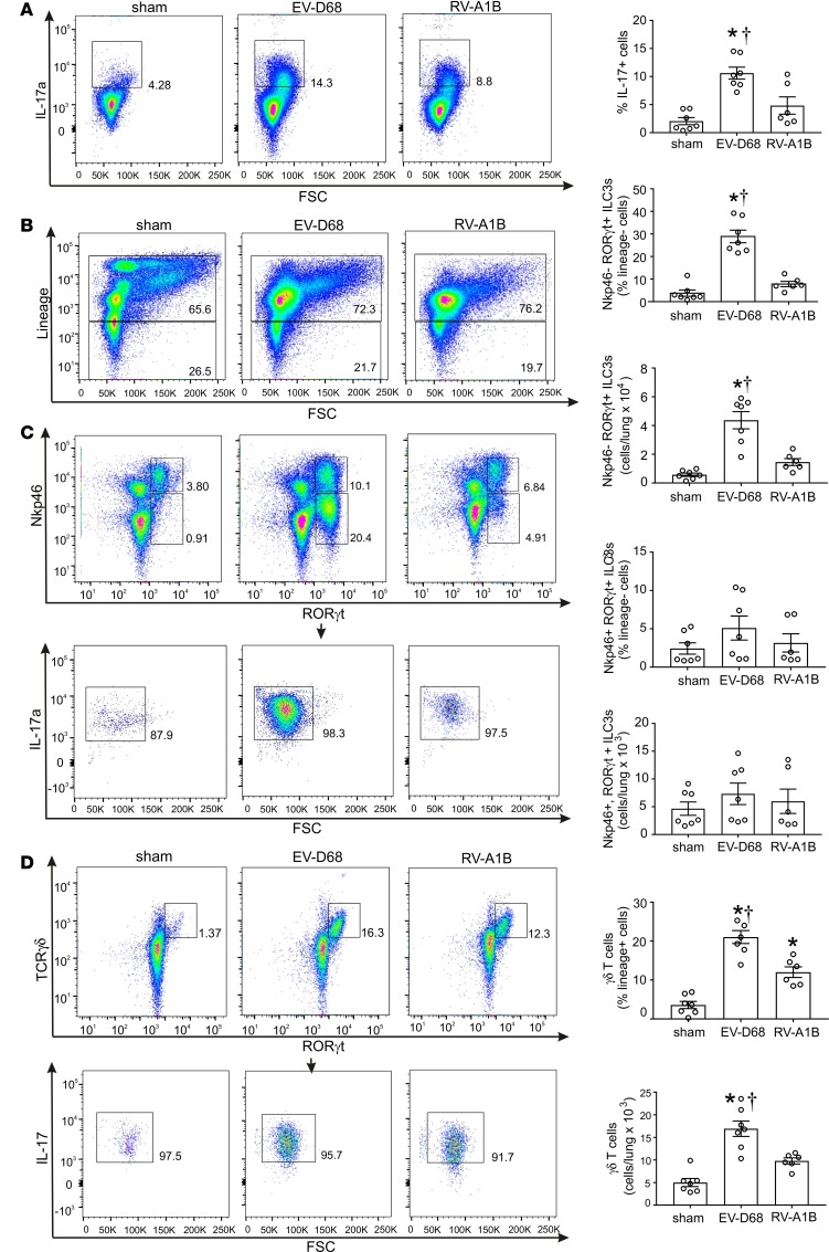
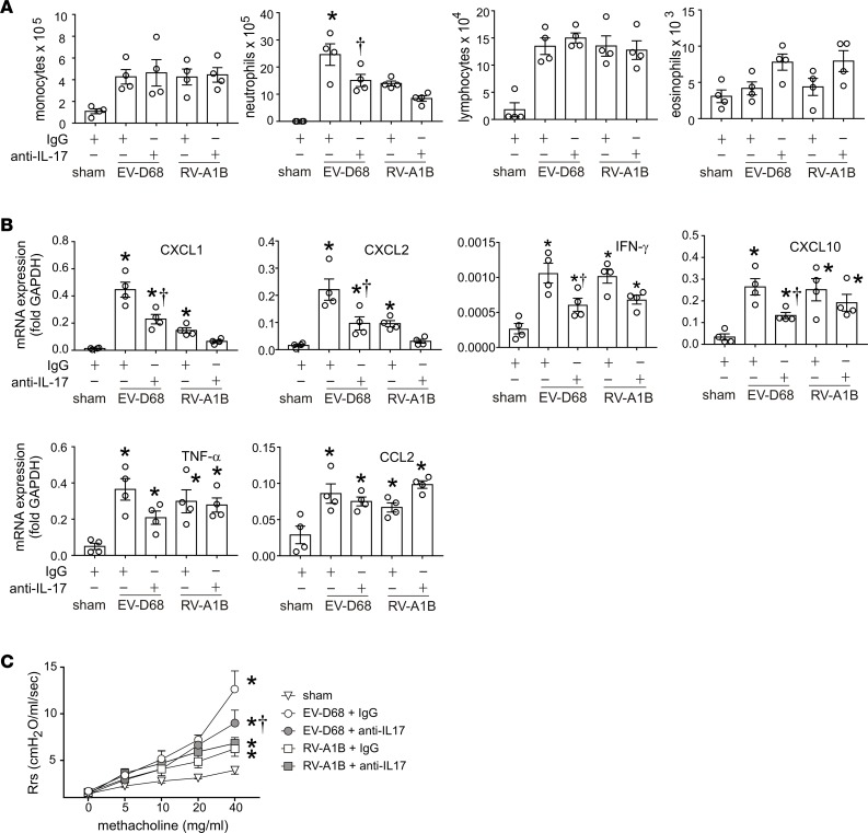
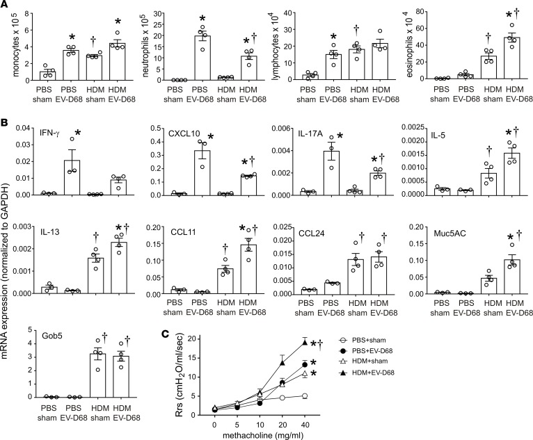
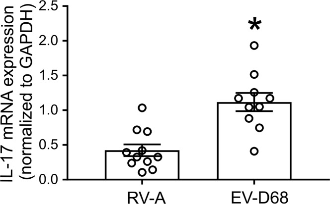
Enterovirus D68 (EV-D68) shares biologic features with rhinovirus (RV). In 2014, a nationwide outbreak of EV-D68 was associated with severe asthma-like symptoms. We sought to develop a mouse model of EV-D68 infection and determine the mechanisms underlying airway disease. BALB/c mice were inoculated intranasally with EV-D68 (2014 isolate), RV-A1B, or sham, alone or in combination with anti-IL-17A or house dust mite (HDM) treatment. Like RV-A1B, lung EV-D68 viral RNA peaked 12 hours after infection. EV-D68 induced airway inflammation, expression of cytokines (TNF-α, IL-6, IL-12b, IL-17A, CXCL1, CXCL2, CXCL10, and CCL2), and airway hyperresponsiveness, which were suppressed by anti-IL-17A antibody. Neutrophilic inflammation and airway responsiveness were significantly higher after EV-D68 compared with RV-A1B infection. Flow cytometry showed increased lineage-, NKp46-, RORγt+ IL-17+ILC3s and γδ T cells in the lungs of EV-D68-treated mice compared with those in RV-treated mice. EV-D68 infection of HDM-exposed mice induced additive or synergistic increases in BAL neutrophils and eosinophils and expression of IL-17, CCL11, IL-5, and Muc5AC. Finally, patients from the 2014 epidemic period with EV-D68 showed significantly higher nasopharyngeal IL-17 mRNA levels compared with patients with RV-A infection. EV-D68 infection induces IL-17-dependent airway inflammation and hyperresponsiveness, which is greater than that generated by RV-A1B, consistent with the clinical picture of severe asthma-like symptoms.

Keywords: Allergy; Asthma; Cytokines; Infectious disease; Pulmonology.


Conflict of interest statement

Conflict of interest: The authors have declared that no conflict of interest exists.

Figures

Figure 1
Figure 1. Viral RNA is detectable in the lungs of EV-D68–treated mice.
(A) Female 8- to 10-week-old BALB/c mice were inoculated with 5 × 10⁶ ePFU of EV-D68 or 5 × 10⁶ PFU of RV-A1B by intranasal instillation, and lungs and nasal washes were examined by RT-PCR for viral RNA at the indicated time points. There was a small significant increase in lung viral RNA copy number between the 4- and 12-hour time point, and RNA was detected up to day 4. Data are shown as mean ± SEM; n =3 mice/group from 3 different experiments; *P < 0.05 by 1-way ANOVA, compared with 4-hour time point. (B) Supernatants from homogenized mouse lungs from sham- or EV-D68–inoculated mice were overlaid onto confluent rhabdomyosarcoma cell monolayers and examined for cytopathic effects. The image on the right shows cytopathic effects of EV-D68. (C) After inoculation, lungs were fixed and processed for immunofluorescence staining using Alexa Fluor 568–labeled anti-VP3 antibody. The image on the right shows EV-D68 in epithelial cells. Images were taken at ×200 magnification.
Figure 2
Figure 2. EV-D68 inoculation increases airway inflammation.
Female 8- to 10-week-old BALB/c mice were treated with sham or 5 × 10⁶ ePFU of EV-D68. The lungs were harvested after the indicated time points and processed for BAL and mRNA expression. (A) Inflammatory cell counts in BAL at indicated time points. (B) mRNA expression analysis of indicated genes at indicated time points. Data are shown as mean ± SEM; n = 3 mice in each group from two different experiments; *P < 0.05 by 1-way ANOVA, compared with sham.
Figure 3
Figure 3. EV-D68 induces greater neutrophil inflammation than RV-A1B.
Female 8- to 10-week-old BALB/c mice were treated with sham, 5 × 10⁶ ePFU of EV-D68, or 5 × 10⁶ PFU of RV-A1B. Mice were sacrificed 48 hours later and analyzed for histology, BAL, and qPCR. In another set of mice, changes in airway resistance to nebulized increasing doses of methacholine (0 to 40mg/ml) were assessed. (A) Images showing the inflammation in lungs 48 hours after treatment with sham, EV-D68, or RV-A1B. Images were taken at ×200 magnification. (B) Analysis of BAL inflammatory cells. (C) qPCR analysis of indicated genes. (D) BAL IL-17 protein levels in the indicated groups. (E) Airway responsiveness to methacholine in the 3 groups of mice. Data are shown as mean ± SEM of 3–5 mice/group from at least 2 different experiments; *P < 0.05 by 1-way ANOVA, compared with sham; †P < 0.05 by 1-way ANOVA, compared with RV-1B.
Figure 4
Figure 4. Flow cytometric analysis of IL-17–producing lung cells.
Female 8- to 10-week-old BALB/c mice were treated with sham, 5 × 10⁶ ePFU of EV-D68, or 5 × 10⁶ PFU of RV-A1B. Forty-eight hours later, lungs were harvested, digested with collagenase, and stained with lineage antibody cocktail, anti-NKp46, anti-TCRγδ, anti-RORγt, anti–IL-17, and Pacific Blue (for dead cells). Cells were washed, fixed, and processed for flow cytometry. (A) Flow cytometry of live IL-17+ cells in sham, EV-D68, and RV-A1B groups. Group mean data are also shown. (B) Dot blots of lineage+ and lineage live cells for the 3 conditions. (C) NKp46 and RORγt staining of lineage cells. Group mean data for NKp46, RORγt+, and NKp46 and RORγt double-positive cells are also shown. NKp46 and RORγt+ double-positive cells are gated for IL-17 staining. (D) TCR γδ and RORγt staining of lineage+ cells. Group mean data are also shown. TCR γδ and RORγt double-positive cells are gated for IL-17 staining. Data are shown as mean ± SEM of 6–7 mice/group from 2 separate experiments; *P < 0.05 by 1-way ANOVA, compared with sham; †P < 0.05 by 1-way ANOVA, compared with RV-1B.
Figure 5
Figure 5. Treatment with anti–IL-17 antibody reduces EV-D68–induced airway responsiveness.
Female 8- to 10-week-old BALB/c mice were treated with sham, 5 × 10⁶ ePFU of EV-D68, or 5 × 10⁶ PFU of RV-A1B alone or in combination with anti–IL-17 antibody or isotype control. Changes in airway resistance to nebulized increasing doses of methacholine were assessed in tracheotomized mice using a Buxco FinePointe plethysmograph. BAL and mRNA analysis was also carried out in similarly treated mice. (A) BAL cell counts for the indicated groups. (B) qPCR analysis of lung mRNA expression. Data are shown as mean ± SEM for 4 mice in each group from a single experiment. *P < 0.05 by ANOVA, compared with RV-1B; †P < 0.05 by ANOVA, compared with IgG. (C) Airway responsiveness to increasing doses of methacholine. n =3–4/group from a single experiment. *P < 0.05 by 2-way ANOVA, compared with sham; †P < 0.05 by 2-way ANOVA, compared with EV-D68 + IgG.
Figure 6
Figure 6. EV-D68 induces type 2 cytokines, mucus genes, and greater airway hyperresponsiveness than RV-1B in allergen-challenged mice.
Female 8- to 12-week-old BALB/c mice were challenged with house dust mite (HDM) and treated with sham, 5 × 10⁶ ePFU of EV-D68, or 5 × 10⁶ PFU of RV-A1B. Lungs were harvested, and RNA was extracted for qPCR. BAL analysis was also carried out in similarly treated mice. A separate set of mice was similarly treated and anesthetized and endotracheally intubated for measurement of airway responsiveness. (A) BAL cell counts for PBS/sham-, PBS/EV-D68–, HDM/sham-, and HDM/EV-D68–treated groups. (B) qPCR analysis of the indicated genes for the 4 groups. Data are shown as mean ± SEM of 3–4 mice/group for a single experiment; *P < 0.05 by ANOVA, compared with sham, †P < 0.05 by ANOVA, compared with PBS. (C) Airway methacholine responsiveness of the indicated treatment groups. n =3–4/group from a single experiment; *P < 0.05 by 2-way ANOVA, compared with sham; †P < 0.05 by 2-way ANOVA, compared with all other groups.
Figure 7
Figure 7. Nasopharyngeal IL-17 mRNA expression in patients with airway obstruction.
RNA was extracted from nasopharyngeal swabs and was subjected to cDNA synthesis, followed by qPCR using EV-D68–, RV-, and IL-17–specific primers. The relative expression of IL-17 in RV-A– and EV-D68–infected patients, normalized to GAPDH, is shown. *P < 0.05 by unpaired t test, compared with RV-A.

References

    1. Oberste MS, et al. Enterovirus 68 is associated with respiratory illness and shares biological features with both the enteroviruses and the rhinoviruses. J Gen Virol. 2004;85(Pt 9):2577–2584. - PubMed
    1. Ishiko H, et al. Human rhinovirus 87 identified as human enterovirus 68 by VP4-based molecular diagnosis. Intervirology. 2002;45(3):136–141. doi: 10.1159/000065866. - DOI - PubMed
    1. Schieble JH, Fox VL, Lennette EH. A probable new human picornavirus associated with respiratory diseases. Am J Epidemiol. 1967;85(2):297–310. doi: 10.1093/oxfordjournals.aje.a120693. - DOI - PubMed
    1. Centers for Disease Control Prevention (CDC) Clusters of acute respiratory illness associated with human enterovirus 68--Asia, Europe, and United States, 2008-2010. MMWR Morb Mortal Wkly Rep. 2011;60(38):1301–1304. - PubMed
    1. Messacar K, Abzug MJ, Dominguez SR. 2014 outbreak of enterovirus D68 in North America. J Med Virol. 2016;88(5):739–745. doi: 10.1002/jmv.24410. - DOI - PubMed

Publication types

MeSH terms

Supplementary concepts