

Practice Guideline
doi: 10.5551/jat.GL2017.
Epub 2018 Aug 22.
Japan Atherosclerosis Society (JAS) Guidelines for Prevention of Atherosclerotic Cardiovascular Diseases 2017
Affiliations
- PMID: 30135334
- PMCID: PMC6143773
- DOI: 10.5551/jat.GL2017
Item in Clipboard
Practice Guideline
Japan Atherosclerosis Society (JAS) Guidelines for Prevention of Atherosclerotic Cardiovascular Diseases 2017
J Atheroscler Thromb.
.
No abstract available
Figures
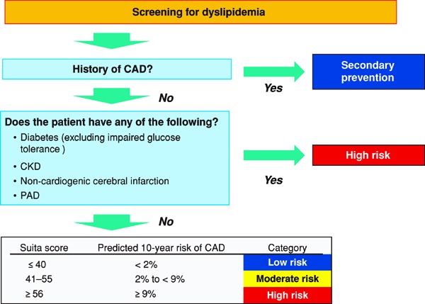
Flowchart Using the Suita Score to Establish LDL-C Management Targets, from the Perspective of CAD Prevention • The Suita score is calculated based on Fig. 2. • Note: For patients diagnosed with FH and those diagnosed with familial type Ⅲ hyperlipidemia, do not use this chart and refer to Chapter 5 (Familial Hypercholesterolemia) and Chapter 6 (Other Types of Primary Dyslipidemias), respectively.
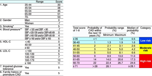
Model for Predicting CAD Onset Using the Suita Score * Ex-smokers should be regarded as nonsmokers. Note that the risk of CAD decreases by almost half 1 year after smoking cessation and drops to the same level as in nonsmokers after 15 years of smoking cessation. ** The current values are used even if the patient is currently undergoing treatment or not. However, counsel the patient while keeping in mind that patients undergoing treatment for hypertension have a higher risk of CAD than those who have the same blood pressure value without undergoing treatment.
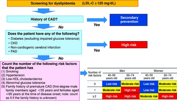
Flowchart for Establishing LDL-C Management Targets from the Perspective of CAD Prevention (Simplified Version Using Risk Factors)
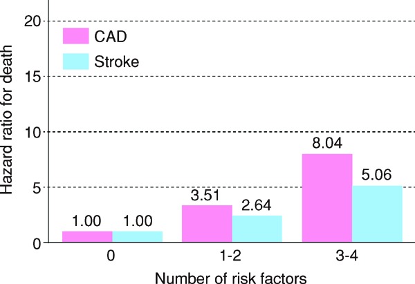
Relationship between the number of concurrent risk factors and death due to CAD and stroke (NIPPON DATA80: 1980–1994) Risk factors: Obesity, hypertension, hyperglycemia, hypercholesterolemia
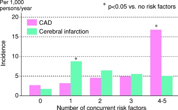
Rerationship between the number of concurrent risk factors and incidences of CAD and cerebral infarction Components of metabolic syndrome: Obesity, impaired glucose tolerance, lipidosis, hyperttention, hyperinsulinemia After adjustment for age, 5-year (1988–1993) follow-up of 1097 men and women aged ≥ 60 years in Hisayama-cho
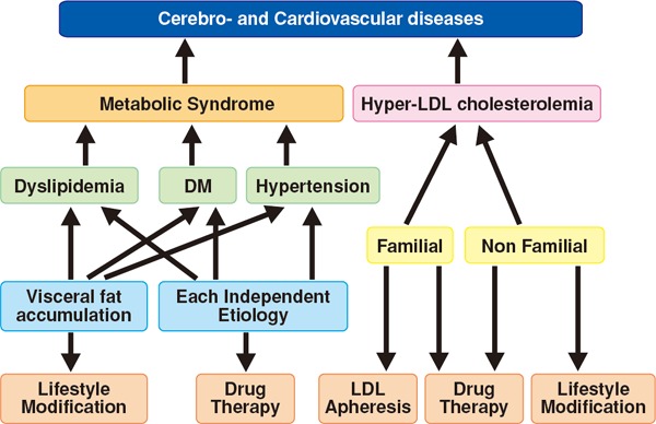
Comprehensive Risks and Risk Factors for Lifestyle Habits Joint Committee for Comprehensive Risk Management Chart for the Prevention of Cerebro- and Cardiovascular Diseases, The Journal of The Japanese Society of Internal Medicine 2015, Vol. 104, No. 4, 824–860
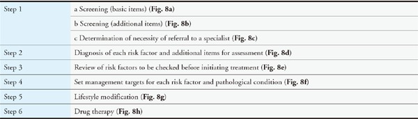
Comprehensive risk assessment and their management in six steps
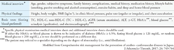
Step 1a Screening (Basic Items)
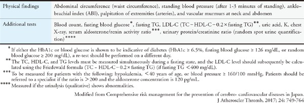
Step 1b Screening (Additional Items)
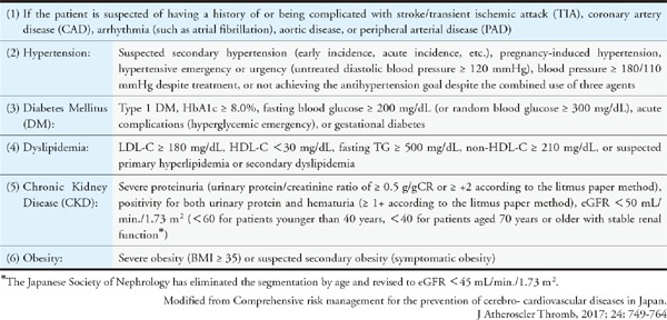
Step 1c Determination of Necessity for Referral to a Specialist
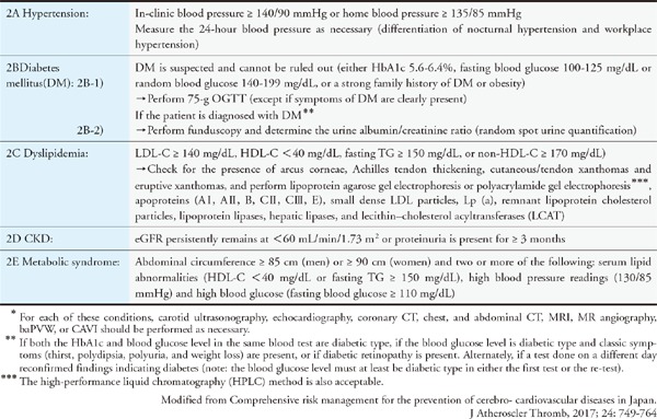
Step 2 Diagnosis of Each Risk Factor and Additional Items for Assessment*
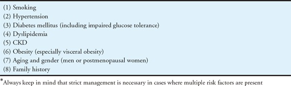
Step 3 Risk Factors to be Reviewed before Initiating Treatment

Step 4 Setting Management Targets Suited to the Risk Factors for Each Pathological Condition*
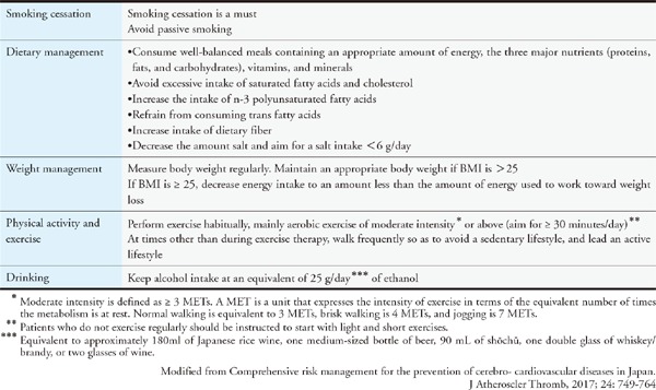
Step 5 Lifestyle modifications
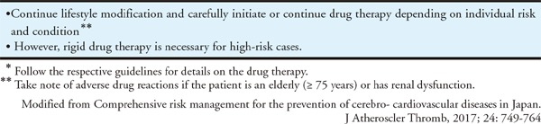
Step 6 Drug Therapy*
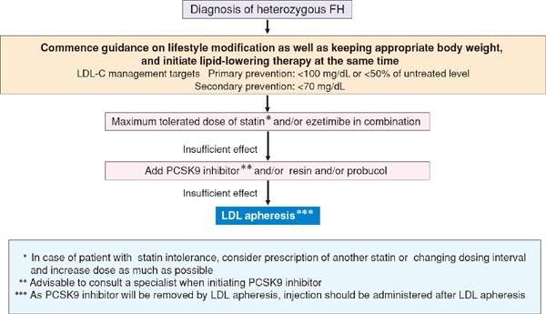
Treatment flow chart for adult (15 years or over) heterozygous FH
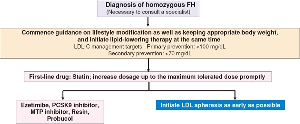
Treatment flow chart for adult (15 years or over) homozygous FH
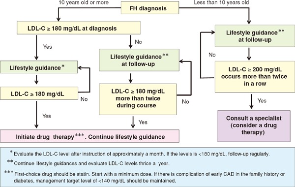
Algorithm for treatment of pediatric FH heterozygote
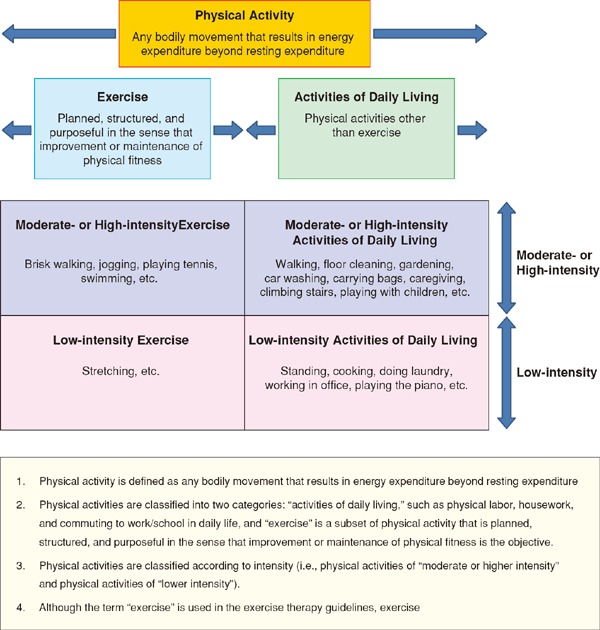
Adapted/modified from: Ministry of Health, Labour and Welfare “Exercise guidelines for health promotion 2006”
References
-
- The joint committee of “The Japan Academy of Neurosonology” and “The Japan Society of Embolus Detection and Treatment” on guideline for nuerosonology. Carotid ultrasound examination. Neurosonology, 2006; 19: 49-67 (in Japanese)
-
- Subcommittee for preparing guidelines for ultrasound diagnosis of carotid artery. Standard method for ultrasound evaluation of carotid artery lesions. Jpn J Med Ultrasonics, 2009; 36: 501-518 (in Japanese)
-
- Subcommittee for preparing guidelines for ultrasound diagnosis of carotid artery. Standard method for ultrasound evaluation of carotid artery lesions 2017. Jpn J Med Ultrasonics, 2018. https://www.jsum.or.jp/committee/diagnostic/pdf/jsum0515_guideline.pdf (in Japanese)
-
- Homma S, Hirose N, Ishida H, Ishii T, Araki G. Carotid plaque and intima-media thickness assessed by b-mode ultrasonography in subjects ranging from young adults to centenarians. Stroke, 2001; 32: 830-835 - PubMed
-
- Nezu T, Hosomi N, Aoki S, Matsumoto M. Carotid intima-media thickness for atherosclerosis. J Atheroscler Thromb, 2016; 23: 18-31 - PubMed