. 2018 Aug 22;8(1):12635.
doi: 10.1038/s41598-018-30895-8.

MicroRNA miR-509 Regulates ERK1/2, the Vimentin Network, and Focal Adhesions by Targeting Plk1

Affiliations

MicroRNA miR-509 Regulates ERK1/2, the Vimentin Network, and Focal Adhesions by Targeting Plk1

Guoning Liao et al. Sci Rep. .

Abstract

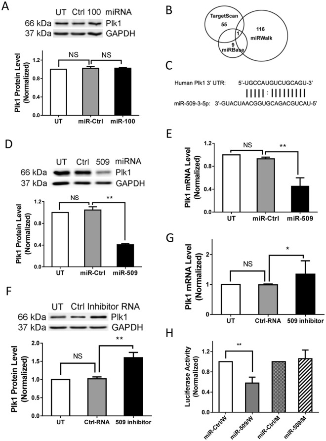
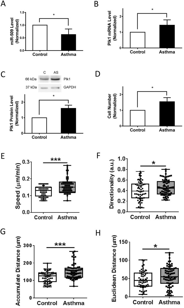
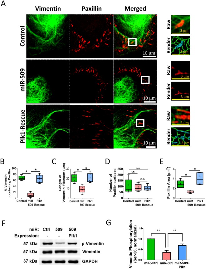
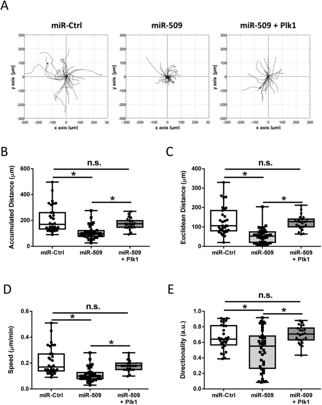
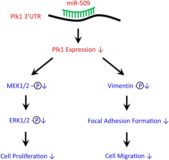
Polo-like kinase 1 (Plk1) has been implicated in mitosis, cytokinesis, and proliferation. The mechanisms that regulate Plk1 expression remain to be elucidated. It is reported that miR-100 targets Plk1 in certain cancer cells. Here, treatment with miR-100 did not affect Plk1 protein expression in human airway smooth muscle cells. In contrast, treatment with miR-509 inhibited the expression of Plk1 in airway smooth muscle cells. Exposure to miR-509 inhibitor enhanced Plk1 expression in cells. Introduction of miR-509 reduced luciferase activity of a Plk1 3'UTR reporter. Mutation of miR-509 targeting sequence in Plk1 3'UTR resisted the reduction of the luciferase activity. Furthermore, miR-509 inhibited the PDGF-induced phosphorylation of MEK1/2 and ERK1/2, and cell proliferation without affecting the expression of c-Abl, a tyrosine kinase implicated in cell proliferation. Moreover, we unexpectedly found that vimentin filaments contacted paxillin-positive focal adhesions. miR-509 exposure inhibited vimentin phosphorylation at Ser-56, vimentin network reorganization, focal adhesion formation, and cell migration. The effects of miR-509 on ERK1/2 and vimentin were diminished in RNAi-resistant Plk1 expressing cells treated with miR-509. Taken together, these findings unveil previously unknown mechanisms that miR-509 regulates ERK1/2 and proliferation by targeting Plk1. miR-509 controls vimentin cytoskeleton reorganization, focal adhesion assembly, and cell migration through Plk1.


Conflict of interest statement

The authors declare no competing interests.

Figures

Figure 1
Figure 1
Plk1 is regulated by miR-509 at mRNA and protein levels in smooth muscle cells. (A) Human airway smooth muscle (HASM) cells were transfected with either 20 nM miR-control (miR-Ctrl) or miR-100, or they were untransfected, for 3 days. Blots of the HASM cells were probed with antibodies against Plk1 and glyceraldehyde-3-phosphate dehydrogenase (GAPDH). Data are mean ± SD (n = 4). NS, not significant. (B) All three online miR search tools predict 3′UTR of human Plk1 as a target of miR-509. (C) Sequence alignment between miR-509 and 3′UTR of human Plk1. (D) Blots of untransfected HASM cells and cells transfected with either miR-Ctrl or miR-509 for 3 days were probed with antibodies against Plk1 and GAPDH. Data are mean ± SD (n = 4, **p < 0.01). (E) HASM cells were transfected with either 20 nM miR-Ctrl or miR-509, or they were untransfected for 3 days. Plk1 mRNA in the cells was assessed by RT-qPCR (reverse transcription quantitative real-time polymerase chain reaction). Data are mean ± SD (n = 4, **p < 0.01). (F) HASM cells were untransfected, or transfected with either 20 nM control RNA or miR-509 inhibitor for 2 days. miR-509 inhibitor is small, chemically modified single-strand RNA molecules designed to specifically bind to and inhibit endogenous miR-509. Blots of the cells were probed with antibodies against Plk1 and GAPDH. Data are mean ± SD (n = 4, **p < 0.01). (G) mRNA of Plk1 in untransfected cells and cells transfected with either 20 nM control RNA or miR-509 inhibitor for 2 days was assessed by RT-qPCR. Data are mean ± SD (n = 7, *p < 0.05). UT, untransfected cells. (H) Relative luciferase activity in cells cotransfected with plasmids encoding either wild-type (W) or mutant (M) Plk1 3′UTR plus miR-Ctrl or miR-509. Data are mean ± SD (n = 5, **p < 0.01). One-way ANOVA was used for statistical analysis.
Figure 2
Figure 2
Asthmatic human airway smooth muscle cells display reduced miR-509 expression, increased Plk1 expression, and enhanced proliferation and migration. (A) miR-509 expression in control and asthmatic HASM cells were determined using the experimental procedure described in Materials and Methods. miR-509 expression is reduced in asthmatic smooth muscle cells (Control cells from 5 donors, asthmatic cells from 4 donors, *p < 0.05). However, expression of Plk1 at mRNA (B) and protein (C) levels is higher in asthmatic HASM cells (Control cells from 5 donors, asthmatic cells from 4 donors, *p < 0.05). (D) Control and asthmatic HASM cells were treated with 10 ng/ml PDGF for 3 days followed by cell counting. The proliferation of asthmatic HASM cells is increased as compared to control cells (Control cells from 5 donors, asthmatic cells from 4 donors, *p < 0.05). (EH). Control and asthmatic HASM cells were replated in 6-well dishes for 5 hours, and migration of these cells was then evaluated by time-lapse microscopy for additional 16 h. NIH ImageJ software was used to analyze cell migration speed, directionality, accumulated distance, and Euclidean distance (*p < 0.05; ***p < 0.01; control cells, n = 58 from 5 donors; asthmatic cells, n = 72 from 4 donors). Student’s t-test was used for statistical analysis.
Figure 3
Figure 3
Role of miR-509 in cell proliferation, c-Abl expression, MEK1/2 and ERK1/2 phosphorylation. (A) HASM cells were transfected with miR-Ctrl or miR-509. One day after transfection, they were stimulated with 10 ng/ml PDGF for additional 1–3 days followed by assessment of cell numbers. Exposure to miR-509 inhibits the PDGF-induced proliferation. Data are mean ± SD (n = 5, **p < 0.01). (B) Treatment with miR-509 does not affect c-Abl protein expression. Blots of HASM cells transfected with either miR-Ctrl or miR-509 for 3 days were probed with antibodies against c-Abl and GAPDH. Data are mean ± SD (n = 5). NS, not significant. (C) HASM cells treated with miR-Ctrl or miR-509 for 3 days, or stable Plk1 (miR-509 resistant) expressing cells were treated with miR-509 for 3 days (rescue cells). Cell extracts were evaluated by immunoblot analysis. Data are mean ± SD (n = 5, **p < 0.01). (D) Representative immunoblots illustrating the role of Plk1 in phosphorylation of MEK1/2 (MEK1: Ser-218/Ser-222; MEK2: Ser-222/Ser-226) and ERK1/2 (ERK1: Thr-202/Tyr-204; ERK2: Thr-185/Tyr-187). HASM cells treated with miR-Ctrl or miR-509, or rescue cells (see above) were stimulated with 10 ng/ml PDGF for 10 min or left unstimulated. MEK1/2 and ERK1/2 phosphorylation in these cells was evaluated by immunoblot analysis. Rescue of Plk1 in miR-509 treated cells restores the PDGF-induced phosphorylation of MEK1/2 and ERK1/2. (E) The phosphorylation levels of MEK1/2 and ERK1/2 in cells stimulated with PDGF are normalized to corresponding unstimulated levels. Data are mean ± SD (n = 7, *p < 0.05; **p < 0.01). (F) Smooth muscle cells were treated with miR-Ctrl or miR-509, or stable Plk1 expressing cells were treated with miR-509. One day after treatment, they were stimulated with 10 ng/ml PDGF for 3 days. The numbers of viable cells were then determined. Rescue of Plk1 in miR-509 treated cells recovers the PDGF-induced proliferation in HASM cells. Data are mean ± SD (n = 5, **p < 0.01). Student’s t-test was used for statistical analysis of (A,B). One-way ANOVA was used for statistical analysis of (C). Two-way ANOVA was used for statistical analysis of (E,F).
Figure 4
Figure 4
Vimentin network organization and focal adhesion size are regulated by miR-509. (A) HASM cells treated with miR-control or miR-509, and rescue cells were stained for vimentin and paxillin. Cell images were taken using a Zeiss LSM880 microscope with Airyscan. The images of vimentin filaments and paxillin staining (>1 µM in length) in cell protrusions were used for Imaris quantitative analysis. White scale bar = 10 μm, yellow scale bar = 3 μm. Imaris software was utilized to 3D-render vimentin filaments and paxillin surfaces. 3D-rendered vimentin is green, paxillin contacting vimentin is cyan, and paxillin alone is red. (B) Distance transformation was utilized to quantify the percent of vimentin filaments contacting paxillin surfaces using Imaris software. (CE) The length of vimentin filaments, paxillin surfaces and paxillin area were quantified using Imaris software. Statistics used in all graphs were one-way ANOVA with Tukey’s post-hoc test, n = 5, *p < 0.05. (F) Representative immunoblots showing the effects of miR-509 and Plk1 on vimentin Ser-56 phosphorylation. Extracts of HASM cells treated with miR-Ctrl, miR-509 or miR-509 plus Plk1 expression construct were immunoblotted with antibodies against phospho-vimentin (Ser-56), total vimentin, and GAPDH. (G) Vimentin phosphorylation is normalized to the level obtained from cells treated with miR-Ctrl. Data are mean ± SD (n = 4, **p < 0.01). One-way ANOVA was used for statistical analysis of B–E and G.
Figure 5
Figure 5
miR-509 negatively regulates smooth muscle cell migration through Plk1. (A) Migratory paths of individual cells treated with miR-Ctrl or miR-509, or rescue cells. Treatment with miR-509 reduces accumulated distance (B), Euclidean distance (C), speed (D) and directionality (E), which is restored in rescue cells. Data are mean values of 31 miR-Ctrl treated cells, 36 miR-509 treated cells and 23 rescue cells. Error bars indicate SD (*p < 0.05). One-way ANOVA was used for statistical analysis of B–E.
Figure 6
Figure 6
Proposed model: miR-509 targets 3′UTR of Plk1 and reduces its expression, which subsequently inhibits the growth factor-induced phosphorylation of MEK1/2 and ERK1/2, and proliferation in smooth muscle cells. Reduced Plk1 expression also diminishes vimentin phosphorylation and organization of the vimentin network, focal adhesion assembly, and migration.

Similar articles

Cited by

References

    1. Tang DD. The Dynamic Actin Cytoskeleton in Smooth Muscle. Advances in pharmacology. 2018;81:1–38. doi: 10.1016/bs.apha.2017.06.001. - DOI - PubMed
    1. Ammit AJ, Panettieri RA., Jr. Airway smooth muscle cell hyperplasia: a therapeutic target in airway remodeling in asthma? Prog.Cell Cycle Res. 2003;5:49–57. - PubMed
    1. Goncharova EA, et al. beta2-adrenergic receptor agonists modulate human airway smooth muscle cell migration via vasodilator-stimulated phosphoprotein. Am. J. Respir. Cell Mol. Biol. 2012;46:48–54. doi: 10.1165/rcmb.2011-0217OC. - DOI - PMC - PubMed
    1. Orsini MJ, et al. MAPK superfamily activation in human airway smooth muscle: mitogenesis requires prolonged p42/p44 activation. Am J Physiol. 1999;277:L479–L488. - PubMed
    1. Petronczki M, Lenart P, Peters JM. Polo on the Rise-from Mitotic Entry to Cytokinesis with Plk1. Dev Cell. 2008;14:646–659. doi: 10.1016/j.devcel.2008.04.014. - DOI - PubMed

Publication types

MeSH terms

LinkOut - more resources