. 2018 Aug 22;8(1):12603.
doi: 10.1038/s41598-018-30770-6.

Glycosylation Significantly Inhibits the Aggregation of Human Prion Protein and Decreases Its Cytotoxicity

Affiliations

Glycosylation Significantly Inhibits the Aggregation of Human Prion Protein and Decreases Its Cytotoxicity

Chuan-Wei Yi et al. Sci Rep. .

Erratum in

Abstract

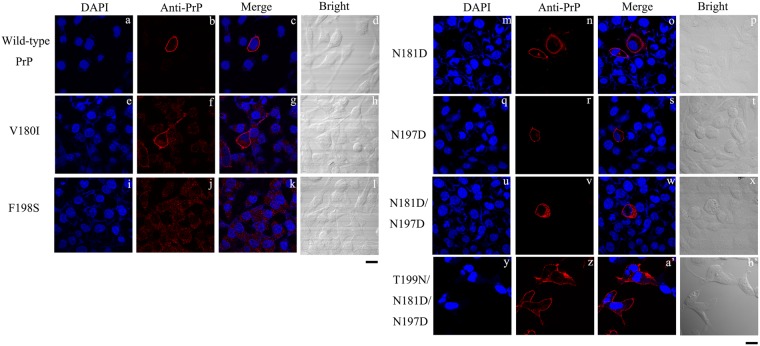
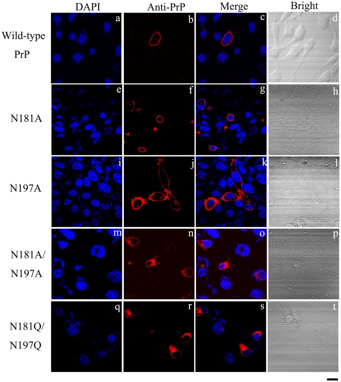
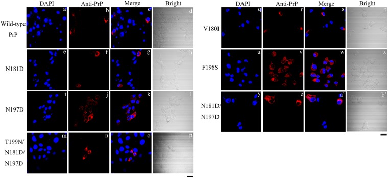
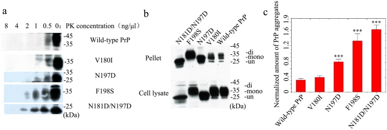
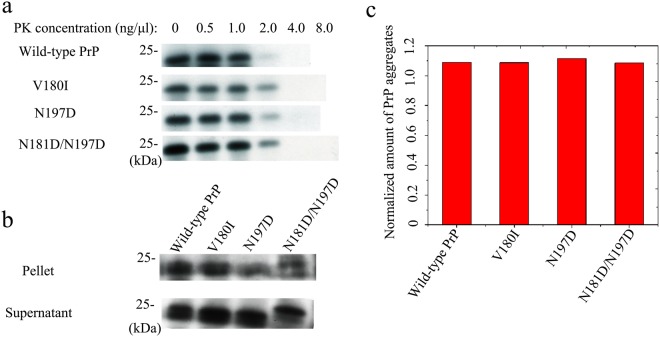
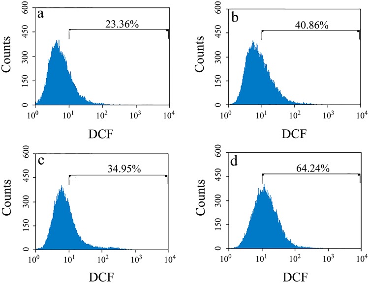
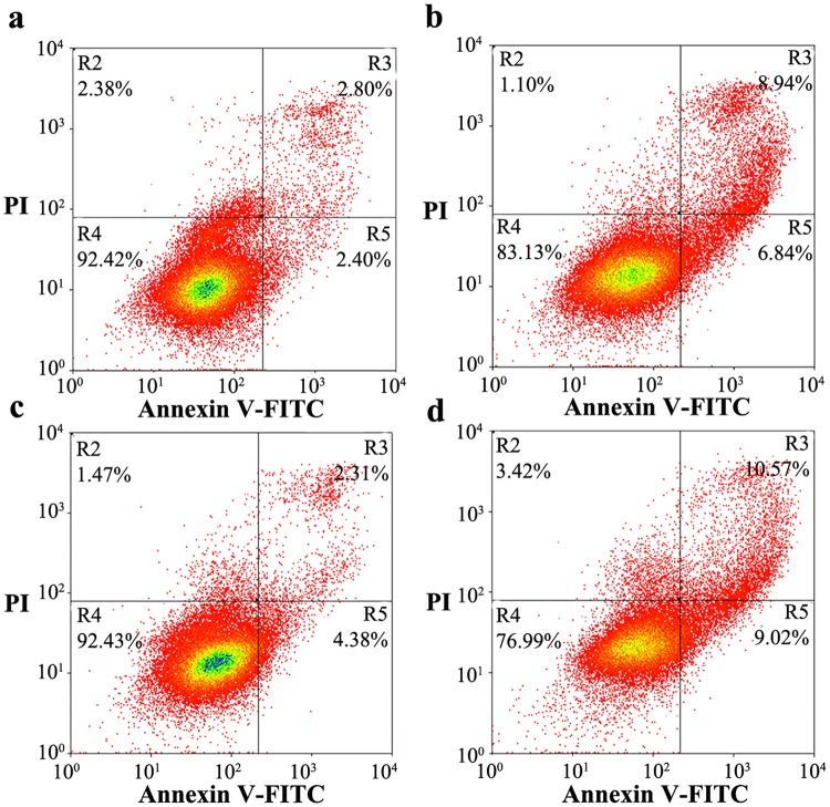
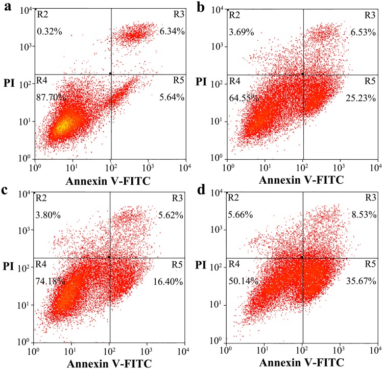
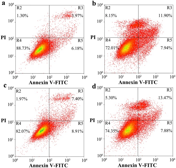
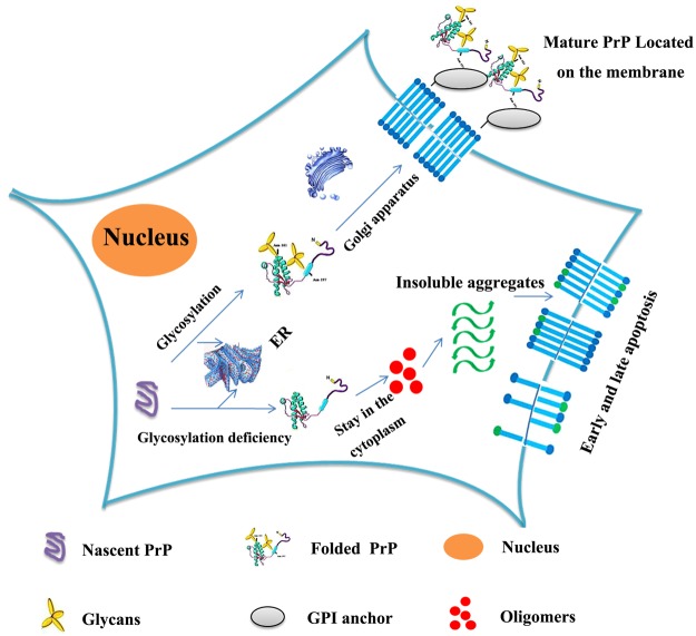
Prion diseases are primarily caused by the misfolding of prion proteins in humans, cattle, sheep, and cervid species. The effects of glycosylation on prion protein (PrP) structure and function have not been thoroughly elucidated to date. In this study, we attempt to elucidate the effects of glycosylation on the aggregation and toxicity of human PrP. As revealed by immunocytochemical staining, wild-type PrP and its monoglycosylated mutants N181D, N197D, and T199N/N181D/N197D are primarily attached to the plasma membrane. In contrast, PrP F198S, a pathological mutant with an altered residue within the glycosylation site, and an unglycosylated PrP mutant, N181D/N197D, primarily exist in the cytoplasm. In the pathological mutant V180I, there is an equal mix of membranous and cytoplasmic PrP, indicating that N-linked glycosylation deficiency impairs the correct localization of human PrP at the plasma membrane. As shown by immunoblotting and flow cytometry, human PrP located in the cytoplasm displays considerably greater PK resistance and aggregation ability and is associated with considerably higher cellular ROS levels than PrP located on the plasma membrane. Furthermore, glycosylation deficiency enhances human PrP cytotoxicity induced by MG132 or the toxic prion peptide PrP 106-126. Therefore, we propose that glycosylation acts as a necessary cofactor in determining PrP localization on the plasma membrane and that it significantly inhibits the aggregation of human PrP and decreases its cytotoxicity.


Conflict of interest statement

The authors declare no competing interests.

Figures

Figure 1
Figure 1
N-linked glycosylation deficiency impairs the correct localization of human PrP at the plasma membrane of RK13 cells. RK13 cells transiently expressing wild-type PrP (a–d), V180I (e–h), F198S (i–l), N181D (m–p), N197D (q–t), N181D/N197D (u–x), and T199N/N181D/N197D (y–b) were cultured for 2 days at 37 °C, fixed, permeabilized, immunostained with the anti-PrP antibody 3F4 and with IgG conjugated to Alexa Fluor 546 (red), stained with DAPI (blue), and observed by confocal microscopy. The scale bars represent 10 μm.
Figure 2
Figure 2
N-linked glycosylation deficiency impairs the correct localization of human PrP at the plasma membrane of RK13 cells. RK13 cells transiently expressing wild-type PrP (a–d), N181A (e–h), N197A (i–l), N181A/N197A (m–p), and N181Q/N197Q (q–t) were cultured for 2 days at 37 °C, fixed, permeabilized, immunostained with the anti-PrP antibody 3F4 and with IgG conjugated to Alexa Fluor 546 (red), stained with DAPI (blue), and observed by confocal microscopy. The scale bars represent 10 μm.
Figure 3
Figure 3
Tunicamycin suppresses glycosylation and results in the translocation of human PrP from the plasma membrane to the cytoplasm of RK13 cells. RK13 cells transiently expressing wild-type PrP (a–d), N181D (e–h), N197D (i–l), T199N/N181D/N197D (m–p), V180I (q-t), F198S (u-x), and N181D/N197D (y-b’) were cultured for 2 days and treated with 5 μg/ml tunicamycin for 36 h at 37 °C, fixed, permeabilized, immunostained with the anti-PrP antibody 3F4 and with IgG conjugated to Alexa Fluor 546 (red), stained with DAPI (blue), and observed by confocal microscopy. The scale bars represent 10 μm.
Figure 4
Figure 4
Human PrP located in the cytoplasm has much greater PK resistance and aggregation ability than PrP located on the plasma membrane of RK13 cells. RK13 cells stably expressing wild-type PrP, V180I, N197D, F198S, or N181D/N197D were cultured for 7 days. Wild-type PrP and its mutants in RK13 cells were digested with various concentrations of PK (from right to left, 0, 0.5, 1.0, 2.0, 4.0, and 8.0 ng/μl) and probed using the anti-PrP antibody 3F4 (a). The normalized amount of insoluble PrP aggregates in the stable cells (c) was calculated as the ratio of the density of insoluble PrP aggregate bands probed by 3F4 to that of the total PrP bands in cell lysates also probed by 3F4 (b). RK13 cells stably expressing wild-type PrP were used as a control. The diglycosylated, monoglycosylated, and unglycosylated bands are referred to as “di-”, “mono-”, and “un-”, respectively, and are annotated in (b). Data on the normalized amounts of insoluble PrP aggregates are expressed as the mean ± S.D. (with error bars) of the values obtained in 3 independent experiments (c). Statistical analyses were performed using Student’s t-test as described in the legend of Fig. 5. The images of wild-type PrP (or V180I) and N197D (or F198S or N181D/N197D) shown were obtained from two different gels using the same exposure time (30 s) (a). The gels and blots of wild-type PrP, V180I, N197D, F198S, and N181D/N197D (b) were cropped from different parts of the same gel with an exposure time of 30 s. Clear delineation with white spaces was used. Full-length blots and gels are presented in Supplementary Figure 5.
Figure 5
Figure 5
Human PrP displays similar PK resistance and aggregation ability after tunicamycin treatment. RK13 cells stably expressing wild-type PrP, V180I, N197D, or N181D/N197D were cultured for 7 days; tunicamycin was then added to the culture medium 48 h before the cells were harvested. Wild-type PrP and its mutants in RK13 cells treated with tunicamycin were digested with various concentrations of PK (from left to right, 0, 0.5, 1.0, 2.0, 4.0, and 8.0 ng/μl) and probed with the anti-PrP antibody 3F4 (a). The normalized amounts of insoluble PrP aggregates in the stable cells treated with tunicamycin (c) were calculated as the ratio of the density of insoluble PrP aggregate bands probed by 3F4 to that of the total PrP bands in cell lysates also probed by 3F4 (b). RK13 cells stably expressing wild-type PrP were used as a control. The gel and blot images of wild-type PrP, V180I, N197D, and N181D/N197D (a,b) shown in the figure were cropped from different parts of the same gel with an exposure time of 30 s. Clear delineation with white spaces is used. Full-length blots and gels are presented in Supplementary Figure 6.
Figure 6
Figure 6
Human PrP located in the cytoplasm of RK13 cells is associated with much higher cellular ROS levels than PrP located on the plasma membrane. RK13 cells stably expressing wild-type PrP (a), V180I (b), N197D (c), or N181D/N197D (d) were cultured for 4 days. The percentage of ROS cells was determined by flow cytometry using the ROS probe DCFH-DA. RK13 cells stably expressing wild-type PrP were used as a control.
Figure 7
Figure 7
N-linked glycosylation deficiency enhances PrP toxicity induced by MG132 in RK13 cells. RK13 cells stably expressing wild-type PrP (a), V180I (b), N197D (c), or the double mutant N181D/N197D (d) were cultured for 3 days and incubated with 1 ng/μl MG132 for 1 day. The percentage of apoptotic cells was determined by flow cytometry. The four quadrants distinguished by annexin V-FITC/PI staining represent viable cells (R4 quadrant), early apoptotic cells (R5 quadrant), late apoptotic cells (R3 quadrant), and operation-damaged cells (R2 quadrant).
Figure 8
Figure 8
N-linked glycosylation deficiency enhances PrP toxicity in RK13 cells induced by the toxic prion peptide PrP 106–126. RK13 cells stably expressing wild-type PrP (a), V180I (b), N197D (c), or the double mutant N181D/N197D (d) were cultured for 3 days and incubated with 60 μM PrP 106–126 for 2 days. The percentage of apoptotic cells was determined by flow cytometry as described in the legend of Fig. 7.
Figure 9
Figure 9
N-linked glycosylation deficiency enhances PrP toxicity in SH-SY5Y cells induced by the toxic prion peptide PrP 106–126. SH-SY5Y cells transiently expressing wild-type PrP (a), V180I (b), N197D (c), or the double mutant N181D/N197D (d) were cultured for 2 days and incubated with 60 μM PrP 106–126 for 2 days. The percentage of apoptotic cells was determined by flow cytometry as described in the legend of Fig. 7.
Figure 10
Figure 10
A hypothetical model showing how glycosylation inhibits the aggregation of human PrP and decreases PrP toxicity in living cells. Under normal circumstances, posttranslational modification of nascent PrP (purple unfolded PrP), including N-linked glycosylation, occurs in the ER; the modified, folded PrP (green folded PrP modified by yellow glycans) then matures in the Golgi apparatus and finally reaches the plasma membrane with the help of the GPI anchor (gray ellipse). However, N-linked glycosylation deficiency causes PrP to remain in the cytoplasm and to have stronger PK resistance and aggregation ability than mature PrP located on the plasma membrane. Glycosylation deficiency also promotes early and late apoptosis induced by PrP oligomers (red balls) and insoluble PrP aggregates (green waves) or by the metabolic consequences of this aggregation, thereby increasing PrP toxicity. Externalized phosphatidylserine is indicated by blue sticks with green balls.

Similar articles

Cited by

References

    1. Prusiner SB. Molecular biology and pathogenesis of prion diseases. Trends Biochem Sci. 1996;21:482–487. doi: 10.1016/S0968-0004(96)10063-3. - DOI - PubMed
    1. Soto C. Prion hypothesis: the end of the controversy? Trends Biochem Sci. 2011;36:151–158. doi: 10.1016/j.tibs.2010.11.001. - DOI - PMC - PubMed
    1. Prusiner S. B. Prions. Proc Natl Acad Sci USA. 1998;95:13363–13383. doi: 10.1073/pnas.95.23.13363. - DOI - PMC - PubMed
    1. Cervenakova L, et al. Phenotype-genotype studies in kuru: implications for new variant Creutzfeldt-Jakob disease. Proc Natl Acad Sci USA. 1998;95:13239–13241. doi: 10.1073/pnas.95.22.13239. - DOI - PMC - PubMed
    1. Rossetti G, et al. Common structural traits across pathogenic mutants of the human prion protein and their implications for familial prion diseases. J Mol Biol. 2011;411:700–712. doi: 10.1016/j.jmb.2011.06.008. - DOI - PubMed

Publication types