

Lysosomal membrane permeabilization causes secretion of IL-1β in human vascular smooth muscle cells
- PMID: 30136196
- PMCID: PMC6133165
- DOI: 10.1007/s00011-018-1178-z
Lysosomal membrane permeabilization causes secretion of IL-1β in human vascular smooth muscle cells
Abstract
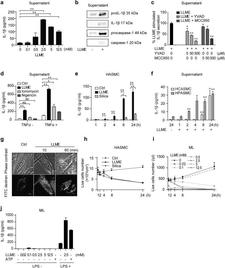
Objective: IL-1β secretion by the inflammasome is strictly controlled and requires two sequential signals: a priming signal and an activating signal. Lysosomal membrane permeabilization (LMP) plays a critical role in the regulation of NLRP3 inflammasome, and generally acts as an activating signal. However, the role of LMP controlling NLRP3 inflammasome activation in human vascular smooth muscle cells (hVSMCs) is not well defined.
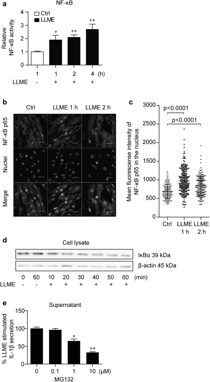
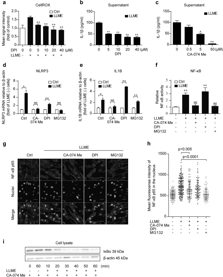
Methods: LMP was induced in hVSMCs by Leu-Leu-O-methyl ester. Cathepsin B was inhibited by CA-074 Me. Cytokine release, mRNA, and protein were quantified by enzyme-linked immunosorbent assay, quantitative PCR, and Western blot, respectively. NF-κB activity was analyzed by immunostaining of the NF-κB p65 nuclear translocation and using the dual-luciferase reporter assay system.
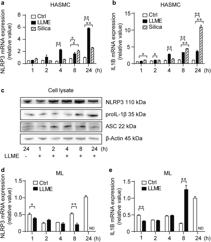
Results: LMP had both priming and activating roles, causing an upregulation of proIL-1β and NLRP3 and the secretion of mature IL-1β from unprimed hVSMCs. LMP activated the canonical NF-κB pathway. The priming effect of LMP was inhibited by CA-074 Me, indicating an upstream role of cathepsin B.
Conclusions: These data support a novel role of LMP as a single stimulus for the secretion of IL-1β from hVSMCs, implying the possibility that hVSMCs are an important initiator of the sterile inflammatory response caused by lysosomal disintegration.
Keywords: Cathepsin B; Human vascular smooth muscle cells (hVSMCs); Inflammasome; Lysosomal membrane permeabilization (LMP); NF-κB.
Conflict of interest statement
The authors have no financial conflicts of interest.
Figures
Similar articles
-
Hydrogen-Rich Saline Attenuated Subarachnoid Hemorrhage-Induced Early Brain Injury in Rats by Suppressing Inflammatory Response: Possible Involvement of NF-κB Pathway and NLRP3 Inflammasome.Mol Neurobiol. 2016 Jul;53(5):3462-3476. doi: 10.1007/s12035-015-9242-y. Epub 2015 Jun 20. Mol Neurobiol. 2016. PMID: 26091790
-
Aloe vera downregulates LPS-induced inflammatory cytokine production and expression of NLRP3 inflammasome in human macrophages.Mol Immunol. 2013 Dec;56(4):471-9. doi: 10.1016/j.molimm.2013.05.005. Epub 2013 Aug 1. Mol Immunol. 2013. PMID: 23911403
-
HBV inhibits LPS-induced NLRP3 inflammasome activation and IL-1β production via suppressing the NF-κB pathway and ROS production.J Hepatol. 2017 Apr;66(4):693-702. doi: 10.1016/j.jhep.2016.12.018. Epub 2016 Dec 24. J Hepatol. 2017. PMID: 28027970
-
The role of lysosomal cysteine cathepsins in NLRP3 inflammasome activation.Arch Biochem Biophys. 2019 Jul 30;670:32-42. doi: 10.1016/j.abb.2019.02.015. Epub 2019 Feb 23. Arch Biochem Biophys. 2019. PMID: 30807742 Review.
-
Behind every smile there's teeth: Cathepsin B's function in health and disease with a kidney view.Biochim Biophys Acta Mol Cell Res. 2022 Apr;1869(4):119190. doi: 10.1016/j.bbamcr.2021.119190. Epub 2021 Dec 27. Biochim Biophys Acta Mol Cell Res. 2022. PMID: 34968578 Review.
Cited by
-
Calciprotein Particles Link Disturbed Mineral Homeostasis with Cardiovascular Disease by Causing Endothelial Dysfunction and Vascular Inflammation.Int J Mol Sci. 2021 Nov 18;22(22):12458. doi: 10.3390/ijms222212458. Int J Mol Sci. 2021. PMID: 34830334 Free PMC article.
-
Mechanisms of demyelination and neurodegeneration in globoid cell leukodystrophy.Glia. 2021 Oct;69(10):2309-2331. doi: 10.1002/glia.24008. Epub 2021 Apr 14. Glia. 2021. PMID: 33851745 Free PMC article. Review.
-
TLR4 regulates vascular smooth muscle cell proliferation in hypertension via modulation of the NLRP3 inflammasome.Am J Transl Res. 2021 Jan 15;13(1):314-325. eCollection 2021. Am J Transl Res. 2021. PMID: 33527026 Free PMC article.
-
Contribution of Particle-Induced Lysosomal Membrane Hyperpolarization to Lysosomal Membrane Permeabilization.Int J Mol Sci. 2021 Feb 25;22(5):2277. doi: 10.3390/ijms22052277. Int J Mol Sci. 2021. PMID: 33668885 Free PMC article.
-
N-Dihydrogalactochitosan Drives Conventional and Alternative Activations of STING to Synergize Type I IFN and IL-1β Productions for Antitumor Immunity.Adv Funct Mater. 2024 Dec 16;34(51):2410079. doi: 10.1002/adfm.202410079. Epub 2024 Sep 9. Adv Funct Mater. 2024. PMID: 39896882
References
-
- Howard AD, Kostura MJ, Thornberry N, Ding GJ, Limjuco G, Weidner J, et al. IL-1-converting enzyme requires aspartic acid residues for processing of the IL-1 beta precursor at two distinct sites and does not cleave 31-kDa IL-1 alpha. J Immunol. 1991;147(9):2964–2969. - PubMed
MeSH terms
Substances
Grants and funding
LinkOut - more resources
Full Text Sources
Other Literature Sources
