

Analyzing Necroptosis Using an RIPK1 Kinase Inactive Mouse Model of TNF Shock
- PMID: 30136236
- PMCID: PMC7250063
- DOI: 10.1007/978-1-4939-8754-2_12
Analyzing Necroptosis Using an RIPK1 Kinase Inactive Mouse Model of TNF Shock
Abstract
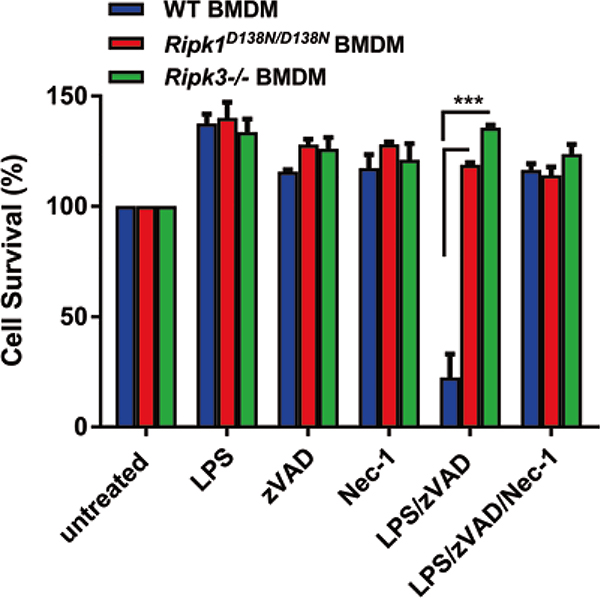
The serine/threonine kinase RIPK1 has numerous biological and pathological functions, mediating prosurvival as well as prodeath apoptotic and necroptotic signaling pathways downstream of various receptors, including death receptors and Toll-like receptors (TLRs). RIPK1 has been implicated in various diseases, including ischemia-reperfusion injury and inflammatory bowel disease (IBD). The recent generation of RIPK1 kinase inactive mice has enabled us to genetically interrogate the role of RIPK1 kinase-mediated necroptosis in disease models. Here, we describe procedures utilizing kinase inactive Ripk1D138N/D138N mice to analyze necroptosis induction in vitro in bone-marrow derived macrophages (BMDMs) and in vivo in a murine model of TNF-induced shock.
Keywords: Bone marrow-derived macrophages; Caspase inhibitor; Cell death; Lipopolysaccharide; Necroptosis; TNF shock.
Figures

References
-
- Kelliher MA, Grimm S, Ishida Y et al. (1998) The death domain kinase RIP mediates the TNF-induced NF-kappaB signal. Immunity 8:297–303 - PubMed
-
- Degterev A, Huang Z, Boyce M et al. (2005) Chemical inhibitor of nonapoptotic cell death with therapeutic potential for ischemic brain injury. Nat Chem Biol 1:112–119 - PubMed
Publication types
MeSH terms
Substances
Grants and funding
LinkOut - more resources
Full Text Sources
Other Literature Sources
Miscellaneous
