

Combined VEGF/PDGF improves olfactory regeneration after unilateral bulbectomy in mice
- PMID: 30136698
- PMCID: PMC6128065
- DOI: 10.4103/1673-5374.238713
Combined VEGF/PDGF improves olfactory regeneration after unilateral bulbectomy in mice
Abstract
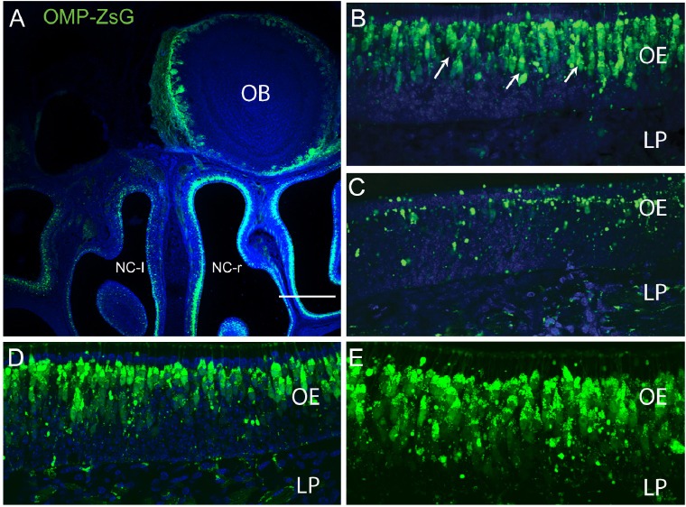
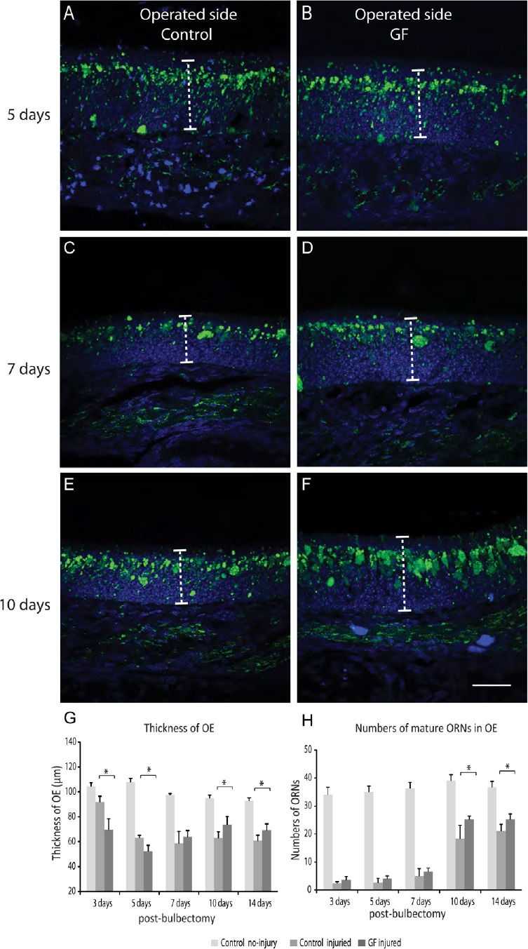
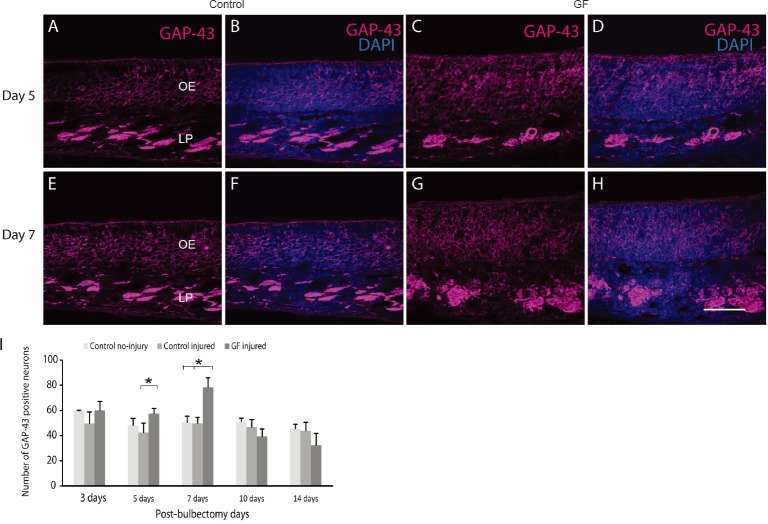
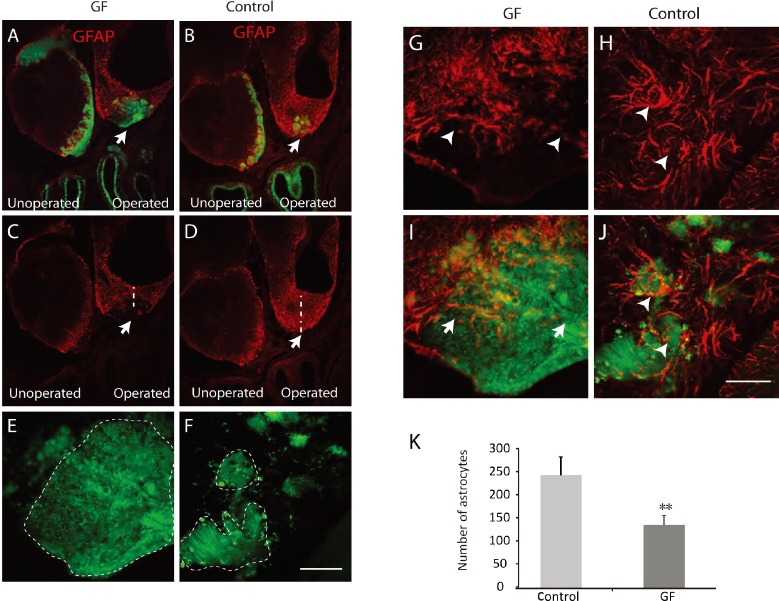
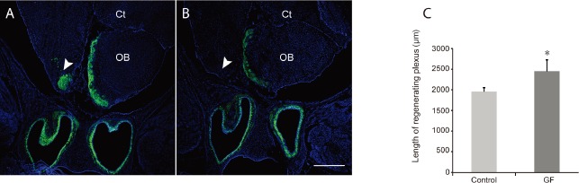
The olfactory receptor neurons lining the nasal cavity have a remarkable capacity to regenerate throughout life. They are replenished continuously and their axons make new connections within the olfactory bulb. However, some factors such as head trauma and skull base surgery damage the olfactory nerve which lead to olfactory dysfunction. Losing the sense of smell has considerable effects on quality of life and life-expectancy. Therefore, there is a clear need to find a treatment for olfactory dysfunction. One such potential treatment is growth factor therapy which showed promising results in the spinal cord and brain injuries. The aim of the present study was to investigate whether combined delivery of two growth factors, vascular endothelial growth factor and platelet-derived growth factor treatment can improve the olfactory neurons regeneration in mice. The degeneration of the olfactory neurons was induced by unilateral bulbectomy. The treatment group received 1.5 µg of the combined growth factors intranasally, while the control injured group received saline. Growth factor treatment significantly increased the number of immature neurons at 5 and 7 days post injury and also the number of mature olfactory neurons at 10 and 14 days post bulbectomy. Regenerating axons extended over a larger volume in the operated cavity in the treatment group compared to control group at 14 days post bulbectomy. The growth factor treatment also significantly reduced astrocytic glia scar in the operated cavity. The results indicate that the combined delivery of the growth factors has the potential to improve olfactory dysfunction.
Keywords: astrocytes; axon; glial scar; growth factors; neuron; olfactory bulb.
Conflict of interest statement
None
Figures
Similar articles
-
Factors that modulate olfactory dysfunction.Neural Regen Res. 2018 Jul;13(7):1151-1155. doi: 10.4103/1673-5374.235018. Neural Regen Res. 2018. PMID: 30028314 Free PMC article. Review.
-
Olfactory glia enhance neonatal axon regeneration.Mol Cell Neurosci. 2010 Nov;45(3):277-88. doi: 10.1016/j.mcn.2010.07.002. Epub 2010 Jul 13. Mol Cell Neurosci. 2010. PMID: 20621189
-
Upregulation of neural growth-associated protein and neural cell adhesion molecule in mouse olfactory epithelium and axons after unilateral removal of the olfactory bulb.Eur Arch Otorhinolaryngol. 1998;255(9):441-5. doi: 10.1007/s004050050095. Eur Arch Otorhinolaryngol. 1998. PMID: 9833210
-
Expression of netrin-1 and netrin-1 receptor, DCC, in the rat olfactory nerve pathway during development and axonal regeneration.Neuroscience. 2002;109(4):643-56. doi: 10.1016/s0306-4522(01)00535-8. Neuroscience. 2002. PMID: 11927147
-
Olfactory ensheathing glia: their contribution to primary olfactory nervous system regeneration and their regenerative potential following transplantation into the injured spinal cord.Brain Res Rev. 2007 Nov;56(1):236-58. doi: 10.1016/j.brainresrev.2007.07.013. Epub 2007 Aug 14. Brain Res Rev. 2007. PMID: 17884174 Review.
Cited by
-
Beyond vessels: unraveling the impact of VEGFs on neuronal functions and structure.J Biomed Sci. 2025 Mar 6;32(1):33. doi: 10.1186/s12929-025-01128-8. J Biomed Sci. 2025. PMID: 40050849 Free PMC article. Review.
-
Role of vascular endothelial growth factor as a critical neurotrophic factor for the survival and physiology of motoneurons.Neural Regen Res. 2023 Aug;18(8):1691-1696. doi: 10.4103/1673-5374.363194. Neural Regen Res. 2023. PMID: 36751781 Free PMC article. Review.
-
A Single Intraventricular Injection of VEGF Leads to Long-Term Neurotrophic Effects in Axotomized Motoneurons.eNeuro. 2020 May 29;7(3):ENEURO.0467-19.2020. doi: 10.1523/ENEURO.0467-19.2020. Print 2020 May/Jun. eNeuro. 2020. PMID: 32371476 Free PMC article.
-
[Research progress in the treatment of sensorineural olfactory dysfunction].Lin Chuang Er Bi Yan Hou Tou Jing Wai Ke Za Zhi. 2021 Apr;35(4):365-370. doi: 10.13201/j.issn.2096-7993.2021.04.019. Lin Chuang Er Bi Yan Hou Tou Jing Wai Ke Za Zhi. 2021. PMID: 33794640 Free PMC article. Review. Chinese.
-
Pharmacovigilance imbalance analysis of VEGFR-TKI-related taste and smell disorders.Sci Rep. 2025 Jan 24;15(1):3118. doi: 10.1038/s41598-025-87678-1. Sci Rep. 2025. PMID: 39856344 Free PMC article.
References
-
- Aiba T, Mori J, Nakai Y. Nerve growth factor (NGF) and its receptor in rat olfactory epithelium. Acta Otolaryngol Suppl. 1993;506:37–40. - PubMed
-
- Chehrehasa F, St John J, Key B. The sorting behaviour of olfactory and vomeronasal axons during regeneration. J Mol Histol. 2005;36:427–436. - PubMed
-
- Chehrehasa F, Key B, St John JA. The cell surface carbohydrate blood group A regulates the selective fasciculation of regenerating accessory olfactory axons. Brain Res. 2008;1203:32–38. - PubMed
-
- Chehrehasa F, Meedeniya AC, Dwyer P, Abrahamsen G, Mackay-Sim A. EdU, a new thymidine analogue for labelling proliferating cells in the nervous system. J Neurosci Methods. 2009;177:122–130. - PubMed
-
- Chehrehasa F, Cobcroft M, Young YW, Mackay-Sim A, Goss B. An acute growth factor treatment that preserves function after spinal cord contusion injury. J Neurotrauma. 2014;31:1807–1813. - PubMed
LinkOut - more resources
Full Text Sources
Other Literature Sources
