Review
. 2019 Apr 1;40(2):447-475.
doi: 10.1210/er.2018-00089.

Structural and Functional Biology of Aldo-Keto Reductase Steroid-Transforming Enzymes

Affiliations
Review

Structural and Functional Biology of Aldo-Keto Reductase Steroid-Transforming Enzymes

Trevor M Penning et al. Endocr Rev. .

Abstract

Aldo-keto reductases (AKRs) are monomeric NAD(P)(H)-dependent oxidoreductases that play pivotal roles in the biosynthesis and metabolism of steroids in humans. AKR1C enzymes acting as 3-ketosteroid, 17-ketosteroid, and 20-ketosteroid reductases are involved in the prereceptor regulation of ligands for the androgen, estrogen, and progesterone receptors and are considered drug targets to treat steroid hormone-dependent malignancies and endocrine disorders. In contrast, AKR1D1 is the only known steroid 5β-reductase and is essential for bile-acid biosynthesis, the generation of ligands for the farnesoid X receptor, and the 5β-dihydrosteroids that have their own biological activity. In this review we discuss the crystal structures of these AKRs, their kinetic and catalytic mechanisms, AKR genomics (gene expression, splice variants, polymorphic variants, and inherited genetic deficiencies), distribution in steroid target tissues, roles in steroid hormone action and disease, and inhibitor design.


Figures

Figure 1.
Figure 1.
Regulation of ligand occupancy of nuclear receptors by HSDs that interconvert active and inactive receptor ligands. HSDs belong to two protein superfamilies, the SDRs and the AKRs. The respective roles of individual enzymes are summarized in Table 1. SIM, selective intracrine modulator; SSRM, selective steroid receptor modulator. Orange circle is steroid hormone receptor. [Reproduced with permission from Penning TM. Hydroxysteroid dehydrogenases and prereceptor regulation of steroid hormone action. Human Reproductive Update. 2003;9(3):193–205.]
Figure 2.
Figure 2.
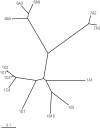
Evolution of the family tree structure of human AKRs. Dendogram adapted from the AKR Superfamily Web site (https://www.med.upenn.edu/akr/) using the multiple sequence alignment program. [Dendogram adapted from the Aldo-Keto Reductase (AKR) Superfamily website (https://www.med.upenn.edu/akr/) using the multiple sequence alignment program. Illustration presentation copyright by the Endocrine Society.]
Figure 3.
Figure 3.
Distribution of human steroid-transforming AKRs in adult male and female target tissues. Expression in adipose tissue is not shown for clarity.
Figure 4.
Figure 4.
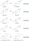
Regulation of steroid receptor ligands by human AKRs. AKR1D1 generates 5β-reduced cholestanes that are precursors to the cholanic bile acids, which then bind and activate the FXR.
Figure 5.
Figure 5.
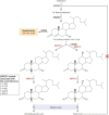
Production of tetrahydrosteroids (THS) by the sequential action of AKRs.
Figure 6.
Figure 6.
(a–c) Typical crystal structures of human AKR1C enzymes adapted from the crystal structure of AKR1C3 in complex with NADP+ and 3′-[(4-nitronaphthalen-1-yl)amino]benzoic acid-BMT4-158 (PDB ID: 4DBS). (a) Ribbon drawing displays the common (α/β)8-barrel motif of the AKRs. The α-helices (in cyan) and β-sheets (in red) of the barrel are indicated. The two helices that are not in the barrel are labeled as H1 and H2. (b) Exhibits the same spatial relationship of conserved active-site residues Asp50, Tyr55, Lys84, and His117 commonly found in human AKR1C isozymes. (c) Positions of the A-loop, B-loop, and C-terminal loop.
Figure 7.
Figure 7.
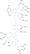
Schema showing binding of the NADP+ cofactor to AKR1C enzymes. The cofactor is in blue. [Reproduced with permission from Jez JM, Bennett MJ, Schlegel BP, et al. Comparative anatomy of the aldo-keto reductase superfamily. BiochemJ. 1997;326(3):625–636.]
Figure 8.
Figure 8.
Steroid binding residues in AKRs. Top, Table showing sequence alignment of steroid-binding residues in AKR1C9 vs the human AKR1C and AKR1D1 enzymes. Note that the steroid-binding residues are predominately in loops A, B, and the C-terminal loop. Bottom, Superposition of AKR1D1 (yellow), AKR1C9 (red), and AKR1C2 (blue) reveals significant conformational differences in loops A, B, and C. [Reproduced with permission from Di Costano L, Drury J, Penning TM, Christianson DW. Crystal structure of human liver Δ4-3-ketosteroid 5β-reductase (AKR1D1) and implications for substrate binding and catalysis. J Biol Chem. 2008;283(24):16830–16839.]
Figure 9.
Figure 9.
Different steroid-binding poses in AKRs. Left, Superimposition of the steroid-binding cavities of AKR1C9⋅NADP+⋅testosterone complex (blue) with the AKR1C2⋅NADP+⋅ursodeoxycholate complex (green). In the first structure, the 3-ketone group of testosterone lies deep in the pocket close to Y55, and the β-face of the steroid and angular methyl groups face W227. This would be a productive binding mode for 3-ketosteroid reduction. In the second structure, ursodeoxycholate has its C24 carboxylate anchored by Y55 and rotation around the steroid long axis from C3 to C17 has occurred. In this structure, the steroid binds backward and upside down relative to testosterone. These alternative binding modes in part explain why human AKR1C enzymes can act as 3-, 17-, and 20-ketosteroid reductases. [Reproduced with permission from Jin Y, Stayrook SE, Albert RH, Penning TM, Lewis M. Crystal structure of human type III 3α-hydroxysteroid dehydrogenase/bile acid binding protein complexed with NAD(P)+ and ursodeoxycholate. Biochemistry. (2001;) 40 (34): 10161–10168. Copyright 2001 American Chemical Society.] Right, Illustration of how AKR1C1 can reduce 3-ketosteroids to 3β-hydroxysteroids and how AKR1C2 can reduce 3-ketosteroids to 3α-hydroxysteroids. In AKR1C1 L54 pushes the α-face of the steroid toward W227 so that hydride transfer occurs to the α-face to produce the 3β-product. In AKR1C2 V54 allows the α-face of the steroid to hug this side of the binding pocket so that hydride transfer will occur to the β-face to produce the 3α-product.
Figure 10.
Figure 10.
Ordered bibi kinetic mechanism of AKRs showing possible inhibitor complexes.
Figure 11.
Figure 11.
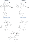
Catalytic mechanisms for AKR enzymes. Top, “Push–pull” mechanism using a diprotic AKR1C enzyme to catalyze ketosteroid reduction and hydroxysteroid oxidation. In the former instance, Y55 has TyrOH2+ character due to its proton relay with H117. In the latter instance, Y55 has phenolate character due to its proton relay with D50 and K84. Bottom, 5β-Reduction of 3-ketosteroids by AKR1D1, where E120 substitutes for H117 and acts as a superacid. E120 also permits the steroid to bind deeper in the pocket to permit hydride transfer to occur at the C5 position. [Reproduced with permission from Di Costano L, Drury J, Penning TM, Christianson DW. Crystal structure of human liver Δ4-3-ketosteroid 5β-reductase (AKR1D1) and implications for substrate binding and catalysis. J Biol Chem. 2008;283(24):16830–16839.]
Figure 12.
Figure 12.
Role of AKR1C2 and AKR1C4 in male virilization. Left panel, Fetal testis Leydig cell steroidogenesis. Only the backdoor pathway to DHT is shown. CYP11A1 represents cholesterol side-chain cleavage enzyme. CYP17A1, 17α-hydroxylase/17,20-lyase; HSDB2, 3β-hydroxysteroid dehydrogenase type 2; HSD17B3, androgenic 17β-hydroxysteroid dehydrogenase type 3; HSD17B6, 17β-hydroxysteroid dehydrogenase type 6; SRD5A, steroid 5α-reductase; StAR, steroid acute regulatory protein. Italics refer to gene names. Right panel, DHT synthesized in the fetal Leydig cells is required for the formation of the male genitalia and virilization. Loss-of-function mutations in AKR1C2 and AKR1C4 prevent the formation of DHT by the backdoor pathway and actively protect the undifferentiated genital anlage from androgens so that the female phenotype predominates. “X” marks the end of the anogenital distance, which is longer in boys.
Figure 13.
Figure 13.
Role of AKR1D1 in bile acid deficiency. Pivotal roles of AKR1D1 and AKR1C4 in human liver bile acid biosynthesis. In AKR1D1 deficiency, lack of feedback inhibition by primary bile acids on the expression of CYP7A1 mediated by FXR leads to diversion of 7α-hydroxy-cholest-4-ene-3-one to hepatoxic allo-bile acids. CYP7A1, steroid 7α-hydroxylase; SRD5A, steroid 5α-reductase type 1. Arrows to chenodeoxycholate and cholic acid represent the multiple steps required to convert C27 cholestanes to C24 cholanes. Italics refer to gene names.
Figure 14.
Figure 14.
Role of AKRs in steroidogenesis in breast cancer. CYP19A1, aromatase; DHEA, dehydroepiandrosterone; HSD3B1, 3β-hydroxysteroid dehydrogenase type 1; HSD17B1, 17β-hydroxysteroid dehydrogenase type 1 (estrogenic 17β-HSD); HSD17B2, 17β-hydroxysteroid dehydrogenase type 2; STS, steroid sulfatase. Italics refer to gene names.
Figure 15.
Figure 15.
Role of AKR1C3 in androgen biosynthesis in CRPC. 3α-Adiol, 5α-androstane-3α,17β-diol; 3β-Adiol, 5α-androstane-3β,17β-diol; 5α-adione, 5α-andostane-3,17-dione; Δ5-Adiol, 5-androstene-3β,17β-diol; CYP17A1, 17α-hydroxylase/17,20-lyase; HSD3B1, 3β-hydroxysteroid dehydrogenase type 1; HSD17B6, 17β-hydroxysteroid dehydrogenase type 6 (also known as RODH); SRD5A, steroid 5α-reductase, type 1 and type 2. Reactions in the box occur in the prostate tumor. Italics refer to gene names.
Figure 16.
Figure 16.
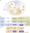
Role of AKR1C3 in PCOS. (a) Schematic representation of the proposed mechanistic link between AE, insulin resistance, and lipotoxicity in PCOS, and (b) graphical representation of the major human androgen biosynthesis pathways. AKR1C3 plays a central gatekeeping role in androgen activation in the classic androgen synthesis pathway and the alternative (backdoor) pathway to 5α-DHT and the 11-oxygenated androgen synthesis pathway. Active androgens capable of activating the androgen receptor are highlighted in blue boxes and white font. [Reproduced with permission from O'Reilly MW, Kempegowda P, Jenkinson C, et al. 11-Oxygenated C19 steroids are the predominant androgens in polycystic ovary syndrome. J Clin Endocrinol Metab. 2017;102:840–848.]

References

    1. Funder JW, Pearce PT, Smith R, Smith AI. Mineralocorticoid action: target tissue specificity is enzyme, not receptor, mediated. Science. 1988;242(4878):583–585. - PubMed
    1. Penning TM. Hydroxysteroid dehydrogenases and pre-receptor regulation of steroid hormone action. Hum Reprod Update. 2003;9(3):193–205. - PubMed
    1. Penning TM, Jin Y, Rizner TL, Bauman DR. Pre-receptor regulation of the androgen receptor. Mol Cell Endocrinol. 2008;281(1–2):1–8. - PMC - PubMed
    1. Labrie F. Intracrinology. Mol Cell Endocrinol. 1991;78:C113–C118. - PubMed
    1. Penning TM, Bauman DR, Jin Y, Rizner TL. Identification of the molecular switch that regulates access of 5α-DHT to the androgen receptor. Mol Cell Endocrinol. 2007;265-266:77–82. - PMC - PubMed

Publication types

MeSH terms