. 2018 Nov 1;41(11):zsy160.
doi: 10.1093/sleep/zsy160.

New insights into the timing and potential mechanisms of respiratory-induced cortical arousals in obstructive sleep apnea

Affiliations

New insights into the timing and potential mechanisms of respiratory-induced cortical arousals in obstructive sleep apnea

Jason Amatoury et al. Sleep. .

Abstract

Study objectives: A negative intrathoracic pressure threshold is one commonly proposed mechanism for triggering respiratory-induced arousals in obstructive sleep apnea (OSA). If so, they should occur during inspiration, shortly after maximal negative pressure swings. Alternatively, respiratory-induced arousals may occur throughout the respiratory cycle if other mechanisms also contribute. However, arousal timing has been minimally investigated. This study aimed to (1) determine the temporal relationship between respiratory-induced arousals and breathing phase and (2) characterize neuromuscular and load compensation responses prior to arousal.

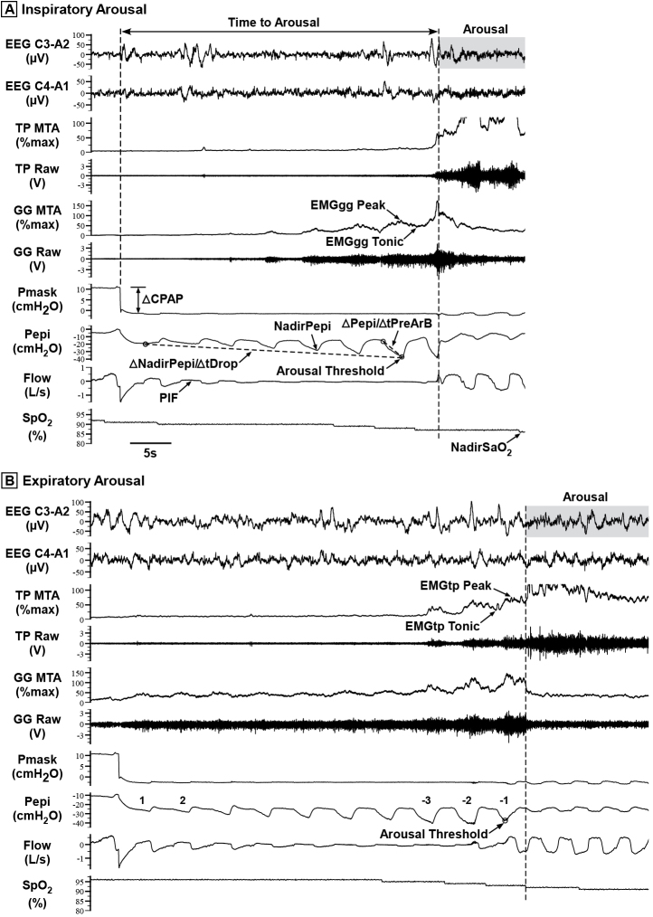
Methods: Fifty-one CPAP-treated OSA patients underwent a sleep physiology study with genioglossus and tensor palatini EMG, nasal mask/pneumotachograph, and epiglottic pressure. Transient CPAP reductions were delivered to induce respiratory-related arousals.

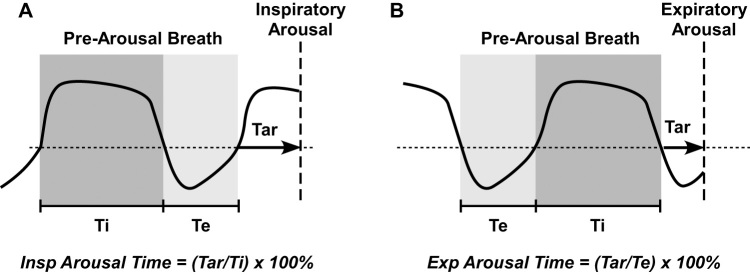
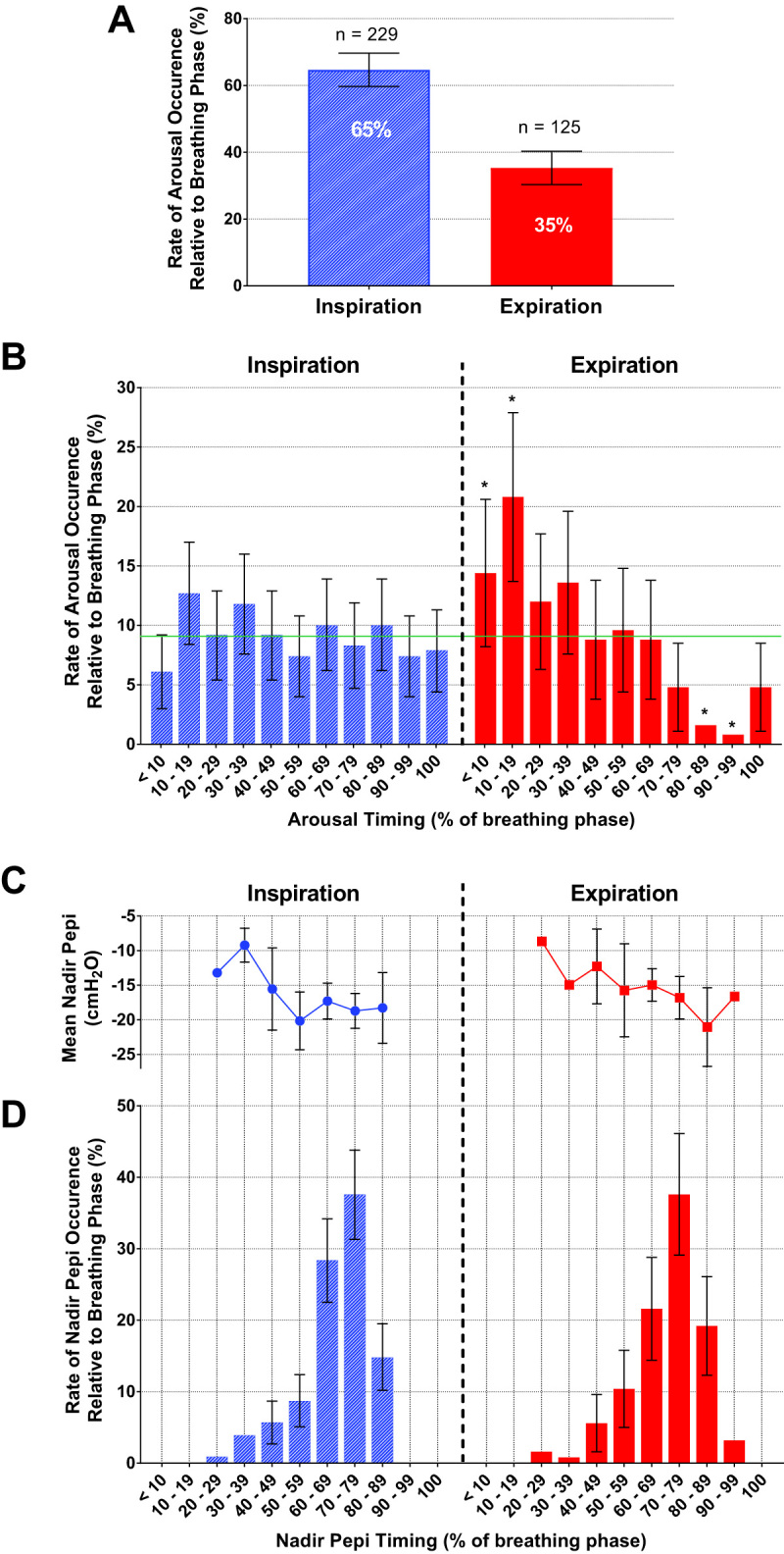
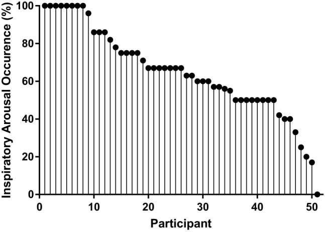
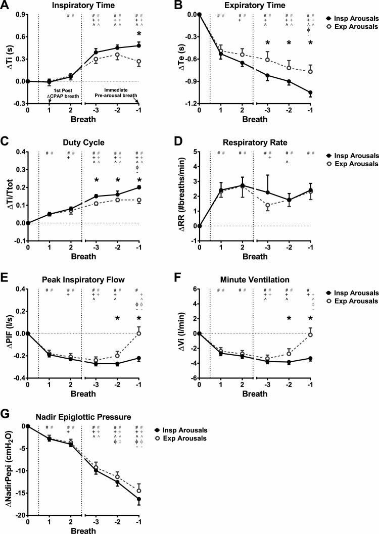
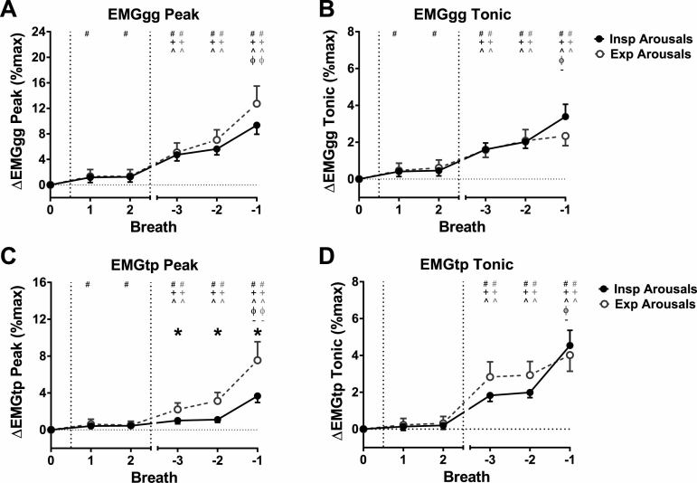
Results: Of 354 arousals, 65(60-70)%[mean(CI)] occurred during inspiration, 35(30-40)% during expiration. Nadir epiglottic pressure occurred 68(66-69)% into inspiration while inspiratory arousals had a uniform distribution throughout inspiration. Expiratory arousals occurred predominantly in early expiration. CPAP reductions initially reduced minute ventilation by ~2.5 liter/min, which was restored immediately prior to expiratory but not inspiratory arousals. Duty cycle just prior to arousal was greater for inspiratory versus expiratory arousals [0.20(0.18-0.21) vs. 0.13(0.11-0.15)Δbaseline, p = 0.001]. Peak tensor palatini EMG was higher for expiratory versus inspiratory arousals during prearousal breaths [7.6(5.8-9.6) vs. 3.7(3.0-4.5)%Δbaseline, p = 0.001], whereas genioglossus and tonic tensor palatini EMG were similar between arousal types.

Conclusions: Over one third of respiratory-induced arousals occur during expiration. These findings highlight the importance of nonpressure threshold mechanisms of respiratory-induced arousals in OSA and suggest that expiratory arousals may be a novel marker of enhanced tensor palatini neuromuscular compensation.


Figures

Figure 1.
Figure 1.
Schematic illustrating calculation of relative inspiratory (A) and expiratory (B) arousal time. Thick vertical dashed line indicates location of arousal onset. Tar = time to arousal from beginning of inspiration (A) or expiration (B). Ti = duration of first complete inspiratory cycle immediately prior to arousal. Te = duration of first complete expiratory cycle immediately prior to arousal. Arousal onset time relative to the start of the corresponding respiratory phase was determined and expressed as a per cent of the prearousal inspiratory or expiratory time. Note that the prearousal breath was used for relative respiratory timing of arousal since the arousal breath is disrupted by the arousal itself, hence altering inspiratory/expiratory durations.
Figure 2.
Figure 2.
Raw data examples showing a transient reduction in CPAP (Pmask) that induced a respiratory-related (A) inspiratory arousal and (B) expiratory arousal. Raw genioglossus (GG) and tensor palatini (TP) EMG (EMGgg and EMGtp) were rectified, moving-time-averaged (100 ms window) and expressed as a %maximum for each participant [4, 23], i.e. GG MTA and TP MTA. Some of the key study parameters quantified are shown, including respiratory arousal threshold, the nadir epiglottic pressure (Pepi), or NadirPepi, immediately preceding arousal [2, 4]; ∆NadirPepi/∆tDrop, rate of change of NadirPepi during the CPAP drop, measured from the first breath following each CPAP reduction to the breath immediately prior to arousal; ∆Pepi/∆tPreArB, rate of change of Pepi during the breath immediately preceding arousal, measured from the breath start to NadirPepi within that breath; time to arousal; and NadirSpO2, the minimum blood arterial oxygen saturation (SpO2) caused by the reduction in CPAP. Respiratory and pharyngeal muscle parameters were quantified for the two breaths following each CPAP reduction (breaths 1 and 2) and where available for three breaths prior to arousal (breaths −3 to −1), including (not all indicated on Figure): PIF = peak inspiratory flow; tidal volume and minute ventilation; Ti = inspiratory time; Te = expiratory time; Ttot = Ti + Te; Ti/Ttot = duty cycle; RR = respiratory rate; EMGgg Peak and Tonic muscle activity; EMGtp Peak and Tonic muscle activity. Note the greater increase in tensor palatini muscle activity and flow prior to the expiratory arousal (B) compared with inspiratory arousal (A).
Figure 3.
Figure 3.
Percent and number (n) of arousals that occur during inspiration and expiration (A) and their distribution with respect to breath timing (B). Mean nadir epiglottic pressure (Pepi) calculated from the inspiratory effort just prior to the arousal breath (C), and nadir Pepi occurrence with respect to breath timing (D). Error bars represent the 95% confidence intervals. Horizontal line (green) indicates the % expected by chance (9.09%) and * indicates a significant difference in arousal probability from chance (B). See Supplementary Material for breakdown of bin and error bar values (including standard error and standard deviation for epiglottic pressure; Supplementary Table S1). Of a total of 354 arousals, 65% occurred during inspiration and 35% occurred during expiration (A). Inspiratory arousals had a uniform distribution throughout inspiration (B) and no temporal relationship with nadir epiglottic pressure, which occurred in the latter stages of inspiration (D). Expiratory arousals occurred predominantly in early expiration (B).
Figure 4.
Figure 4.
Proportion of inspiratory arousals for all 51 participants.
Figure 5.
Figure 5.
Respiratory timing and ventilatory parameters for inspiratory (Insp) arousals (closed symbols) and expiratory (Exp) arousals (open symbols) for the first two breaths following CPAP reduction (Breaths 1 and 2), and the three breaths prior to arousal during the CPAP reduction (Breaths −3 to −1; Breath −1 is immediately prior to arousal), expressed as a change (∆) from baseline therapeutic CPAP (Breath 0; average of 60 s immediately prior to CPAP reduction). (A) Inspiratory time (Ti), (B) expiratory time (Te), (C) duty cycle (Ti/Ttot; Ttot = Ti + Te), (D) respiratory rate (RR), (E) peak inspiratory flow (PIF), (F) minute ventilation (Vi), and (G) nadir epiglottic pressure (NadirPepi). Data are estimated marginal mean (confidence interval). *p < 0.05 for Insp vs. Exp arousals. Between breath comparisons are shown for Insp and Exp arousals: # p < 0.05 vs. Breath 0, + p < 0.05 vs. Breath 1, ^ p < 0.05 vs. Breath 2, ϕ p < 0.05 vs. Breath −3, – p < 0.05 vs. Breath −2. For the breath immediately prior to arousal following CPAP reduction (Breath −1), Exp arousals have lower increases in Ti and Ti/Ttot and lower reductions in Te compared with Insp arousals. RR increases are immediate following CPAP reduction for both Insp and Exp arousals and are mostly maintained at that level for the duration of the CPAP drop. PIF and Vi return to baseline levels on Breath −1 for Exp arousals, though remain reduced for Insp arousals. There were no differences between Insp and Exp arousals on Breaths 1 and 2 for any of the parameters. Negative NadirPepi progressively increases from Breaths 1 to -1. There are no differences in NadirPepi changes between Insp and Exp arousals for all analyzed breaths (G).
Figure 6.
Figure 6.
Peak and tonic muscle activity for the genioglossus (EMGgg Peak and EMGgg Tonic) (A and B) and tensor palatini (EMGtp Peak and EMGtp Tonic) (C and D) muscles for inspiratory (Insp) (closed symbols) and expiratory (Exp) (open symbols) arousals. Data [expressed as estimated marginal mean (confidence interval)] are for the first two breaths following CPAP reduction (Breaths 1 and 2), and the three breaths prior to arousal (Breaths −3 to −1; Breath −1 is immediately prior to arousal), expressed as a change (∆) from baseline therapeutic CPAP (Breath 0). There were greater increases in EMGtp Peak for Exp arousals compared with Insp arousals. *p < 0.05 for Insp vs. Exp arousals. # p < 0.05 vs. Breath 0, + p < 0.05 vs. Breath 1, ^ p < 0.05 vs. Breath 2, ϕ p < 0.05 vs. Breath −3, – p < 0.05 vs. Breath −2.

References

    1. Phillipson EA, et al. Arousal: the forgotten response to respiratory stimuli. Am Rev Respir Dis. 1978;118(5):807–809. - PubMed
    1. Eckert DJ, et al. Arousal from sleep: implications for obstructive sleep apnea pathogenesis and treatment. J Appl Physiol (1985). 2014;116(3):302–313. - PubMed
    1. Younes M. Role of arousals in the pathogenesis of obstructive sleep apnea. Am J Respir Crit Care Med. 2004;169(5):623–633. - PubMed
    1. Eckert DJ, et al. Defining phenotypic causes of obstructive sleep apnea. Identification of novel therapeutic targets. Am J Respir Crit Care Med. 2013;188(8):996–1004. - PMC - PubMed
    1. Edwards BA, et al. Clinical predictors of the respiratory arousal threshold in patients with obstructive sleep apnea. Am J Respir Crit Care Med. 2014;190(11):1293–1300. - PMC - PubMed

Publication types

MeSH terms