

Diffusion-weighted magnetic resonance imaging to assess diffuse renal pathology: a systematic review and statement paper
- PMID: 30137580
- PMCID: PMC6106641
- DOI: 10.1093/ndt/gfy163
Diffusion-weighted magnetic resonance imaging to assess diffuse renal pathology: a systematic review and statement paper
Abstract
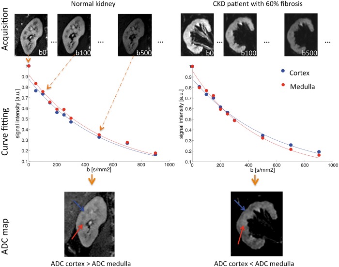
Diffusion-weighted magnetic resonance imaging (DWI) is a non-invasive method sensitive to local water motion in the tissue. As a tool to probe the microstructure, including the presence and potentially the degree of renal fibrosis, DWI has the potential to become an effective imaging biomarker. The aim of this review is to discuss the current status of renal DWI in diffuse renal diseases. DWI biomarkers can be classified in the following three main categories: (i) the apparent diffusion coefficient-an overall measure of water diffusion and microcirculation in the tissue; (ii) true diffusion, pseudodiffusion and flowing fraction-providing separate information on diffusion and perfusion or tubular flow; and (iii) fractional anisotropy-measuring the microstructural orientation. An overview of human studies applying renal DWI in diffuse pathologies is given, demonstrating not only the feasibility and intra-study reproducibility of DWI but also highlighting the need for standardization of methods, additional validation and qualification. The current and future role of renal DWI in clinical practice is reviewed, emphasizing its potential as a surrogate and monitoring biomarker for interstitial fibrosis in chronic kidney disease, as well as a surrogate biomarker for the inflammation in acute kidney diseases that may impact patient selection for renal biopsy in acute graft rejection. As part of the international COST (European Cooperation in Science and Technology) action PARENCHIMA (Magnetic Resonance Imaging Biomarkers for Chronic Kidney Disease), aimed at eliminating the barriers to the clinical use of functional renal magnetic resonance imaging, this article provides practical recommendations for future design of clinical studies and the use of renal DWI in clinical practice.
Figures
References
-
- Stehling MK, Turner R, Mansfield P.. Echo-planar imaging: magnetic resonance imaging in a fraction of a second. Science 1991; 254: 43–50 - PubMed
-
- Damasio MB, Tagliafico A, Capaccio E. et al. Diffusion-weighted MRI sequences (DW-MRI) of the kidney: normal findings, influence of hydration state and repeatability of results. Radiol Med 2008; 113: 214–224 - PubMed
-
- Wang WJ, Pui MH, Guo Y. et al. MR diffusion tensor imaging of normal kidneys. J Magn Reson Imaging 2014; 40: 1099–1102 - PubMed
-
- Sigmund EE, Vivier PH, Sui D. et al. Intravoxel incoherent motion and diffusion-tensor imaging in renal tissue under hydration and furosemide flow challenges. Radiology 2012; 263: 758–769 - PubMed
Publication types
MeSH terms
Substances
LinkOut - more resources
Full Text Sources
Other Literature Sources
Medical
Research Materials
