. 2018 Aug 23;14(8):e1007558.
doi: 10.1371/journal.pgen.1007558. eCollection 2018 Aug.

The small GTPase RAB-35 defines a third pathway that is required for the recognition and degradation of apoptotic cells

Affiliations

The small GTPase RAB-35 defines a third pathway that is required for the recognition and degradation of apoptotic cells

Ryan Haley et al. PLoS Genet. .

Abstract

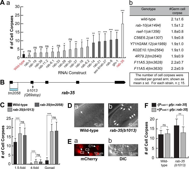
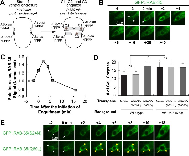
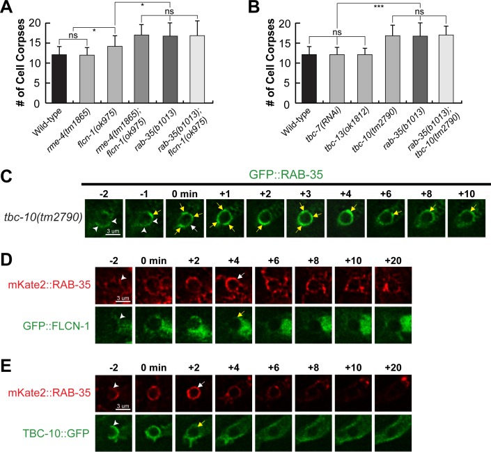
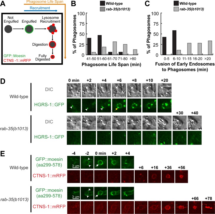
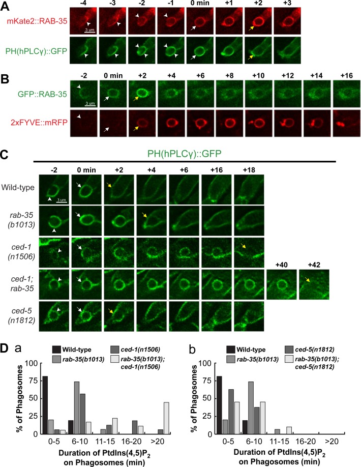
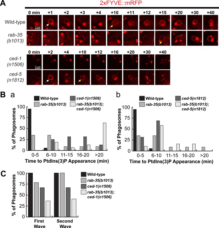
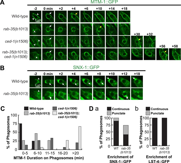
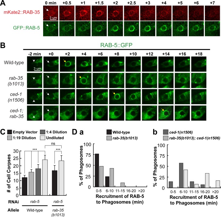
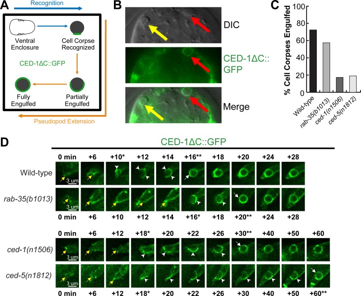
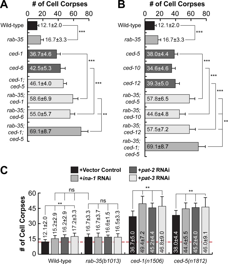
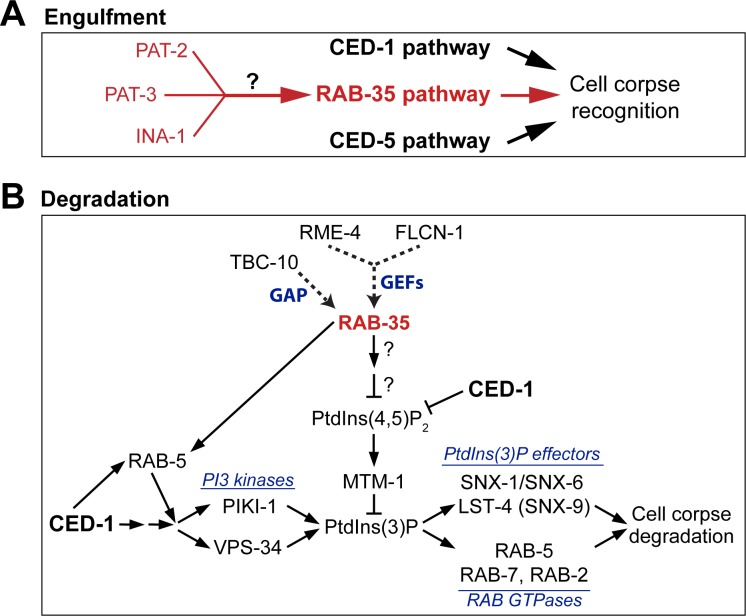
In metazoans, apoptotic cells are swiftly engulfed by phagocytes and degraded inside phagosomes. Multiple small GTPases in the Rab family are known to function in phagosome maturation by regulating vesicle trafficking. We discovered rab-35 as a new gene important for apoptotic cell clearance from a genetic screen targeting putative Rab GTPases in Caenorhabditis elegans. We further identified TBC-10 as a putative GTPase-activating protein (GAP), and FLCN-1 and RME-4 as two putative Guanine Nucleotide Exchange Factors (GEFs), for RAB-35. We found that RAB-35 was required for the efficient incorporation of early endosomes to phagosomes and for the timely degradation of apoptotic cell corpses. More specifically, RAB-35 promotes two essential events that initiate phagosome maturation: the switch of phagosomal membrane phosphatidylinositol species from PtdIns(4,5)P2 to PtdIns(3)P, and the recruitment of the small GTPase RAB-5 to phagosomal surfaces. These functions of RAB-35 were previously unknown. Remarkably, although the phagocytic receptor CED-1 regulates these same events, RAB-35 and CED-1 appear to function independently. Upstream of degradation, RAB-35 also facilitates the recognition of apoptotic cells independently of the known CED-1 and CED-5 pathways. RAB-35 localizes to extending pseudopods and is further enriched on nascent phagosomes, consistent with its dual roles in regulating apoptotic cell-recognition and phagosome maturation. Epistasis analyses indicate that rab-35 acts in parallel to both of the canonical ced-1/6/7 and ced-2/5/10/12 clearance pathways. We propose that RAB-35 acts as a robustness factor, defining a novel pathway that aids these canonical pathways in both the recognition and degradation of apoptotic cells.


Conflict of interest statement

The authors have declared that no competing interests exist.

Figures

Fig 1
Fig 1. rab-35 functions during apoptotic cell clearance in C. elegans.
(A) The numbers of germ cell corpses were scored in 48-hour post-L4 adult gonads. A minimum of 15 animals were scored. Germ cell corpses were counted (a) after RNAi treatment of 17 C. elegans genes encoding RAB proteins, and (b) in wild-type and mutant alleles of rab-10 and 7 additional candidate rab genes. (B) rab-35 gene structure. Black rectangles mark exons. The blue rectangle indicates the location of the deletion in the tm2058 allele. The arrow marks the position of the nonsense mutation carried in the b1013 allele. (C) The numbers of cell corpses were scored at various developmental stages: 1.5-fold embryos, 4-fold embryos, and the 48-hour post-L4 adult gonad. For each data point, at least 15 animals were scored. The bars and error bars indicate mean value and standard deviation (sd), respectively. (D) Differential interference contrast (DIC) microscopy images of adult gonads. White arrows mark germ cell corpses. (E) The ventral surface of a rab-35(b1013) embryo that expresses MFG-E8::mcherry was visualized using both the mCherry (a) and DIC (b) channels at ~330 minutes post-first cleavage. White arrows mark the presence of MFG-E8::mcherry on C1, C2, and C3. (F) The gfp::rab-35 transgene expressed in engulfing cells is able to rescue the rab-35 mutant phenotype. The mean numbers of cell corpses in 1.5-fold stage embryos in strains carrying or not carrying Pced-1gfp::rab-35 were presented in the bar graph. For each data point, at least 15 animals were scored. Error bars represent sd. (A, C, F) Brackets above the bars indicate the samples that are compared by the Student t-test. p-values are summarized as such: *, 0.001 < p < 0.05; **, 0.00001 < p <0.001; ***, p <0.00001; ns, no significant difference.
Fig 2
Fig 2. RAB-35 is localized to extending pseudopods and further enriched on nascent phagosomes.
All GFP reporters are expressed in engulfing cells under the control of Pced-1. (A) Diagram illustrating the features that help us visualize ventral enclosure and apoptotic cell clearance. The start of ventral enclosure is defined as the moment the two ventral hypodermal cells (ABpraapppp and ABplaapppp) start extending to the ventral midline. Both the position of cell corpses C1, C2, and C3 (brown dots) as well as the identity of their engulfing cells are shown. (B) Time-lapse recording of GFP::RAB-35 during the engulfment and degradation of cell corpse C3 in a wild-type embryo. “0 min”: the moment a nascent phagosome is just formed. Arrowheads mark the extending pseudopods. One arrow marks the nascent phagosome. (C) Graph showing the relative GFP::RAB-35 signal intensity over time on the surface of pseudopods and the phagosome shown in B. The GFP signal intensity was measured on the phagosomal surface and in the surrounding cytoplasm every 2 minutes, starting from the “-4 min” time point. The phagosomal / cytoplasmic signal ratio over time was presented. Data is normalized relative to the signal ratio at the “-4 min” time point. (D) Bar graph presenting the mean numbers and sd (error bars) of cell corpses scored in 1.5-fold stage wild-type or rab-35(b1013) mutant embryos, in the presence or absence of transgenes overexpressing GFP::RAB-35(S24N) or GFP::RAB-35(Q69L). For each data point, at least 15 animals were scored. Brackets above the bars indicate the samples that are compared by the Student t-test. p-values are summarized as such: *, 0.001 < p < 0.05; **, 0.00001 < p <0.001; ***, p <0.00001; ns, no significant difference. (E) Time-lapse images exhibiting the localization of GFP::RAB-35(S24N) and GFP::RAB-35(Q69L) during the engulfment and degradation of C3. “0 min” is the moment a nascent phagosome is just formed. Arrowheads mark extending pseudopods. A white arrow marks the nascent phagosome. Regions with enriched GFP::RAB-35(Q69L) signal on the phagosomal membrane are marked by yellow arrows.
Fig 3
Fig 3. FLCN-1 and RME-4 are the candidate GEFs and TBC-10 is a candidate GAP for RAB-35.
(A and B) Bar graph displaying results of the epistasis analysis between rab-35 and genes that encode candidate GEF (A) and GAP (B) proteins for RAB-35. The mean numbers of cell corpses in 1.5-fold stage wild-type and various single and double mutant strains are presented. For each data point, at least 15 animals were scored. Error bars indicate sd. Brackets above the bars indicate the samples that are compared by the Student t-test. p-values are summarized as such: *, 0.001 < p < 0.05; **, 0.00001 < p <0.001; ***, p <0.00001; ns, no significant difference. (C-E): Time-lapse recording of the engulfment of C3 and the beginning of phagosome maturation in the tbc-10(tm2790) mutant (C) and wild-type (D-E) embryos. Reporters are indicated on the side. “0 min” is the moment a nascent phagosome is just formed. Arrowheads indicate extending pseudopods. (C) A white arrow marks the nascent phagosome. Regions on the phagosomal membrane with enriched GFP::RAB-35 signal are marked by yellow arrows. (D) White and yellow arrows each mark the burst of RAB-35 signal on and the loss of FLCN-1 signal from the phagosome, respectively. (E) White and yellow arrows each mark the burst of RAB-35 signal on and the loss of TBC-10 from the phagosome, respectively.
Fig 4
Fig 4. rab-35 mutants exhibit delays in the recruitment of early endosomes, but not lysosomes, to phagosomes.
(A) Diagram outlining the experiment strategy to measure the life span of a phagosome. GFP::moesin serves to mark the “0 min” time point when a phagosome is just formed, while CTNS-1::mRFP acts to track the recruitment and fusion of lysosomes to the phagosome as well as to label the phagosome during degradation. (B) Histogram displaying the life span of phagosomes bearing C1, C2, and C3 in wild-type and rab-35(b1013) embryos. The life span of a phagosome is defined as the time interval between the “0 min” time point and the time point when a phagosome shrinks to one-third of its radius at “0 min”. For each genotype, at least 15 phagosomes were scored. (C) Histogram displaying the range of time it takes for early endosomes to be recruited to the phagosomal surface in wild-type and rab-35(b1013) embryos. Phagosomes bearing C1, C2, and C3 were scored. The time interval between “0 min” and the time point when the accumulating early endosomes first form a continuous ring around a phagosome is measured and exhibited. For each genotype, at least 15 phagosomes were scored. (D) Time-lapse images monitoring the recruitment of early endosomes (reporter: HGRS-1::GFP) to the phagosomal surface after a phagosome forms (the “0 min” time point). The cell corpse (white arrows) is visualized using DIC microscopy. Arrowheads indicate HGRS-1 puncta on the phagosome. The GFP ring, when it is first completed around the phagosome, is labeled with a yellow arrow. (E) Time-lapse images showing the engulfment and degradation of a phagosome bearing C3 and the recruitment of lysosomes to phagosomal surfaces, using GFP::moesin as the pseudopod reporter and CTNS-1::mRFP as a lysosomal and phagosomal marker. “0 min” is the moment when a phagosome (white arrow) is just formed. Arrowheads indicate extending pseudopods.
Fig 5
Fig 5. RAB-35 is enriched on phagosomal surfaces during the PtdIns(4,5)P2 to PtdIns(3)P shift and contributes to the PtdIns(4,5)P2 removal from nascent phagosomes.
(A-C) Time-lapse images during and after the formation of a phagosome carrying C3 in wild-type embryos. “0 min” is the moment when a phagosome is just formed. Arrowheads indicate extending pseudopods. White arrows mark the nascent phagosome. (A) Reporters: Pced-1 mKate2::rab-35 and the PtdIns(4,5)P2 marker Pced-1 PH(hPLCγ)::gfp. Yellow arrows mark the moment when both the gain of mKate2::RAB-35 and the loss of PH(hPLCγ)::GFP from the phagosomal surface is observed. (B) Reporters: Pced-1 gfp::rab-35 and the PtdIns(3)P marker Pced-1 2xFYVE::mRFP. Yellow arrows mark the moment when both GFP::RAB-35 and 2xFYVE::mRFP are rapidly enriched on the phagosomal surface. (C) Reporter: Pced-1 PH(hPLCγ)::gfp. Yellow arrows mark the first time point when PtdIns(4,5)P2 is no longer observed on the phagosome surface. (D) Histograms displaying the range of time it takes for the disappearance of PtdIns(4,5)P2 from the surface of phagosomes bearing C1, C2, and C3 in embryos of various genotypes. The time interval between the formation of a nascent phagosome (“0 min”) and the first time point when the PH(hPLCγ)::GFP signal is no longer enriched on the phagosomal surface are displayed. For each genotype, at least 15 phagosomes were scored.
Fig 6
Fig 6. rab-35 and ced-1 function in parallel to produce PtdIns(3)P on the phagosomal membrane.
(A) Time-lapse images during and after the formation of a phagosome carrying C3 in embryos of different genotypes expressing Pced-12xFYVE::mRFP. “0 min” is the moment when a phagosome is just formed. White and yellow arrows indicate the time point when the 1st and 2nd waves of PtdIns(3)P appear on the phagosome, respectively. (B) Histogram displaying the range of time it takes for the 1st peak of PtdIns(3)P to appear on phagosomes bearing C1, C2, or C3 in embryos of various genotypes, starting counting from the “0 min” time point, as shown in (A). For each genotype, at least 15 phagosomes were scored. (C) The frequency of appearance of the 1st and 2nd peaks of PtdIns(3)P on phagosomes carrying C1, C2, and C3 in embryos of various genotypes. For each genotype, at least 15 phagosomes were scored.
Fig 7
Fig 7. The rab-35(b1013) mutation impairs MTM-1 removal from, and SNX-1 recruitment to, phagosomal surfaces.
(A and B) Time-lapse images during and after the formation of a phagosome carrying C3 in embryos of different genotypes expressing Pced-1 mtm-1::gfp (A) or Pced-1 snx-1::gfp (B), respectively. “0 min” is the moment when a phagosome is just formed. Arrowheads indicate extending pseudopods. White and yellow arrows on phagosomes in (A) indicate the time points when MTM-1::GFP first and last appear on the phagosome surface, respectively. White arrows in (B) mark the regions on the phagosomal surface that have an enriched GFP signal. (C) Histogram displaying the range of time that MTM-1 persists on phagosomes in wild-type and rab-35(b1013) embryos. Phagosomes bearing cell corpses C1, C2, and C3 were scored. This time interval is defined as that between the “0 min” time point and the first time point that MTM-1 is no longer found on the phagosome. For each genotype, at least 15 phagosomes were scored. (D) The efficiency of recruitment of SNX-1::GFP (a) and LST-4::GFP (b) to the surface of phagosomes carrying C1, C2, and C3 was scored in various genotypes. SNX-1::GFP is either distributed onto the entire phagosomal surface evenly (“continuous”) or attached to phagosomal surfaces as puncta (“punctate”) (a), whereas LST-4::GFP is enriched onto phagosomes only in the “continuous” pattern (b). For each genotype, at least 15 phagosomes were scored.
Fig 8
Fig 8. rab-35 functions upstream of and promotes the phagosomal localization of rab-5.
(A) Time-lapse images starting from the completion of phagosome formation (“0 min” time point) of a phagosome carrying C3 in a wild-type embryo co-expressing Pced-1 mKate2::rab-35 and Pced-1 gfp::rab-5. An arrow in each strip marks the nascent phagosome. (B) Time-lapse images during and after the formation (“0 min” time point) of a phagosome carrying C3 in a wild-type embryo expressing Pced-1 gfp::rab-5. Arrowheads indicate extending pseudopods. White and yellow arrows mark the nascent phagosome and the phagosome onto which RAB-5 is first seen localized to, respectively. (C) Epistasis analysis results performed between rab-5 and rab-35. rab-5 was inactivated by feeding worms with different dilutions of E. coli carrying the RNAi construct. The numbers of cell corpses were scored in the 1.5-fold stage F1 embryos. For each data point, at least 15 embryos were scored. Mean ± sd are presented. *, 0.001 < p < 0.05; **, 0.00001 < p <0.001; ***, p <0.00001; ns, no significant difference. (D) Histograms displaying the range of time it takes for the appearance of RAB-5 on the surface of phagosomes bearing C1, C2, and C3 in embryos of various genotypes. The time interval between the “0 min” time point and the first time point that the GFP::RAB-5 signal is observed enriched on the phagosomal surface of at least 15 phagosomes for each genotype are displayed.
Fig 9
Fig 9. RAB-35, CED-1, and CED-5 function in parallel to recognize cell corpses.
(A) Diagram outlining how the moments of cell corpse recognition and internalization are determined utilizing the CED-1ΔC::GFP reporter. The moment of recognition is defined as the first time point GFP is seen enriched in a region in contact between the engulfing and dying cell, with the moment of ventral enclosure used as a reference point (“0 min”). The time span between the moment of recognition and the moment when the nascent phagosome forms is the period of internalization. (B) Images of part of a 1.5-fold stage embryo expressing Pced-1ced-1ΔC::gfp. Cell corpses are identified under DIC optics. Red arrows mark an engulfed cell corpse, which is surrounded by a CED-1ΔC::GFP ring. Yellow arrows marks an unengulfed cell corpse, which lacks CED-1ΔC::GFP on its surface. (C) The fraction of cell corpses that have been engulfed (surrounded by a CED-1ΔC::GFP ring) in 1.5-fold to 2-fold stage embryos of various genotypes are presented as bars. For each genotype, at least 15 embryos were scored. (D) Time-lapse images before and after the formation of a phagosome carrying C3 in embryos of different genotypes expressing Pced-1ced-1ΔC::gfp. “0 min” is the moment ventral enclosure initiates. Arrowheads indicate extending pseudopods. A white arrow marks the nascent phagosome. Yellow arrows label the extending ventral hypodermal cell ABpraapppp. For each genetic background, a single asterisk marks the moment when recognition is first observed, while the double asterisks mark the moment when the nascent phagosome is formed.
Fig 10
Fig 10. rab-35 in an engulfment pathway independent of both the ced-1 and ced-5 pathways yet includes the integrins.
Results of epistasis analysis performed between rab-35 and (A) members of the ced-1/-6/-7 pathway, (B) members of the ced-2/-5/-10/-12 pathway, and (C) genes encoding C. elegans integrins. The average numbers of cell corpses observed in 1.5-stage embryos of various genotypes are listed and presented as bars. Error bars indicate sd. For each strain, at least 15 animals were scored. Student t-test was used for data analysis: *, 0.001 < p < 0.05; **, 0.00001 < p <0.001; ***, p <0.00001; ns, no significant difference. (A) Null alleles [rab-35(b1013), ced-1(n1506), and ced-6(n2095)] were used. (B) Null alleles [ced-5(n1812) and ced-12(n3261)] and a severe loss-of-function allele ced-10(n1993) were used. (C) C. elegans integrin genes ina-1, pat-2, and cdc-42 were inactivated in wild-type, rab-35(b1013), ced-1(n1506), and ced-5(n1812) backgrounds by RNAi. A dashed line represents the mean number of cell corpses observed in untreated wild-type embryos.
Fig 11
Fig 11. Model explaining RAB-35’s action in the clearance of apoptotic cells.
Diagram illustrating the roles of RAB-35 in the engulfment (A) and degradation (B) of cell corpses in C. elegans. See Discussion for more details. RAB-35 functions in parallel with the ced-1/-6/-7/dyn-1 and ced-2/-5/-10/-12 pathways in the recognition of cell corpses but in the same pathway as ina-1, pat-2, and pat-3 (A). In addition, RAB-35 functions in parallel to the CED-1 pathway during phagosome maturation (B). On the surface of nascent phagosomes, RAB-35 stimulates the turnover of both PtdIns(4,5)P2 and its effector MTM-1, a PI-3 phosphatase, on the phagosomal surfaces. In addition, RAB-35 helps to recruit RAB-5, which promotes the production of PtdIns(3)P. Together, these two events promote the production of phagosomal PtdIns(3)P. PtdIns(3)P in turn promotes the recruitment of its downstream effectors, driving the progression of phagosome maturation and cell corpse degradation. rab-35 mutants are defective in the recruitment of SNX-1 but not LST-4/SNX-9 to phagosomes, perhaps due to that other factors may be involved in their recruitment. Question marks indicate that how RAB-35 regulates PtdIns(4,5)P2 turnover remains to be investigated.

Comment in

References

    1. Lockshin RA, Williams CM. Programmed cell death—II. Endocrine potentiation of the breakdown of the intersegmental muscles of silkmoths. J Insect Physiol. 1964. August 1;10(4):643–9.
    1. Kerr JFR, Wyllie AH, Currie AR. Apoptosis: A Basic Biological Phenomenon with Wide-ranging Implications in Tissue Kinetics. Br J Cancer. 1972. August;26(4):239–57. - PMC - PubMed
    1. Elliott MR, Ravichandran KS. Clearance of apoptotic cells: implications in health and disease. J Cell Biol. 2010. June 28;189(7):1059–70. 10.1083/jcb.201004096 - DOI - PMC - PubMed
    1. Nagata S. Apoptosis and Clearance of Apoptotic Cells. Annu Rev Immunol [Internet]. 2018;36(1). Available from: 10.1146/annurev-immunol-042617-053010 - DOI - PubMed
    1. Sulston JE, Schierenberg E, White JG, Thomson JN. The embryonic cell lineage of the nematode Caenorhabditis elegans. Dev Biol. 1983. November 1;100(1):64–119. - PubMed

Publication types

MeSH terms

LinkOut - more resources