Review
. 2018 Aug 22;99(4):640-663.
doi: 10.1016/j.neuron.2018.07.002.

Parcellating Cerebral Cortex: How Invasive Animal Studies Inform Noninvasive Mapmaking in Humans

Affiliations
Review

Parcellating Cerebral Cortex: How Invasive Animal Studies Inform Noninvasive Mapmaking in Humans

David C Van Essen et al. Neuron. .

Abstract

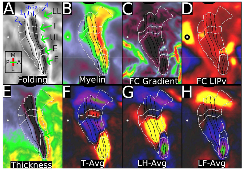
The cerebral cortex in mammals contains a mosaic of cortical areas that differ in function, architecture, connectivity, and/or topographic organization. A combination of local connectivity (within-area microcircuitry) and long-distance (between-area) connectivity enables each area to perform a unique set of computations. Some areas also have characteristic within-area mesoscale organization, reflecting specialized representations of distinct types of information. Cortical areas interact with one another to form functional networks that mediate behavior, and each area may be a part of multiple, partially overlapping networks. Given their importance to the understanding of brain organization, mapping cortical areas across species is a major objective of systems neuroscience and has been a century-long challenge. Here, we review recent progress in multi-modal mapping of mouse and nonhuman primate cortex, mainly using invasive experimental methods. These studies also provide a neuroanatomical foundation for mapping human cerebral cortex using noninvasive neuroimaging, including a new map of human cortical areas that we generated using a semiautomated analysis of high-quality, multimodal neuroimaging data. We contrast our semiautomated approach to human multimodal cortical mapping with various extant fully automated human brain parcellations that are based on only a single imaging modality and offer suggestions on how to best advance the noninvasive brain parcellation field. We discuss the limitations as well as the strengths of current noninvasive methods of mapping brain function, architecture, connectivity, and topography and of current approaches to mapping the brain's functional networks.

Keywords: architectonics; brain; connectivity; cortical areas; function; networks; neuroanatomy; nonhuman primate; parcellation; topography.


Conflict of interest statement

Declaration of Interests

The authors declare no competing interests.

Figures

Figure 1.
Figure 1.
A. A parcellation of mouse cortex containing 41 cortical areas and including seven subareas of primary somatosensory (SSp): barrel field [SSp-bfd], nose and mouth [SSp-nm], lower jaw [SSp-lj], upper limb [SSp-ul], lower limb [SSp-ll], trunk [SSp-tr], and unassigned [SSp-un] (Gămănut et al., 2018). The illustrated section shows genetically encoded fluorescent “tdTomato” labeling in parvalbumin expressing interneurons (PVtdT). Other architectonic markers used for this parcellation include cytochrome oxidase (CO), M2 muscarinic acetylcholine receptor, and vesicular glutamate transporter VGluT2. B. Visuotopic organization of 9 extrastriate visual areas revealed by triple-color anterograde tracer injections (Wang and Burkhalter, 2007). A slightly different parcellation for mouse visuotopic areas was reported by (Garrett et al., 2014) using intrinsic optical imaging. C. A 19 × 47 area+subarea quantitative connectivity matrix based on retrograde tracers injected into 18 areas, with two sub-areas for SSp, (Gămănut et al., 2018). Green entries indicate the injected area. Reproduced with permission from Wang and Burkhalter, 2007 and Gămănut et al., 2018.
Figure 2.
Figure 2.
Schematic representation of 24 retinotopic areas in owl monkey visual cortical areas overlaid on a myelin-stained flatmount. Purple and yellow-green represent areas with opposite field signs. Retinotopy of areas M, ventral VP and VA, and ITi, and IT r were taken from Allman and Kaas (1975), Newsome and Allman (1980), and (Weller and Kaas, 1987). Note that Angelucci and Rosa (Angelucci and Rosa, 2015) proposed an alternative parcellation, with VP+ and DLp-grouped into area VLP for the owl monkey as well as the marmoset (analogous to a “V3” occupying much but not all of the cortex outside of V2), as might also be consistent with the discordant case of Sereno et al. (Sereno et al., 2015). Reproduced, with permission, from Sereno et al., (Sereno et al., 2015).
Figure 3.
Figure 3.
Color-biased fMRI activations (blue and cyan) found near face patches (orange and red), displayed on an inflated right hemisphere surface rotated up to show the ventral surface. Abbreviations for face patches (orange): PL, posterior lateral; ML, middle lateral; MF, middle fundus; AF, anterior fundus; AL, anterior lateral; AM, anterior medial; for color patches, PVc, posterior ventral color; CLc, central lateral color; and ‘c’ after other abbreviations stands for color., Reproduced, with permission, from (Lafer-Sousa and Conway, 2013)
Figure 4.
Figure 4.
The HCP’s multi-modal parcellation, version 1.0 (HCP_MMP1.0). The 180 areas delineated and identified in both left and right hemispheres are displayed on inflated and flattened cortical surfaces. Black outlines indicate areal borders. Colors indicate the extent to which the areas are associated in the resting state with auditory (red), somatosensory (green), visual (blue), task positive (towards white), or task negative (towards black) groups of areas. The bottom right illustrates the 3D color space used in the figure. Reproduced with permission from Glasser et al. (Glasser et al., 2016a). Data at http://balsa.wustl.edu/WN56
Figure 5
Figure 5
shows areas (black borders) and subregions (white borders) of the sensorimotor strip, displayed on cortical flatmaps of the right hemisphere. The intersection of these two sets of borders represent subareas. A. Folding maps with abbreviations for lower limb (LL), trunk (T), upper limb (UL), eye (E), and face (F). B. Myelin map, with contrast adjusted to illustrate correspondence with subareas, e.g. a reproducible dip in myelin content between the upper limb and face subregions of area 3b and between the upper limb, eye, and face subareas of area 4 (Glasser et al., 2016a). Areal boundaries for areas 4, 3a, 3b, 1, and 2 are black. C. Resting state functional connectivity (FC) gradients that were used to define the subregions using the semiautomated border drawing approach. D. Functional connectivity from the heavily myelinated LIPv seed (black circle, which has functional connectivity with some parts of the sensorimotor strip. E. Thickness map. F, G, and H show the task fMRI contrasts for moving the tongue (T-AVG), left hand (LH-AVG), and left foot (LF-AVG). Data at https://balsa.wustl.edu/PLx0.
Figure 6
Figure 6
shows the topographic organization of the left hemisphere language network (column 1, d=40 ICA RSN). Four seeds are placed along the anterior-posterior axis of area PSL in the left hemisphere (marked by white arrows from posterior to anterior). The functional connectivity pattern shows corresponding changes in the other major nodes of the language network, including 55b, SFL, and 44. The pattern is present when seeded in either hemisphere, but stronger in the left hemisphere and is also present if any of the four highlighted areas is used as the seed. Data at https://balsa.wustl.edu/7Z3k.
Figure 7.
Figure 7.
A. TFMs related to the right hand sensorimotor network from task fMRI data concatenated across the 7 HCP tasks (left) and resting-state fMRI data (right). Each includes relevant portions of primary motor and sensory areas, SII, pre-motor cortex, insular cortex, supplementary and cingulate motor areas, plus the thalamus, the caudate and putamen, two distinct hotspots in the cerebellum, and in the case of the face component, cranial nerve nuclei in the brainstem. Aside from the motor task, none of these other task-specific task temporal ICA components matched resting state components very closely. B. TFMs related to the lateralized language network. Data at https://balsa.wustl.edu/6w32.

References

    1. Abdollahi RO, Kolster H, Glasser MF, Robinson EC, Coalson TS, Dierker D, Jenkinson M, Van Essen DC, and Orban GA. (2014) Correspondences between retinotopic areas and myelin maps in human visual cortex. Neuroimage. 99, 509–524. - PMC - PubMed
    1. Allman JM, and Kaas JH. (1974) The organization of the second visual area (V II) in the owl monkey: a second order transformation of the visual hemifield. Brain Res 76, 247–265. - PubMed
    1. Allman JM, and Kaas JH. (1975) The dorsomedial cortical visual area: a third tier area in the occipital lobe of the owl monkey (Aotus trivirgatus). Brain Res 100, 473–487. - PubMed
    1. Allman JM, and Kaas JH. (1976) Representation of the visual field on the medial wall of occipital-parietal cortex in the owl monkey. Science. 191, 572–575. - PubMed
    1. Amunts K, Lepage C, Borgeat L, Mohlberg H, Dickscheid T, Rousseau ME, Bludau S, Bazin PL, Lewis LB, Oros-Peusquens AM, Shah NJ, Lippert T, Zilles K, and Evans AC. (2013) BigBrain: an ultrahigh-resolution 3D human brain model. Science. 340, 1472–1475. - PubMed

Publication types

LinkOut - more resources