. 2018 Aug 23;9(1):3380.
doi: 10.1038/s41467-018-05931-w.

Structure and mechanism of cancer-associated N-acetylglucosaminyltransferase-V

Affiliations

Structure and mechanism of cancer-associated N-acetylglucosaminyltransferase-V

Masamichi Nagae et al. Nat Commun. .

Abstract

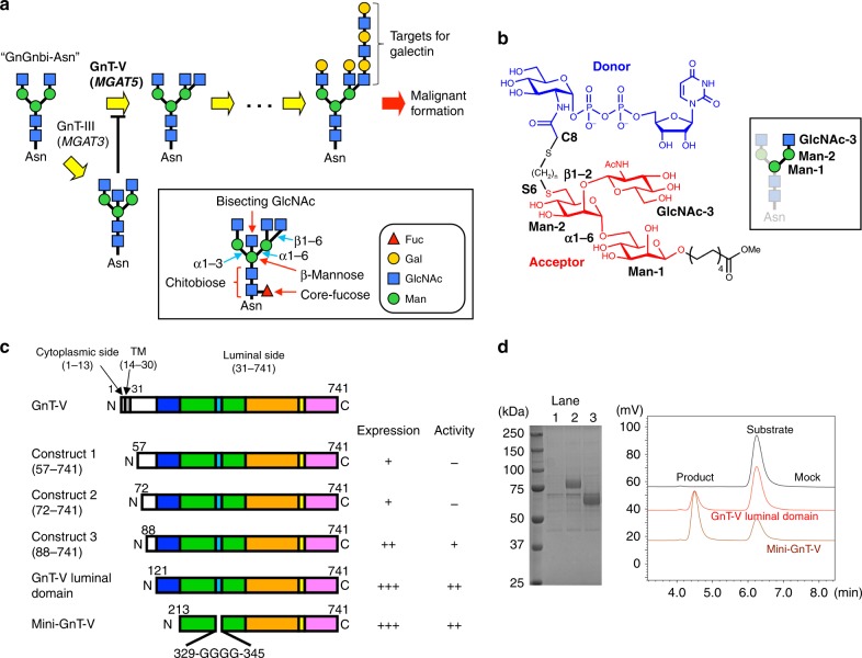
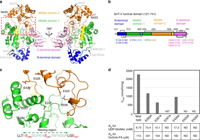
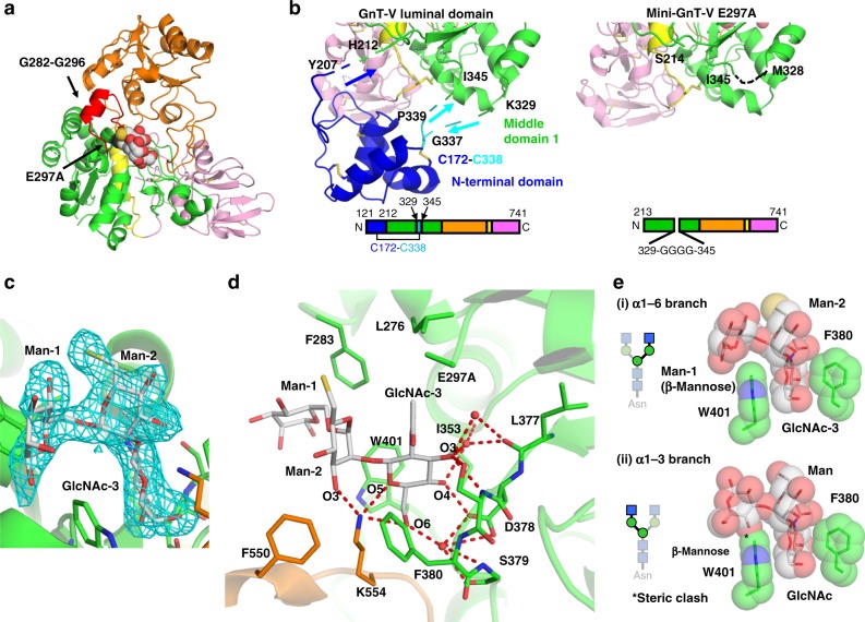
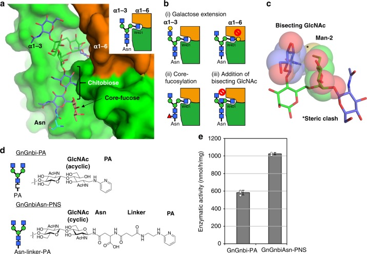
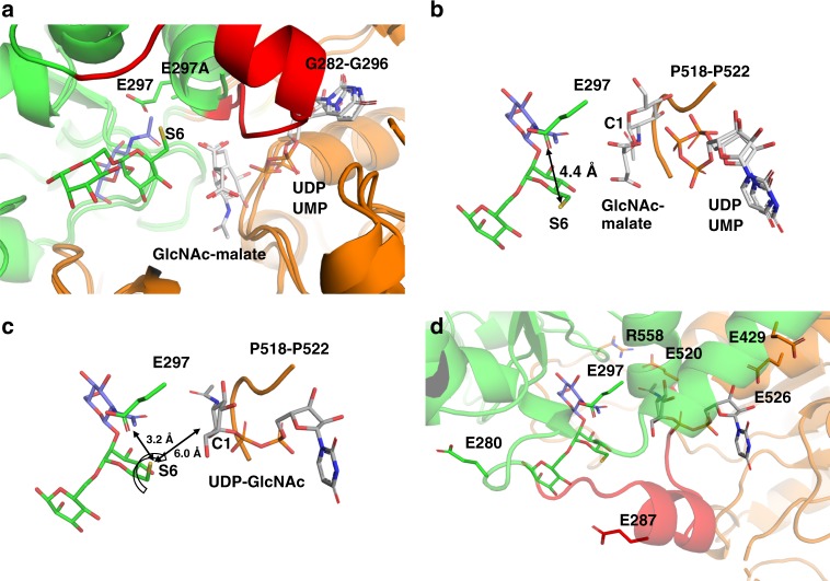
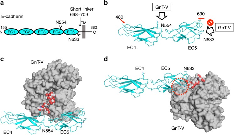
N-acetylglucosaminyltransferase-V (GnT-V) alters the structure of specific N-glycans by modifying α1-6-linked mannose with a β1-6-linked N-acetylglucosamine branch. β1-6 branch formation on cell surface receptors accelerates cancer metastasis, making GnT-V a promising target for drug development. However, the molecular basis of GnT-V's catalytic mechanism and substrate specificity are not fully understood. Here, we report crystal structures of human GnT-V luminal domain with a substrate analog. GnT-V luminal domain is composed of a GT-B fold and two accessary domains. Interestingly, two aromatic rings sandwich the α1-6 branch of the acceptor N-glycan and restrain the global conformation, partly explaining the fine branch specificity of GnT-V. In addition, interaction of the substrate N-glycoprotein with GnT-V likely contributes to protein-selective and site-specific glycan modification. In summary, the acceptor-GnT-V complex structure suggests a catalytic mechanism, explains the previously observed inhibition of GnT-V by branching enzyme GnT-III, and provides a basis for the rational design of drugs targeting N-glycan branching.


Conflict of interest statement

The authors declare no competing interests.

Figures

Fig. 1
Fig. 1
GnT-V transfers β1-6 branch on N-glycan via C-terminal region. a The β1-6 branch formation of N-glycan catalyzed by GnT-V. The substrate of GnT-V is GlcNAc-terminated biantennary glycan termed “GnGnbi-Asn”. The β1-6 branch formation leads to further LacNAc extension, which is the target for galectin. The β1–4 branch at β-mannose catalyzed by GnT-III, named as bisecting GlcNAc, hampers β1-6 branch formation. Monosaccharide symbols follow the symbol nomenclature for glycans (SNFG) system. Nomenclature of sugar residues (red arrows) and glycosidic linkages (cyan arrows) are indicated in boxes. b Chemical structure of bisubstrate-type inhibitor of GnT-V used for co-crystallization. Donor and acceptor substrates are colored with blue and red, respectively. Three sugar residues (GlcNAcβ1-2Manα1-6Man) of acceptor moiety are labeled. In this study, the inhibitor of which the linker length is (n = 3) and Ki value is 18 μM was used. Acceptor trisaccharide corresponds to α1-6 branch of biantennary glycan (GnGnbi) as shown in the box. c Domain architecture of full length and truncated constructs of human GnT-V. Expression levels and catalytic activities of truncated constructs are indicated on the right of these constructs. d Enzymatic activity of GnT-V luminal domain and mini-GnT-V. The two constructs were expressed in COS-7 cells and purified through Ni2+-column, followed by SDS-PAGE with CBB staining (left) or activity assays (right). Lane 1, mock, lane 2, GnT-V luminal domain, and lane 3, mini-GnT-V. The eluates from Ni2+-column were incubated with acceptor substrate GnGnbi-PA and donor substrate UDP-GlcNAc, and the reaction mixtures were analyzed by HPLC
Fig. 2
Fig. 2
Overall structure and catalytic residues of GnT-V luminal domain. a Overall structure of GnT-V luminal domain is shown in ribbon model. The N-terminal domain (A128-Y207 in blue and G337-P339 in cyan), middle domain 1 (H212-K329 and I345-L424 in green and E606-H627 in yellow), middle domain 2 (G425-Y605 in orange), and C-terminal domain (G628-L741 in pink) are shown in ribbon models. The disulfide bonds and N-glycan attached on N433 are indicated with rod models and labeled. Putative catalytic center of GnT-V is indicated with a red dotted box. Among three putative N-glycans (N334, N433, and N447), only GlcNAc residue of N433 is assigned in the final model. Three N-glycosylation sites are indicated. The three missing loops between N-terminal domain and middle domain 1 are also indicated. b Schematic representation of disulfide bond pattern of GnT-V luminal domain. Cysteine residues, which form disulfide bonds, are also indicated. c Structural positions of six glutamates (E280, E287, E297, E429, E520, and E526) in GnT-V in respect of mutational experiments are shown in rod model. E280 and E287 are located in the missing loop region (K279-P294) shown as green dotted line. Sequence of missing loop region is shown in lower panel. The side chain of R558, which forms a salt bridge with E526 is also shown in rod model. d Kinetic parameters of wild-type and mutated GnT-V luminal domains. Vmax values are shown in bar graph representation. Km values for UDP-GlcNAc (donor substrate) and GnGnbi-PA (acceptor substrate) are shown in lower panel. aND not detected
Fig. 3
Fig. 3
Structure of mini-GnT-V E297A in complex with bisubstrate-type inhibitor. a Crystal structure of mini-GnT-V E297A in complex with bisubstrate-type inhibitor. Trisaccharide is shown in sphere model. Highly mobile region (G282-G296) and E297A are colored with red and black, respectively, and indicated. b Close-up views of domain boundary between N-terminal domain and middle domain 1 in GnT-V luminal domain (left panel) and mini-GnT-V E297A (right panel). Key residues of these two structures are labeled. Domain architectures are shown in bottom. c Omit map contoured at 3.0 σ level around acceptor binding site is shown in cyan mesh. Carbohydrate and amino-acid residues are shown in rod models. d Close-up view of interaction with acceptor trisaccharide. Direct and water-mediated interaction network is depicted with red dotted lines. Three water molecules, which link glycan and mini-GnT-V are conserved in the two complexes. Details of interaction network are also summarized in Supplementary Table 2. e Two aromatic residues, F380 and W401, restrict the conformation of trisaccharide and define the branch specificity. Two aromatic rings and trisaccharide residues are shown in rod and semi-transparent sphere models. The interaction of α1-6 branch observed in crystal structure (i) and docking model of α1-3 branch (ii) are shown in upper and lower panels, respectively. Steric clash is shown in asterisk
Fig. 4
Fig. 4
Substrate specificity of GnT-V toward various types of N-glycans. a Docking model of N-glycan bearing α1-3 branch, chitobiose, core fucose, and asparagine. Sugar residues and GnT-V are shown in rod and surface models, respectively. Schematic drawing of interaction mode is also shown in right panel. Structure of N-glycan was built based on the atomic structures of biantennary glycans (PDB codes: 5XFI and 4BM7). b Schematic drawing of interaction modes in various types of N-glycans. (i) Galactose extension at two branches, (ii) core fucosylation, and (iii) addition of bisecting GlcNAc are shown. Structural details of these interactions are also shown in Supplementary Figures 7 and 8. c Superposition of bisecting GlcNAc onto trisaccharide structure shown in rod model. Bisecting GlcNAc and Man-2 are also shown in semi-transparent spheres. Steric conflict is indicated with asterisk. d Two differently labeled biantennary N-glycans applied to enzymatic assays. The oligosaccharide-type glycan (GnGnbi-PA) and asparagine-type glycan (GnGnbi-Asn-PNS) are shown in upper and lower panels, respectively. Schematic drawing of labeled glycans and close-up view of chemical structures around chitobiose unit of two substrates are also shown in left panel. e Enzymatic activity of GnT-V luminal domain toward the two types of substrates (100 pmol each per reaction) is shown. Activity was repeatedly measured (n = 3) using the same enzymes and substrates (n = 3). The graph shows means ± S.D
Fig. 5
Fig. 5
Putative ternary complex structure of wild-type GnT-V and substrates. a Structural superposition of GnT-V luminal domain (apo form), mini-GnT-V in complex with acceptor, BaBshA-UDP complex (PDB code: 3MBO) and BaBshA-UMP-GlcNAc-malate complex (PDB code: 5D00). b Close-up view of ligand binding site. The acceptor trisaccharide (mini-GnT-V-acceptor complex), UDP, UMP, and GlcNAc-malate (BaBshA ligand complexes) are shown. The distance between E297 and S6 of Man-2 is also indicated. c Ternary complex model of wild-type GnT-V, acceptor trisaccharide and UDP-GlcNAc. The position of UDP-GlcNAc was inferred by superposing the structure of UDP-GlcNAc extracted from GnT-I-UDP-GlcNAc complex (PDB code: 1FOA) onto UDP moiety of BaBshA complex. The S6 of Man-2 is also rotated toward E297. The distances between S6 of Man-2 and E297 or C1 of UDP-GlcNAc are indicated. d Six glutamates selected for mutational experiments (Fig. 2c) are shown in the ternary complex
Fig. 6
Fig. 6
Hypothetical interaction mode between GnT-V and E-cadherin. a Schematic representation of human E-cadherin. E-cadherin is a type I membrane protein and has five ectodomains (EC1–5). Two N-glycans at N554 and N633 are highlighted. EC5 connects to transmembrane helix via short linker region (698-709). b Crystal structure of murine E-cadherin EC4-EC5 ectodomains (PDB code: 3Q2V) is shown in ribbon model. Two N-glycosylation sites, N554 and N633, are shown in rod models. c Docking models of GnT-V and N-glycan at N554 of E-cadherin EC4-EC5 ectodomain. GnT-V, N-glycan and E-cadherin are shown in surface, sphere and ribbon models, respectively. d Docking models of GnT-V and N-glycan at N633 of E-cadherin EC4-EC5 ectodomains. GnT-V, N-glycan and E-cadherin are shown in surface, sphere and ribbon models, respectively. Steric clash is indicated with red dotted circle. The three figures, bd, are depicted from the same view angle

Similar articles

Cited by

References

    1. Varki A. Biological roles of glycans. Glycobiology. 2017;27:3–49. doi: 10.1093/glycob/cww086. - DOI - PMC - PubMed
    1. Moremen KW, Tiemeyer M, Nairn AV. Vertebrate protein glycosylation: diversity, synthesis and function. Nat. Rev. Mol. Cell Biol. 2012;13:448–462. doi: 10.1038/nrm3383. - DOI - PMC - PubMed
    1. Takeuchi H, Haltiwanger RS. Role of glycosylation of Notch in development. Semin. Cell. Dev. Biol. 2010;21:638–645. doi: 10.1016/j.semcdb.2010.03.003. - DOI - PMC - PubMed
    1. Kleene R, Schachner M. Glycans and neural cell interactions. Nat. Rev. Neurosci. 2004;5:195–208. doi: 10.1038/nrn1349. - DOI - PubMed
    1. Ohtsubo K, Marth JD. Glycosylation in cellular mechanisms of health and disease. Cell. 2006;126:855–867. doi: 10.1016/j.cell.2006.08.019. - DOI - PubMed

Publication types

MeSH terms