. 2018 Aug 23;8(1):12655.
doi: 10.1038/s41598-018-31023-2.

Chromatin de-condensation by switching substrate elasticity

Affiliations

Chromatin de-condensation by switching substrate elasticity

Morgane Rabineau et al. Sci Rep. .

Abstract

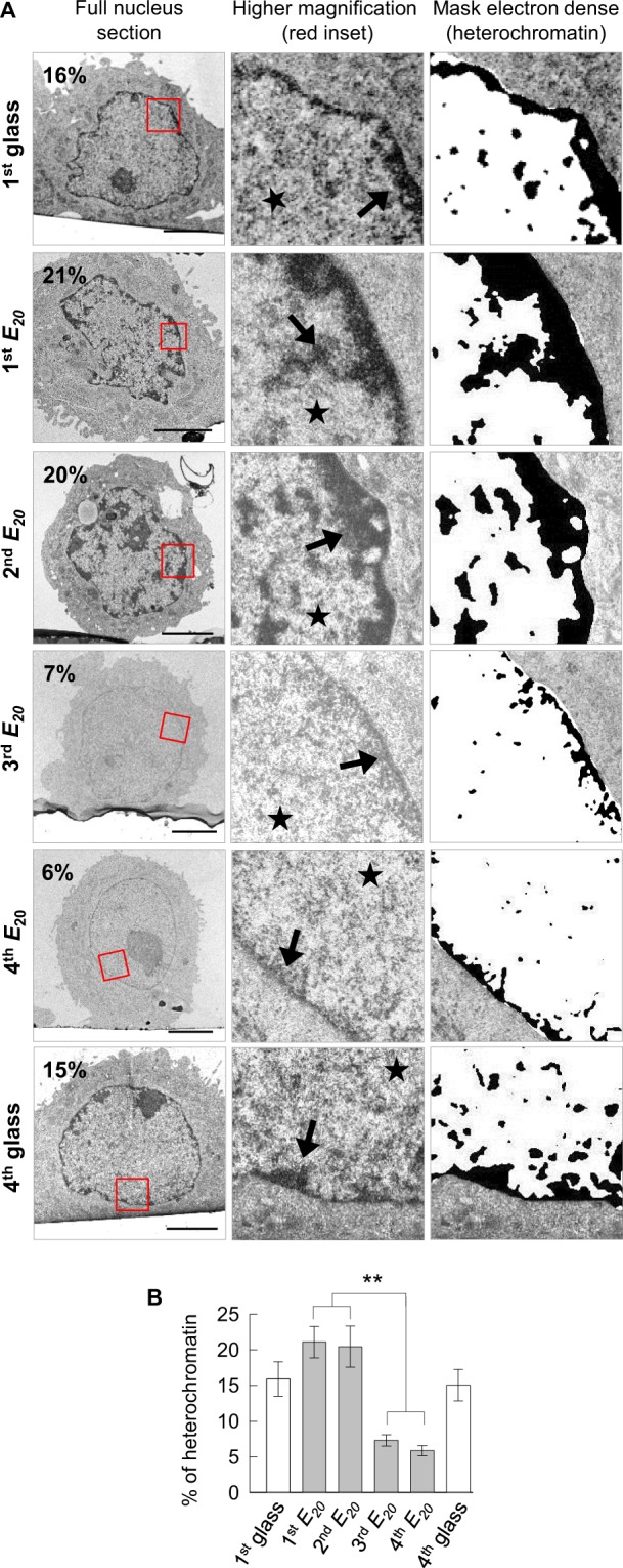
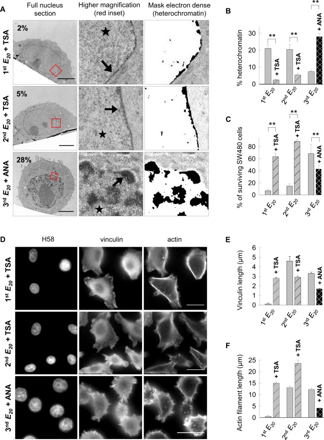
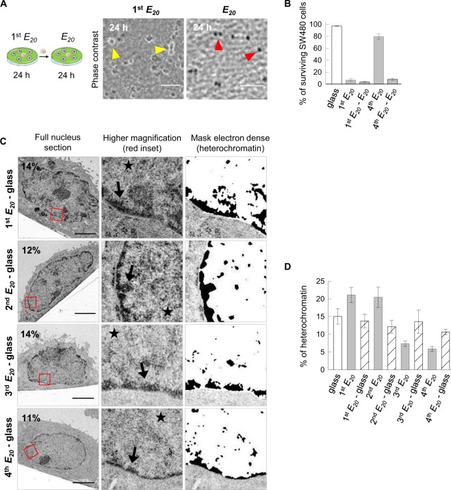
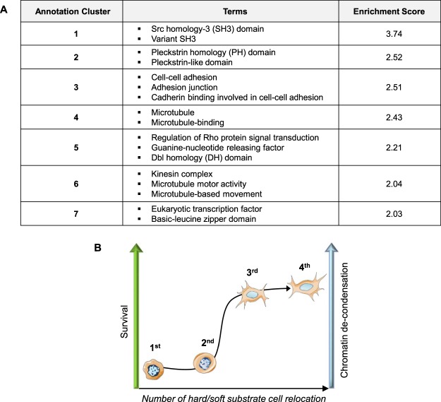
Mechanical properties of the cellular environment are known to influence cell fate. Chromatin de-condensation appears as an early event in cell reprogramming. Whereas the ratio of euchromatin versus heterochromatin can be increased chemically, we report herein for the first time that the ratio can also be increased by purely changing the mechanical properties of the microenvironment by successive 24 h-contact of the cells on a soft substrate alternated with relocation and growth for 7 days on a hard substrate. An initial contact with soft substrate caused massive SW480 cancer cell death by necrosis, whereas approximately 7% of the cells did survived exhibiting a high level of condensed chromatin (21% heterochromatin). However, four consecutive hard/soft cycles elicited a strong chromatin de-condensation (6% heterochromatin) correlating with an increase of cellular survival (approximately 90%). Furthermore, cell survival appeared to be reversible, indicative of an adaptive process rather than an irreversible gene mutation(s). This adaptation process is associated with modifications in gene expression patterns. A completely new approach for chromatin de-condensation, based only on mechanical properties of the microenvironment, without any drug mediation is presented.


Conflict of interest statement

The authors declare no competing interests.

Figures

Figure 1
Figure 1
Conditions and timing of the experiments.
Figure 2
Figure 2
Cell survival and vinculin assembly are influenced by switching the substrate elasticity. (A) Percentage of surviving SW480 cells based on the S1 from three pooled independent experiments (error bars represent s.e.m.). (B) Representative images of cells cultured for 24 h on glass, 1st E20, 2nd E20 and 3rd E20 immunolabelled with anti-vinculin counterstained with Hoechst 33258 and phalloidin. Scale bars: 10 µm. (C) Quantitative data for cell areas (in µm2), based on B, were determined using ImageJ. We used 200–300 cells for each condition (glass, 1st E20, 2nd E20 and 3rd E20). (D) Quantitative data for nuclear areas (in µm2) based on B were determined using ImageJ. We used 200–300 cells for each condition. (E) Quantification of vinculin spots (in µm) based on B by measuring 20 contacts per cell in 20 cells for each condition. (F) Quantification of actin stress fibers (in µm) using ImageJ based on B by measuring the length of 20 fibers per cell in 20 cells for each condition. (C–F) Results for three independent experiments are shown (error bars represent s.e.m.).
Figure 3
Figure 3
Cell motility influenced by switching the substrate elasticity. Motility of SW480 cells after successive seeding on soft substrate. (A) Time-lapse monitoring representative images of cells cultured on glass, 2nd E20 and 3rd E20 for 20 h. Inset: enlargement of the white dotted panel. Arrowhead: coloured cellular trajectory. (B) Cell trajectories from A by NIS-Elements 3D tracking. (C) Number of cells analysed, percentage of cells in class 1 and class 2, and mean speed of these cells, from A. The results from two pooled independent experiments for each condition.
Figure 4
Figure 4
Chromatin de-condensation by switching the substrate elasticity (A) Representative ultrastructural images of cells after 24 h of culture on 1st glass, 1st E20, 2nd E20, 3rd E20, 4th E20, and 4th glass. Scale bars: 5 µm, arrow: heterochromatin, star: euchromatin, (B) Percentage of heterochromatin in the nucleus of approximately 20 cells for each condition, from A, for two pooled independent experiments (error bars represents s.e.m.).
Figure 5
Figure 5
Cell survival is chromatin conformation-dependent (A) Representative ultrastructural images of cells after 24 h of culture on the 1st E20 + TSA, 2nd E20 + TSA and 3rd E20 + ANA. Scale bars: 5 µm, arrow: heterochromatin, star: euchromatin. (B) Percentage of heterochromatin in the nucleus of approximately 20 cells for each condition, from A, for two pooled independent experiments (error bars represents s.e.m.). (C) Percentage of surviving SW480 cells from S4 for two or three pooled independent experiments (error bars represent s.e.m.). (D) Representative images of cells cultured for 24 h on 1st E20 + TSA, 2nd E20 + TSA and 3rd E20 + ANA immunolabelled with anti-vinculin andcounterstained with Hoechst 33258 and phalloidin. Scale bars: 10 µm. (E) Quantification of vinculin spots (in µm) based on D by measuring 20 contacts per cell in 20 cells for each condition. (F) Quantification of actin stress (in µm) using ImageJ based on D by measuring the length of 20 actin stress fibers per cell in 20 cells for each condition. (D–F) Results for three independent experiments (error bars represent s.e.m.).
Figure 6
Figure 6
Cell motility is chromatin conformation-dependent. (A) Time-lapse monitoring representative images of cells cultured on 1st E20 + TSA, 2nd E20 + TSA and 3rd E20 + ANA for 20 h. Inset: enlargement of the white dotted panel, arrowhead: coloured cellular trajectory. (B) Cell trajectories, from A, by NIS-Elements 3D tracking. (C) Number of cells analysed, percentage of cells in class 1 and class 2 and mean speed of these cells, from A. The Results from two pooled independent experiments for each condition. Class 1 speed of 1st E20 + TSA vs 2nd E20 + TSA vs 3rd E20 + ANA: **.
Figure 7
Figure 7
Reversible adaptation of chromatin de-condensation. (A) Representative images acquired by phase contrast microscopy: 1st E20 cells cultured for 24 h on E20. Yellow arrowhead: refractive objects corresponding to surviving cells; E20 cells cultured on the 1st E20 for 24 h and transferred to new E20 for 24 h without an intermediate step on glass. Red arrowhead: non-refractive objects corresponding to necrotic cells. Scale bars: 100 µm. (B) Percentage of surviving SW480 cells from S4D for two independent experiments (the error bars represent the s.e.m.). (C) Representative ultrastructural images of cells cultured on 1st E20 - glass, 2nd E20 - glass, 3rd E20 - glass and 4th E20 - glass. Scale bars: 5 µm. (D) Percentage of heterochromatin in the nucleus of approximately 20 cells for each condition from C for two pooled independent experiments (error bars represents s.e.m.).
Figure 8
Figure 8
Functional clustering annotation of the differentially expressed genes. (A) Seven clusters identified with DAVID 6.8 with an enrichment score >2. (B) Cartoon summarizing the influence of the number of hard/soft substrate SW480 cell relocation on survival and chromatin conformation (hard = glass, soft = E20).

References

    1. Clevers H. The cancer stem cell: premises, promises and challenges. Nat. Med. 2011;17:313–319. doi: 10.1038/nm.2304. - DOI - PubMed
    1. Lambert AW, Pattabiraman DR, Weinberg RA. Emerging Biological Principles of Metastasis. Cell. 2017;168:670–691. doi: 10.1016/j.cell.2016.11.037. - DOI - PMC - PubMed
    1. Laugesen A, Helin K. Chromatin repressive complexes in stem cells, development, and cancer. Cell Stem Cell. 2014;14:735–751. doi: 10.1016/j.stem.2014.05.006. - DOI - PubMed
    1. Badeaux AI, Shi Y. Emerging roles for chromatin as a signal integration and storage platform. Nat. Rev. Mol. Cell Biol. 2013;14:211–224. doi: 10.1038/nrm3545. - DOI - PMC - PubMed
    1. Dillon N, Festenstein R. Unravelling heterochromatin: competition between positive and negative factors regulates accessibility. Trends Genet. 2002;18:252–258. doi: 10.1016/S0168-9525(02)02648-3. - DOI - PubMed

Publication types