

Flatfoot and normal foot a comparative analysis of the stress shielding
- PMID: 30140126
- PMCID: PMC6104143
- DOI: 10.1016/j.jor.2018.08.002
Flatfoot and normal foot a comparative analysis of the stress shielding
Erratum in
-
Erratum regarding missing Declaration of Competing Interest statements in previously published articles.J Orthop. 2020 Dec 15;24:293. doi: 10.1016/j.jor.2020.12.006. eCollection 2021 Mar-Apr. J Orthop. 2020. PMID: 33994702 Free PMC article.
Abstract
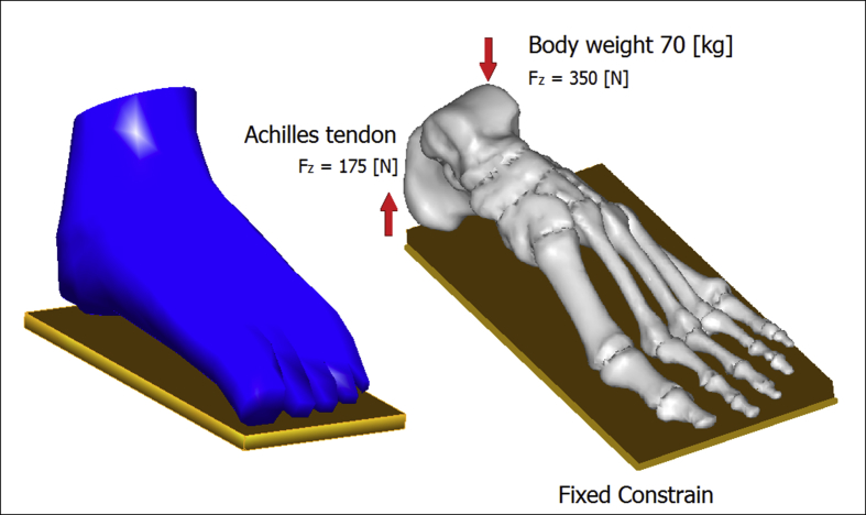
Objective: this study aims to develop a comprehensive 3D FE model of the foot to investigate the effect of soft tissue stiffness on the plantar pressure distributions and the internal load transfer between bony structures.
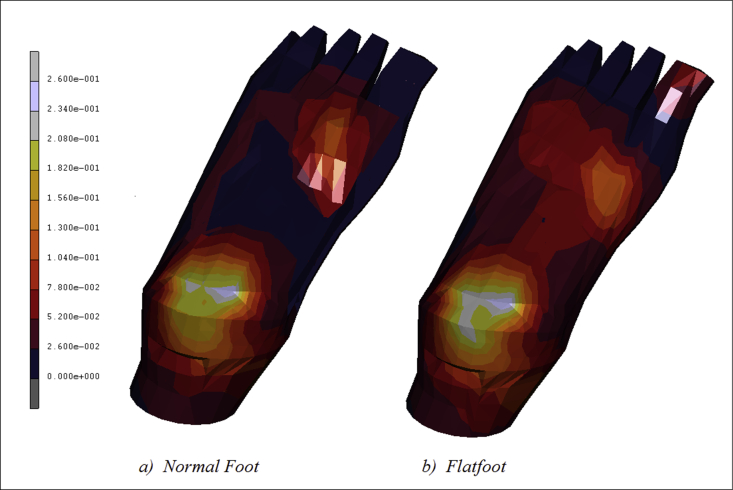
Methods: the stress shielding occurring on the plantar surface of a flatfoot was investigated and compared with the mechanical behavior of a healthy foot, trough baropodometric analyses and the FE models.
Results: the flatfoot evidences a more intensive stress-shielding map with significant values of pressure acting on the medial plantar fascia.
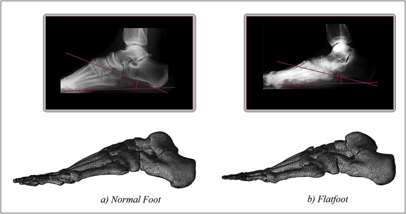
Conclusion: Clinically and radiographically, symptomatic adult flatfoot is a complex abnormality involving all three dimensions and multiple joints within the foot.
Keywords: CAD; FE analysis; Foot model.
Figures


References
-
- Di Giovanni J.E., Smith S.D. Normal biomechanics of the adult rearfoot: a radiographic analysis. J Am Podiatry Assoc. 1976;66(11):812–824. - PubMed
-
- Figura M.A., Smith S.D. Frontal plane deformity of the subtalar joint in flexible flat foot: a preliminary study. J Am Podiatry Assoc. 1976;66(11):867–872. - PubMed
-
- McCormack A.P., Ching R.P., Sangeorzan B.J. Biomechanics of procedures used in adult flatfoot deformity. Foot Ankle Clin. 2001;6(1):15–23. - PubMed
-
- Lewis G.S. The Pennsylvania State University; State College, PA: 2008. Computational Modeling of the Mechanics of Flatfoot Deformity and its Surgical Corrections. Ph.D. dissertation.
-
- Kitaoka H.B., Luo Z., An K. Three-dimensional analysis of flatfoot deformity: cadaver study. Foot Ankle Int. 1998;19(7):447–451. - PubMed
LinkOut - more resources
Full Text Sources
Other Literature Sources
Medical
Miscellaneous
