

The Free Radical Diseases of Prematurity: From Cellular Mechanisms to Bedside
- PMID: 30140369
- PMCID: PMC6081521
- DOI: 10.1155/2018/7483062
The Free Radical Diseases of Prematurity: From Cellular Mechanisms to Bedside
Abstract
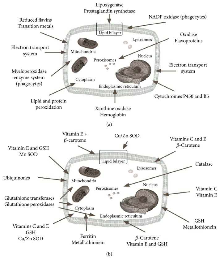
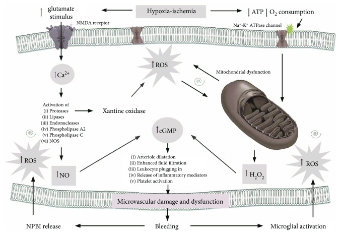
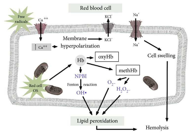
During the perinatal period, free radicals (FRs) are involved in several physiological roles such as the cellular responses to noxia, the defense against infectious agents, the regulation of cellular signaling function, and the induction of a mitogenic response. However, the overproduction of FRs and the insufficiency of an antioxidant mechanism result in oxidative stress (OS) which represents a deleterious process and an important mediator of damage to the placenta and the developing fetus. After birth, OS can be magnified by other predisposing conditions such as hypoxia, hyperoxia, ischemia, hypoxia ischemia-reperfusion, inflammation, and high levels of nonprotein-bound iron. Newborns are particularly susceptible to OS and oxidative damage due to the increased generation of FRs and the lack of adequate antioxidant protection. This impairment of the oxidative balance has been thought to be the common factor of the so-called "free radical related diseases of prematurity," including retinopathy of prematurity, bronchopulmonary dysplasia, intraventricular hemorrhage, periventricular leukomalacia, necrotizing enterocolitis, kidney damage, and oxidative hemolysis. In this review, we provide an update focused on the factors influencing these diseases refining the knowledge about the role of OS in their pathogenesis and the current evidences of such relationship. Mechanisms governing FR formation and subsequent OS may represent targets for counteracting tissue damage.
Figures
References
Publication types
MeSH terms
Substances
LinkOut - more resources
Full Text Sources
Other Literature Sources
