

Gambogic acid inhibits LPS-induced macrophage pro-inflammatory cytokine production mainly through suppression of the p38 pathway
- PMID: 30140411
- PMCID: PMC6098961
- DOI: 10.22038/IJBMS.2018.23897.5995
Gambogic acid inhibits LPS-induced macrophage pro-inflammatory cytokine production mainly through suppression of the p38 pathway
Abstract
Objectives: In traditional Chinese medicine, gamboge can detoxify bodies, kill parasites, and act as a hemostatic agent. Recent studies have demonstrated that gambogic acid (GBA) suppressed inflammation in arthritis, and also presented antitumor effect. Thus, this study investigated the new biological properties of GBA on macrophages.
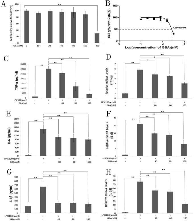
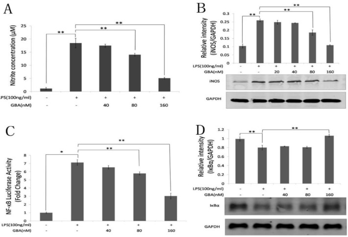
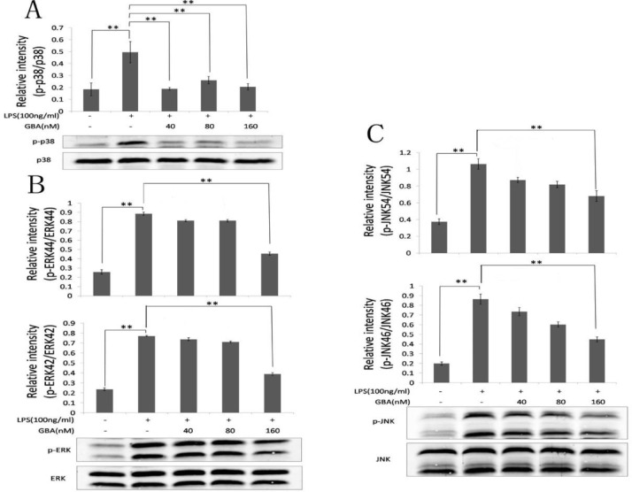
Materials and methods: RAW 264.7 cells were pretreated with GBA at different concentrations (10, 20, 40, 80, 160, 320 nM) for 24 hrs, and then cell viability was measured using Cell Counting Kit (CCK)-8 assays. Pro-inflammatory cytokines such as TNF-α, IL-6 and IL-1β were determined using ELISA kits and qPCR. Then nitrite concentration was calculated according to a standard curve. At last, the effect of GBA on MAPK and NF-κB signaling pathways was assessed by western blot and luciferase reporter gene assay.
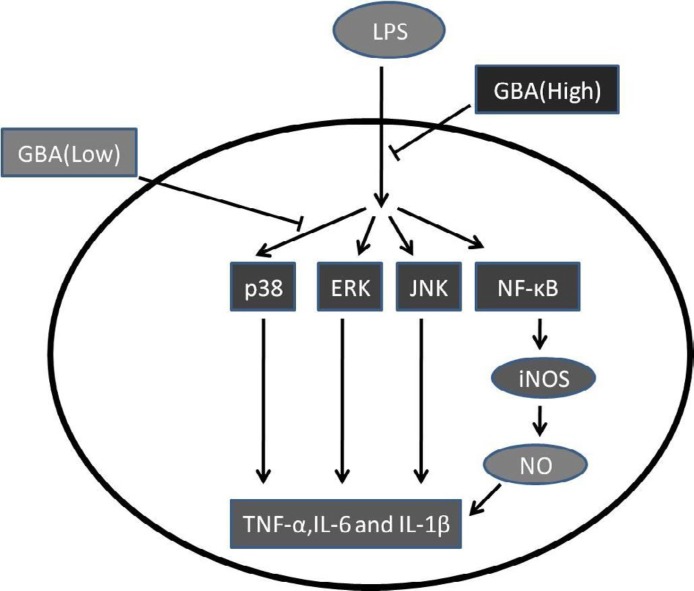
Results: GBA (IC50: 260 nM) suppressed the TNF-α, IL-6 and IL-1β expression induced by lipopolysaccharide (LPS) in RAW 264.7 cells. The expression of TNF-α, IL-6 and IL-1β decreased to 30-50% and 70-75% in the high-dose (160 nM) and low-dose (40 and 80 nM) GBA groups, respectively. Furthermore, the nitric oxide (NO) production and the activation of NF-κB, ERK, and JNK pathways were significantly reduced in high-dose (160 nM) GBA only, while p38 pathway was inhibited at both low (40 and 80 nM) and high (160 nM) concentration of GBA.
Conclusion: These data suggested that GBA inhibited LPS-induced production of pro-inflammatory cytokines including TNF-α, IL-6 and IL-1β mainly through the suppression of the p38 pathway.
Keywords: Anti-inflammatory agents; Gambogic acid; MAPK; NF-κB; RAW 264.7 cells; p38.
Figures
Similar articles
-
Anti-proliferation and anti-inflammation effects of corilagin in rheumatoid arthritis by downregulating NF-κB and MAPK signaling pathways.J Ethnopharmacol. 2022 Feb 10;284:114791. doi: 10.1016/j.jep.2021.114791. Epub 2021 Oct 29. J Ethnopharmacol. 2022. PMID: 34737112
-
Anti-inflammatory effect of the six compounds isolated from Nauclea officinalis Pierrc ex Pitard, and molecular mechanism of strictosamide via suppressing the NF-κB and MAPK signaling pathway in LPS-induced RAW 264.7 macrophages.J Ethnopharmacol. 2017 Jan 20;196:66-74. doi: 10.1016/j.jep.2016.12.007. Epub 2016 Dec 15. J Ethnopharmacol. 2017. PMID: 27989509
-
Pinocembrin attenuates lipopolysaccharide-induced inflammatory responses in Labeo rohita macrophages via the suppression of the NF-κB signalling pathway.Fish Shellfish Immunol. 2016 Sep;56:459-466. doi: 10.1016/j.fsi.2016.07.038. Epub 2016 Aug 2. Fish Shellfish Immunol. 2016. PMID: 27492123
-
Nitidine chloride inhibits LPS-induced inflammatory cytokines production via MAPK and NF-kappaB pathway in RAW 264.7 cells.J Ethnopharmacol. 2012 Oct 31;144(1):145-50. doi: 10.1016/j.jep.2012.08.041. Epub 2012 Sep 3. J Ethnopharmacol. 2012. PMID: 22971898
-
Anti-inflammatory effects of ursodeoxycholic acid by lipopolysaccharide-stimulated inflammatory responses in RAW 264.7 macrophages.PLoS One. 2017 Jun 30;12(6):e0180673. doi: 10.1371/journal.pone.0180673. eCollection 2017. PLoS One. 2017. PMID: 28665991 Free PMC article.
Cited by
-
Gambogic acid affects high glucose-induced apoptosis and inflammation of retinal endothelial cells through the NOX4/NLRP3 pathway.Ann Transl Med. 2023 Feb 28;11(4):168. doi: 10.21037/atm-22-6591. Epub 2023 Feb 16. Ann Transl Med. 2023. Retraction in: Ann Transl Med. 2023 Dec 20;11(12):424. doi: 10.21037/atm-2023-26. PMID: 36923084 Free PMC article. Retracted.
-
Naturally occurring anti-cancer compounds: shining from Chinese herbal medicine.Chin Med. 2019 Nov 6;14:48. doi: 10.1186/s13020-019-0270-9. eCollection 2019. Chin Med. 2019. PMID: 31719837 Free PMC article. Review.
-
Expression and role of ABIN1 in sepsis: In vitro and in vivo studies.Open Med (Wars). 2020 Dec 4;16(1):33-40. doi: 10.1515/med-2021-0008. eCollection 2021. Open Med (Wars). 2020. Retraction in: Open Med (Wars). 2022 Jun 11;17(1):1064. doi: 10.1515/med-2022-0503. PMID: 33364432 Free PMC article. Retracted.
References
-
- Bartold PM, Cantley MD, Haynes DR. Mechanisms and control of pathologic bone loss in periodontitis. Periodontology. 2010;53:55–69. - PubMed
-
- Adams DO, Hamilton TA. The cell biology of macrophage activation. Annu Rev Immunol. 1984;2:283–318. - PubMed
-
- Lu YC, Yeh WC, Ohashi PS. LPS/TLR4 signal transduction pathway. Cytokine. 2008;42:145–151. - PubMed
-
- Kim KN, Heo SJ, Yoon WJ, Kang SM, Ahn G, Yi TH, et al. Fucoxanthin inhibits the inflammatory response by suppressing the activation of NF-kappaB and MAPKs in lipopolysaccharide-induced RAW 2647 macrophages. Eur J Pharmacol. 2010;649:369–375. - PubMed
LinkOut - more resources
Full Text Sources
Research Materials
Miscellaneous