Squamous Cell Carcinoma in a Calyceal Diverticulum Detected by Percutaneous Nephroscopic Biopsy
- PMID: 30140478
- PMCID: PMC6081520
- DOI: 10.1155/2018/3508537
Squamous Cell Carcinoma in a Calyceal Diverticulum Detected by Percutaneous Nephroscopic Biopsy
Abstract
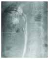
A 73-year-old woman was referred to our department with a complaint of asymptomatic gross hematuria. Dynamic computed tomography revealed a complicated (Bosniak type IIF) cyst in the upper pole of her right kidney, which was diagnosed as a calyceal diverticulum. The diagnosis was confirmed by ureteroscopy. The diverticulum was filled with a soft protein matrix that was difficult to completely remove from the inner surface of the calyceal diverticulum. Endoscopy combined with intrarenal surgery (ECIRS) was performed to completely remove the matrix. Percutaneous nephroscopy further revealed papillary lesions on the surface of the diverticulum, confirmed as squamous cell carcinoma on pathological assessment. A laparoscopic right radical nephroureterectomy was performed, with curative intent. Pathological assessment confirmed a high-grade squamous cell carcinoma with renal parenchymal invasion (pT3). Although carcinomas in a calyceal diverticulum are highly uncommon, when present, these tend to be high-grade neoplasms that deeply invade the parenchymal wall. As the effective management of these lesions is difficult, early-stage diagnosis is required for curative treatment. We report the case of squamous cell carcinoma in a calyceal diverticulum that was difficult to diagnose on preoperative computed tomography, urinal cytology examination, and ureteroscopy but was found during ECIRS.
Figures


Similar articles
-
Invasive urothelial carcinoma within a calyceal diverticulum associated with renal stones: A case report.Oncol Lett. 2015 Oct;10(4):2439-2441. doi: 10.3892/ol.2015.3590. Epub 2015 Aug 11. Oncol Lett. 2015. PMID: 26622866 Free PMC article.
-
Type 2 calyceal diverticulum with an unusual appearance in the lower pole of the kidney.J Radiol Case Rep. 2022 Jun 30;16(6):12-17. doi: 10.3941/jrcr.v16i6.4334. eCollection 2022 Jun. J Radiol Case Rep. 2022. PMID: 35875367 Free PMC article.
-
Pediatric calyceal diverticulum treatment: An experience with endoscopic and laparoscopic approaches.J Pediatr Urol. 2015 Aug;11(4):172.e1-6. doi: 10.1016/j.jpurol.2015.04.013. Epub 2015 May 21. J Pediatr Urol. 2015. PMID: 26052004
-
Challenges in the diagnosis of calyceal diverticulum: A report of two cases and review of the literature.J Xray Sci Technol. 2019;27(6):1155-1167. doi: 10.3233/XST-190549. J Xray Sci Technol. 2019. PMID: 31476195 Review.
-
Surgical management of the calyceal diverticulum.Curr Opin Urol. 2003 May;13(3):255-60. doi: 10.1097/00042307-200305000-00015. Curr Opin Urol. 2003. PMID: 12692451 Review.
Cited by
-
Is the presence of upper tract transitional cell carcinoma in a calyceal diverticulum a risk factor for early metastasis? A case report and review of the literature.SAGE Open Med Case Rep. 2024 Sep 30;12:2050313X241288341. doi: 10.1177/2050313X241288341. eCollection 2024. SAGE Open Med Case Rep. 2024. PMID: 39399581 Free PMC article.
-
A case report of renal calyceal diverticulum with hypertension in children and review of literature.BMC Pediatr. 2022 Jan 11;22(1):35. doi: 10.1186/s12887-021-03081-5. BMC Pediatr. 2022. PMID: 35016649 Free PMC article. Review.
-
A Case Report of Calyceal Diverticulum: Differential Diagnosis for Organ-Preserving Operations.Front Surg. 2021 Sep 16;8:731796. doi: 10.3389/fsurg.2021.731796. eCollection 2021. Front Surg. 2021. PMID: 34604297 Free PMC article.
References
-
- Adachi T., Ezaki K., Funai K. A case of transitional cell carcinoma in a pyelocaliceal diverticulum. Hinyokika Kiyo. Acta Urologica Japonica. 1989;35(8):1383–1386. - PubMed
-
- Yoshimura K., Yoshida H., Kawase N., Taki Y. A case of transitional cell carcinoma and milk of calcium in a pyelocalyceal diverticulum. Hinyokika Kiyo. Acta Urologica Japonica. 1998;44(9):649–652. - PubMed
Publication types
LinkOut - more resources
Full Text Sources
Other Literature Sources
Research Materials

