

doi: 10.18240/ijo.2018.08.28.
eCollection 2018.
Choroidal neovascularization post macular surgery: a case series
Affiliations
- PMID: 30140652
- PMCID: PMC6090133
- DOI: 10.18240/ijo.2018.08.28
Item in Clipboard
Choroidal neovascularization post macular surgery: a case series
Int J Ophthalmol.
.
No abstract available
Figures
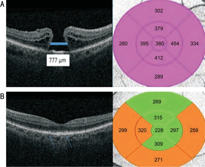
A: Right eye full thickness macular hole in P1. The thickness map of the right macula is also attached next to the macular hole image; B: P1's left eye showing deposition of drusen (blue arrows) at the macular area without any obvious evidence of CNV. The thickness map of the left macula is also attached.

A: Right eye OCT post macular surgery in P1. Note the presence of intra-retinal fluid (white arrow) and pigment epithelial detachment (blue arrow). The macular thickness map shows an increase in the central retinal thickness after the macular hole repair; B: OCT after the intravitreal injection of dexamethasone implant showing persistence of CME (white arrow) and pigment epithelial detachment (blue arrow). There was a slight increase in the central retinal thickness as demonstrated by the thickness map.

A: Early area of hyperfluorescence in P1's right macula (blue arrow) demonstrating a predominantly classic sub-foveal CNV; B: Area of late staining increasing in size and intensity over time (white arrow); C, D: Persistent intra-retinal fluid on OCT scans in P1 despite treatment with dexamethasone implant (grey arrow) and presence of pigment epithelial detachment indicating the presence of a predominantly classic sub-foveal CNV (green arrow).

Note the presence of persistent intra-retinal fluid post ranibizumab×3 (April 2017 to June 2017) and aflibercept×1 (July 2017). As there was no response to intravitreal injection of anti-VEGF therapeutic agents, treatment was ceased. The central retinal thickness was persistently high despite treatment.
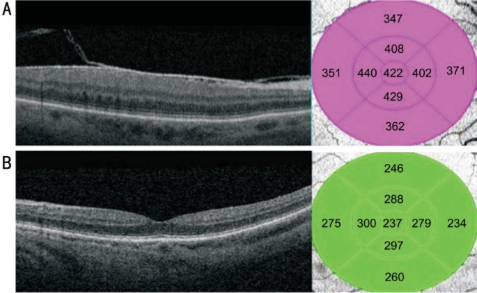
A: OCT scan from P2's right fundus. ERM noted but no signs of exudative maculopathy. Increased central retinal thickness due to the presence of ERM; B: OCT scan from P2's left fundus also shows no signs of AMD. Normal macular thickness.

A: OCT scan of P2's right fundus showing a grey well circumscribed sub-foveal CNV (blue arrow) with presence of sub-retinal and intra-retinal fluid; B: Presence of an active type 2 classic sub-foveal CNV (white arrows); C: Persistence of CNV and fluid despite treatment with anti-VEGF therapeutic agents. The central retinal thickness has increased after the ERM peel.

A: OCT scan of P3's right fundus showing an inactive sub-foveal CNV (thick blue arrow) with presence of foveal atrophy as demonstrated on the thickness map; B: OCT scan of P3's left fundus. Note the presence of a thick ERM causing macular schisis. Also note the presence of drusen (thin blue arrows) suggesting the concomitant presence of dry AMD.

A: OCT scan of P3's left fundus showing a large macular hemorrhage with a fibrovascular pigment epithelial detachment (white arrows) after ERM peeling; B: Late phase of the FFA of P3's left fundus. Note the presence of masking (green arrow) corresponding with the area of the macular hemorrhage. Also note the presence of late staining (red arrow) suggesting the concomitant presence of dry AMD changes; C: After 3 injections of aflibercept in 4 weekly intervals, the macular hemorrhage has reduced in size, but the fibrovascular pigment epithelial detachment (thick blue arrow) is still present and there are also still tiny pockets of intra-retinal fluid (thin blue arrow).
References
-
- Sources for macular degeneration: facts & figures. [Accessed on 28 Feb 2018]. Available at: http://www.brightfocus.org/sources-macular-degeneration-facts-figures.
-
- Wong WL, Su X, Li X, Cheung CM, Klein R, Cheng CY, Wong TY. Global prevalence of age-related macular degeneration and disease burden projection for 2020 and 2040: a systematic review and meta-analysis. Lancet Glob Health. 2014;2(2):e106–e116. - PubMed
-
- Pascolini D, Mariotti SP. Global estimates of visual impairment: 2010. Br J Ophthalmol. 2012;96(5):614–618. - PubMed
-
- Patel AK, Newcomb CW, Liesegang TL, Pujari SS, Suhler EB, Thorne JE, Foster CS, Jabs DA, Levy-Clarke GA, Nussenblatt RB, Rosenbaum JT, Sen HN, Artornsombudh P, Kothari S, Kempen JH, Systemic Immunosuppressive Therapy for Eye Diseases Research Group Risk of retinal neovascularisation in cases of uveitis. Ophthalmology. 2016;123(3):646–654. - PMC - PubMed
LinkOut - more resources
Full Text Sources
Other Literature Sources