

Deuterium metabolic imaging (DMI) for MRI-based 3D mapping of metabolism in vivo
- PMID: 30140744
- PMCID: PMC6105304
- DOI: 10.1126/sciadv.aat7314
Deuterium metabolic imaging (DMI) for MRI-based 3D mapping of metabolism in vivo
Abstract
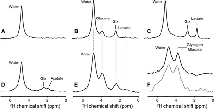
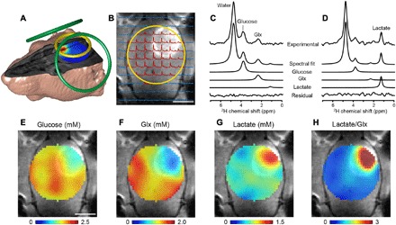
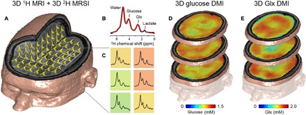
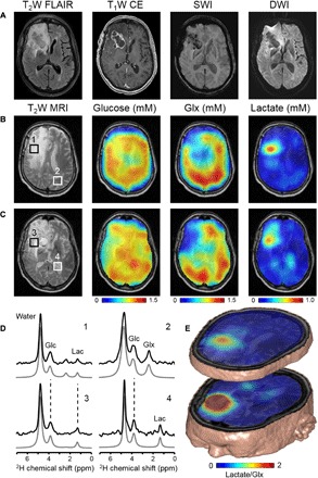
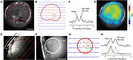
Currently, the only widely available metabolic imaging technique in the clinic is positron emission tomography (PET) detection of the radioactive glucose analog 2-18F-fluoro-2-deoxy-d-glucose (18FDG). However, 18FDG-PET does not inform on metabolism downstream of glucose uptake and often provides ambiguous results in organs with intrinsic high glucose uptake, such as the brain. Deuterium metabolic imaging (DMI) is a novel, noninvasive approach that combines deuterium magnetic resonance spectroscopic imaging with oral intake or intravenous infusion of nonradioactive 2H-labeled substrates to generate three-dimensional metabolic maps. DMI can reveal glucose metabolism beyond mere uptake and can be used with other 2H-labeled substrates as well. We demonstrate DMI by mapping metabolism in the brain and liver of animal models and human subjects using [6,6'-2H2]glucose or [2H3]acetate. In a rat glioma model, DMI revealed pronounced metabolic differences between normal brain and tumor tissue, with high-contrast metabolic maps depicting the Warburg effect. We observed similar metabolic patterns and image contrast in two patients with a high-grade brain tumor after oral intake of 2H-labeled glucose. Further, DMI used in rat and human livers showed [6,6'-2H2]glucose stored as labeled glycogen. DMI is a versatile, robust, and easy-to-implement technique that requires minimal modifications to existing clinical magnetic resonance imaging scanners. DMI has great potential to become a widespread method for metabolic imaging in both (pre)clinical research and the clinic.
Figures
References
-
- Zilberter Y., Zilberter M., The vicious circle of hypometabolism in neurodegenerative diseases: Ways and mechanisms of metabolic correction. J. Neurosci. Res. 95, 2217–2235 (2017). - PubMed
-
- Morató L., Bertini E., Verrigni D., Ardissone A., Ruiz M., Ferrer I., Uziel G., Pujol A., Mitochondrial dysfunction in central nervous system white matter disorders. Glia 62, 1878–1894 (2014). - PubMed
-
- Öz G., Alger J. R., Barker P. B., Bartha R., Bizzi A., Boesch C., Bolan P. J., Brindle K. M., Cudalbu C., Dinçer A., Dydak U., Emir U. E., Frahm J., González R. G., Gruber S., Gruetter R., Gupta R. K., Heerschap A., Henning A., Hetherington H. P., Howe F. A., Hüppi P. S., Hurd R. E., Kantarci K., Klomp D. W. J., Kreis R., Kruiskamp M. J., Leach M. O., Lin A. P., Luijten P. R., Marjańska M., Maudsley A. A., Meyerhoff D. J., Mountford C. E., Nelson S. J., Necmettin Pamir M., Pan J. W., Peet A. C., Poptani H., Posse S., Pouwels P. J. W., Ratai E.-M., Ross B. D., Scheenen T. W. J., Schuster C., Smith I. C. P., Soher B. J., Tkáč I., Vigneron D. B., Kauppinen R. A.; MRS Consensus Group , Clinical proton MR spectroscopy in central nervous system disorders. Radiology 270, 658–679 (2014). - PMC - PubMed
Publication types
MeSH terms
Substances
Grants and funding
LinkOut - more resources
Full Text Sources
Other Literature Sources
Medical
