. 2018 Sep 17;10(9):555-565.
doi: 10.1039/c8ib00111a.

A high-throughput microtissue platform to probe endothelial function in vitro

Affiliations

A high-throughput microtissue platform to probe endothelial function in vitro

Alexandra L Crampton et al. Integr Biol (Camb). .

Abstract

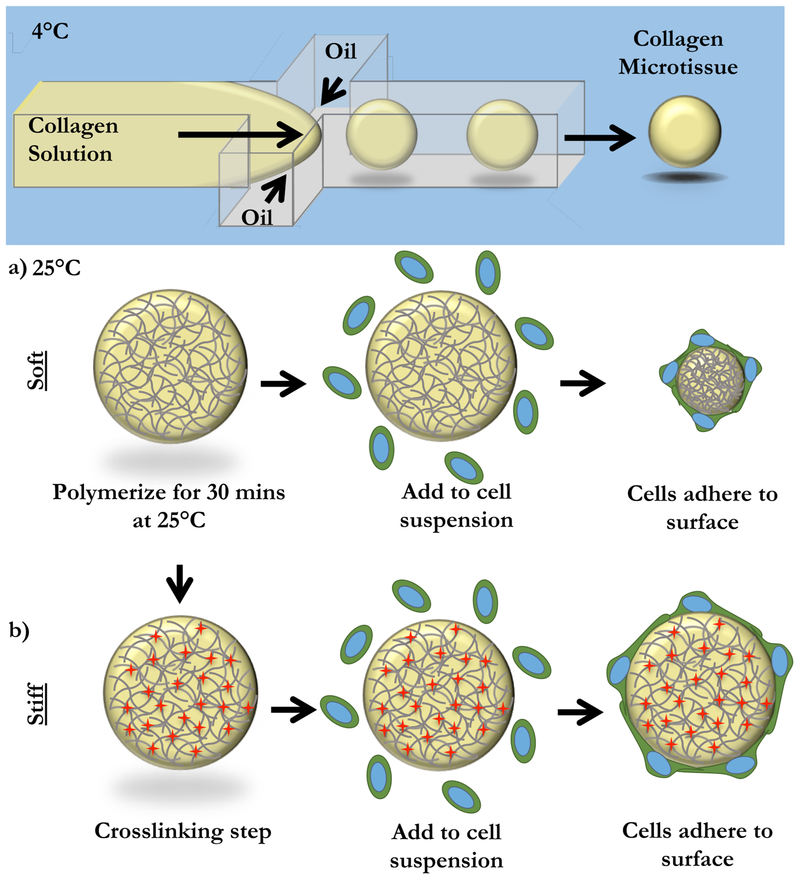
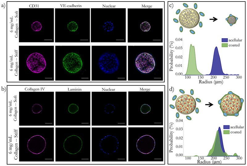
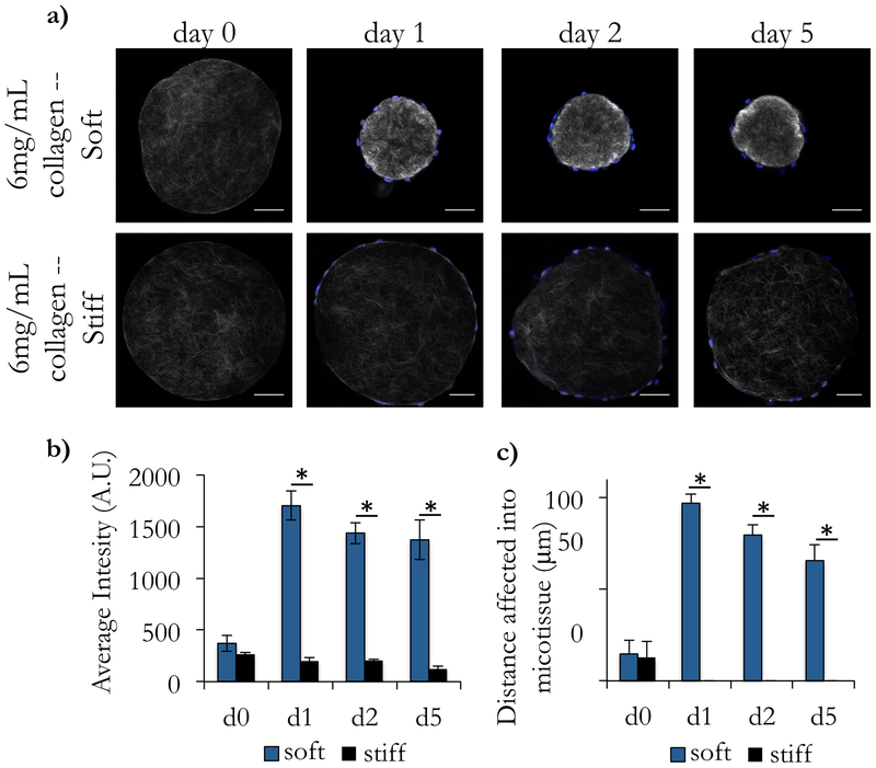
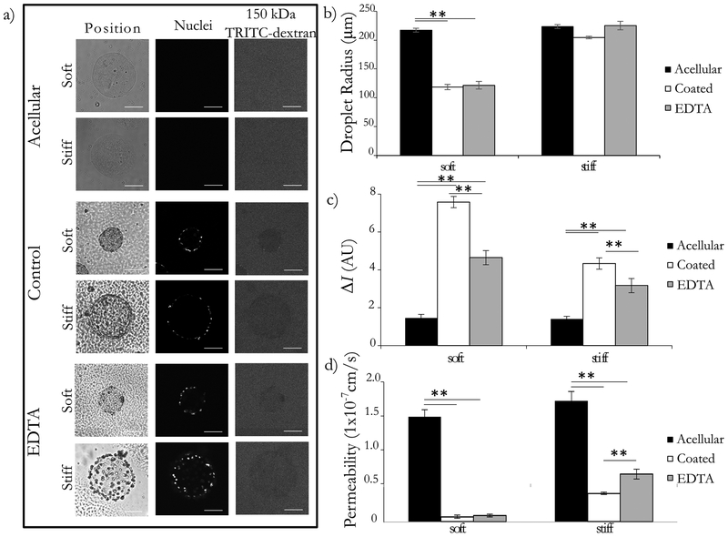
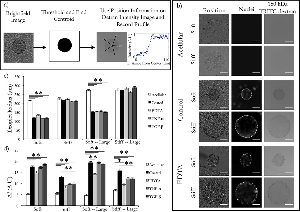
A critical role of vascular endothelium is as a semi-permeable barrier, dynamically regulating the flux of solutes between blood and the surrounding tissue. Existing platforms that quantify endothelial function in vitro are either significantly throughput limited or overlook physiologically relevant extracellular matrix (ECM) interactions and thus do not recapitulate in vivo function. Leveraging droplet microfluidics, we developed a scalable platform to measure endothelial function in nanoliter-volume, ECM-based microtissues. In this study, we describe our high-throughput method for fabricating endothelial-coated collagen microtissues that incorporate physiologically relevant cell-ECM interactions. We showed that the endothelial cells had characteristic morphology, expressed tight junction proteins, and remodeled the ECM via compaction and deposition of basement membrane. We also measured macromolecular permeability using two optical modalities, and found the cell layers: (1) had permeability values comparable to in vivo measurements and (2) were responsive to physiologically-relevant modulators of endothelial permeability (TNF-α and TGF-β). This is the first demonstration, to the authors' knowledge, of high-throughput assessment (n > 150) of endothelial permeability on natural ECM. Additionally, this technology is compatible with standard cell culture equipment (e.g. multi-well plates) and could be scaled up further to be integrated with automated liquid handling systems and automated imaging platforms. Overall, this platform recapitulates the functions of traditional transwell inserts, but extends application to high-throughput studies and introduces new possibilities for interrogating cell-cell and cell-matrix interactions.


Conflict of interest statement

Conflicts of Interest:

There are no conflicts to declare.

Figures

Figure 1:
Figure 1:. Fabrication of Endothelial-coated Collagen Microtissues.
Liquid 6 mg/mL collagen I microtissues were generated using a chilled flow-focusing microfluidic device. Microtissues were collected and polymerized off-chip at 25°C for 30 minutes. (a) In the case of “soft” microtissues, we did not need to further manipulate the collagen microtissues. To coat with cells, microtissues were mixed with a single-cell suspension of endothelial cells. In this “soft” mode, cells compacted the collagen microtissues. (b) To prevent compaction, we created “stiff” microtissues. After polymerization, microtissues were crosslinked with formalin. “Stiff” microtissues were then washed thoroughly and coated by incubating with a single cell suspension of endothelial cells. Cells created a monolayer on the surface of the microtissues, but did not change the overall size of these “stiff” microtissues.
Figure 2:
Figure 2:. Endothelial cells formed confluent monolayers and remodel ECM-based microtissues.
Both soft and stiff microtissues were coated with endothelial cells and cultured for 5 days. (a) On both soft and stiff microtissues, cells create confluent monolayers. Cells in all conditions have characteristic morphology and tight-junction expression, visualized with CD31 and VE-cadherin. (b) Using immunofluorescence, we observed that the endothelial cells deposited Collagen IV and Laminin on the surface of the collagen constructs for both the soft and stiff conditions. We qualitatively observed that (c) coating soft matrices with endothelial cells resulted in compaction, but (d) stiff matrices did not have a significant size change. We quantified the projected area of the droplets before and after coating to quantify this result and found the population shift for the compaction to be extremely statistically significant, but observed no statistically significant change for the stiff matrices. Scale bars 100 μm.
Figure 3:
Figure 3:. Second harmonic generation imaging reveals local remodeling of collagen architecture.
Acellular collagen droplets were coated with endothelial cells and cultured for 5 days. (a) At intermediate timepoints, droplets were collected and imaged using Second Harmonic Generation (SHG) to visualize the collagen fibers. (b) We observed that the average intensity of the soft microtissues on d1, d2, and d5 after coating with endothelial cells was significantly higher than acellular as well as the corresponding timepoints for the stiff tissues. In (a), we observed a dense ring of collagen at the surface of the microtissues. (c) To quantify this, we took linescans from the center of the microtissues to the surface and recorded the image intensity along the line. We repeated this 5 times for each droplet and reported a moving average with standard error of these linescans for each condition. (d) Using these intensity profiles, we measured the distance into the droplet that displayed large differences in intensity (defined as 5-fold brighter than the baseline average on the d0 measurements). We found that the stiff droplets showed very little remodeling of the interior of the droplet, but the soft constructs had large changes in intensity up to 97 μm from the surface of the droplet. Scale bars 100 μm.
Figure 4:
Figure 4:. Microtissue endothelial barrier function is comparable to in vivo permeability.
Endothelial cells were cultured on the surface of soft and stiff collagen microtissues for 5 days. Tissues were collected and some were treated with 5mM EDTA for 30 minutes. Constructs were placed in a 12.5 ug/mL dye bath and incubated for 20 minutes. (a) Constructs were imaged with brightfield and optical sectioning. Brightfield imaging shows the location and geometry of the microtissues, nucelear stain confirms the presence of ceslls, and the movement 150kDa TRITC-Dextran was visualized with flourescence microscopy. We qualitatively observe that acellular constructs do not impede dye movement, whereas the control condition showed exclusion of the dye from the interior of the microtissue for both the soft and stiff conditions. The EDTA treated condition appeared to have an intermediated phenotype. Backround noise was reduced in representative images for clarity. (b) We report the average droplet radius with standard error, showing that the soft constructs were compacted significantly, and the stiff constructs were largely unchanged in diameter. (c) Comparing the intensity inside and outside the droplet (ΔI), we found that ΔI was small for acellular constructs, and largest for coated constructs for both soft and stiff microtissues. We found that the removal of the cell layer resulted in an intermediate ΔI for both stiffnesses. (d) Converting the ΔI to the permeability using Eq. (3), we found the acellular constructs to have the largest permeability, and the coated controls to have a permeability that was statistically significantly smaller for both cases. For the EDTA treated group, we found that this increased the average permeability for the stiff constructs in a statistically significant manner. There was a slight increase in the average also for the soft constructs, but this difference was not statistically significant. We used these permeability values to calculate the contribution of the ECM and the cell layers individually and found the permeability of the cell layer to be on the order of 1×10⁻⁸ cm/s. assay. All comparisons completed with one-way ANOVA with post-hoc Tukey HSD test; p<0.01 = **. Scale bars 100 μm.
Figure 5:
Figure 5:. Endothelial barrier function can be assessed with widefield imaging for high throughput studies.
Endothelial cells were cultured on the surface of collagen microtissues for 5 days before assessing permeability. Constructs were soaked in 12.5 ug/mL bath of 150 kDa fluorescent dextran. Constructs were then imaged with widefield microscopy for the brightfield and fluorescent channels. (a) To assess permeability, we used brightfield images to detect the centroid and edges of each microtissues and collected linescans from the center of the droplet to 100 pixels outside the edge of the construct. (b) Representative images from acellular, control HUEC-coated, and 5 mM EDTA treated microtissues are shown. (c) When comparing the average microtissue radius (shown with standard error) for each condition, we observed consistent compaction of soft microtissues, and little change in size of stiff microtissues. (d) To assess permeability, we calculated the difference in intensity between the outside and inside of the droplet (ΔI). We observed that the acellular constructs were more saturated with dye in all construct conditions for both sizes of dye. However, the cell monolayer demonstrated semi-permeable qualities, which was especially visible on the stiff constructs. We found the addition of the cell layer increased ΔI for all conditions (p<0.01). In the large-soft and both stiff conditions, the modulation of the cell layer with EDTA resulted in a statistically significant decrease in ΔI. When we tested inflammatory cytokines on our platform, TNF-α and TGF-β resulted in statistically signficant decreases in ΔI (relative to the coated control) for both sizes of stiff microtissues. From this, we concluded that the stiff microtissues were better suited for the macromolecular permeability assay (one-way ANOVA with post-hoc Tukey HSD test; p<0.01 = **, p<0.05 = *). Scale bars 100 μm.

References

    1. Sukriti S, Tauseef M, Yazbeck P, Mehta D. Mechanisms Regulating Endothelial Permeability. Pulm Circ. 2014. December 1;4(4):535–51. - PMC - PubMed
    1. Sukriti S, Tauseef M, Yazbeck P, Mehta D. Mechanisms regulating endothelial permeability. Pulm Circ. 2014;4(4):535–51. - PMC - PubMed
    1. Mantovani A, Bussolino F, Dejana E. Cytokine regulation of endothelial cell function. FASEB J. 1992. May 1;6(8):2591–9. - PubMed
    1. Friedman M, Byers SO. Endothelial permeability in atherosclerosis. Arch Pathol. 1963;76:99–105. - PubMed
    1. Veress B, Bálint A, Kóczé A, Nagy Z, Jellinek H. Increasing aortic permeability by atherogenic diet. Atherosclerosis. 1970. May 1;11(3):369–71. - PubMed

Publication types

Substances

LinkOut - more resources