. 2018 Nov 1;315(5):H1477-H1485.
doi: 10.1152/ajpheart.00204.2018. Epub 2018 Aug 24.

Shear stress augments mitochondrial ATP generation that triggers ATP release and Ca2+ signaling in vascular endothelial cells

Affiliations

Shear stress augments mitochondrial ATP generation that triggers ATP release and Ca2+ signaling in vascular endothelial cells

Kimiko Yamamoto et al. Am J Physiol Heart Circ Physiol. .

Abstract

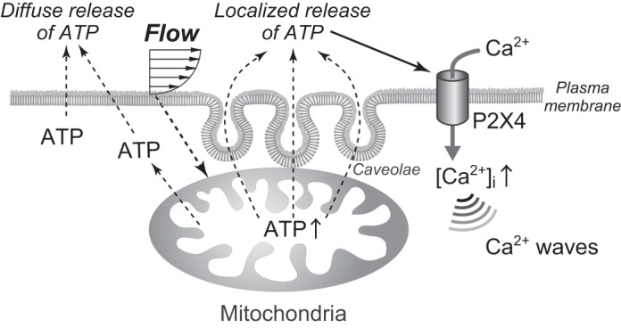
Vascular endothelial cells (ECs) sense and transduce hemodynamic shear stress into intracellular biochemical signals, and Ca2+ signaling plays a critical role in this mechanotransduction, i.e., ECs release ATP in the caveolae in response to shear stress and, in turn, the released ATP activates P2 purinoceptors, which results in an influx into the cells of extracellular Ca2+. However, the mechanism by which the shear stress evokes ATP release remains unclear. Here, we demonstrated that cellular mitochondria play a critical role in this process. Cultured human pulmonary artery ECs were exposed to controlled levels of shear stress in a flow-loading device, and changes in the mitochondrial ATP levels were examined by real-time imaging using a fluorescence resonance energy transfer-based ATP biosensor. Immediately upon exposure of the cells to flow, mitochondrial ATP levels increased, which was both reversible and dependent on the intensity of shear stress. Inhibitors of the mitochondrial electron transport chain and ATP synthase as well as knockdown of caveolin-1, a major structural protein of the caveolae, abolished the shear stress-induced mitochondrial ATP generation, resulting in the loss of ATP release and influx of Ca2+ into the cells. These results suggest the novel role of mitochondria in transducing shear stress into ATP generation: ATP generation leads to ATP release in the caveolae, triggering purinergic Ca2+ signaling. Thus, exposure of ECs to shear stress seems to activate mitochondrial ATP generation through caveola- or caveolin-1-mediated mechanisms. NEW & NOTEWORTHY The mechanism of how vascular endothelial cells sense shear stress generated by blood flow and transduce it into functional responses remains unclear. Real-time imaging of mitochondrial ATP demonstrated the novel role of endothelial mitochondria as mechanosignaling organelles that are able to transduce shear stress into ATP generation, triggering ATP release and purinoceptor-mediated Ca2+ signaling within the cells.

Keywords: ATP; calcium signaling; endothelial cells; mitochondria; shear stress.


Figures

Fig. 1.
Fig. 1.
Monitoring of mitochondrial ATP levels in human pulmonary artery endothelial cells. A: distribution of the fluorescence resonance energy transfer-based ATP biosensor mitAT1.03. This was correctly located in mitochondria stained with MitoTracker deep red FM. Cell nuclei were stained with Hoechst 33342, and the cell margin obtained visualized under a phase-contrast microscope is shown by the dashed lines. B: time course of the fluorescence intensity of cyan fluorescent protein (CFP; blue) and yellow fluorescent protein (YFP; red) inside the regions of interest. Treatment of cells with the ATP synthase inhibitor oligomycin (10 µg/ml) induced a decrease in YFP intensity and an increase in CFP intensity. The YFP-to-CFP ratio therefore decreased, indicating a decrease in ATP levels. Treatment of cells with an inhibitor of glycolysis [2-deoxy-d-glucose (2-DG); 10 mM] had no effect on the YFP-to-CFP ratio. C: ATP sensitivity of purified AT1.03. The plot of YFP-to-CFP ratios against Mg-ATP concentrations showed that the YFP-to-CFP ratio increased in an ATP concentration-dependent manner in the range of 0–10 mM. Oligomycin (10 µg/ml) had no effect on the ATP sensitivity of AT1.03. a.u., Arbitrary units.
Fig. 2.
Fig. 2.
Effects of shear stress on mitochondrial ATP levels. A: pseudocolor images of the yellow fluorescent protein (YFP)-to-cyan fluorescent protein (CFP) ratio before, during, and after the application of shear stress. The colors represent the ATP levels indicated by the scale. Shear stress (3 dyn/cm2) increased ATP levels over the entire mitochondria. The time course of the YFP-to-CFP ratio showed that the ATP level rapidly increased in response to shear stress, remained at the increased level, and then returned to the control level after the shear stress ceased. B: response to the repeated application of shear stress. There was an increase in mitochondrial ATP levels in response to each application of shear stress. Similar findings were observed in many cells. C: effects of hydrostatic pressure on mitochondrial ATP levels. Hydrostatic pressure (40 mmHg) had no effect on ATP levels. In contrast, there was a marked increase in ATP levels in response to shear stress. D: intensity dependency of the shear stress-induced changes in mitochondrial ATP levels. ATP levels in the mitochondria increased further as the intensity of shear stress increased. E: bar graph showing the results of a quantitative analysis of the shear stress-induced changes in ATP levels. Values are means ± SD of the data obtained in 15 cells. *P < 0.05; **P < 0.01.
Fig. 3.
Fig. 3.
Effects of inhibitors of glycolysis and mitochondrial oxidative phosphorylation on the shear stress-induced increase in ATP generation. A: temporal changes in mitochondrial ATP levels. Shear stress induced an increase in ATP levels in cells treated with the glycolysis inhibitor 2-deoxy-d-gllucose (2-DG; 10 mM) but not in cells treated with an ATP synthase inhibitor (oligomycin, 10 µg/ml), a mitochondrial oxidative phosphorylation uncoupler [carbonyl cyanide m-chlorophenylhydrazone (CCCP); 0.4 µg/ml], and a mitochondrial electron transport inhibitor (rotenone, 5 µM). This indicated that shear stress activated the function of mitochondrial oxidative phosphorylation. Neither the addition of EGTA (1 mM) into the culture medium, which chelates extracellular Ca2+, nor treatment of endothelial cells (ECs) with MitoTEMPOL (50 μM), which inactivates mitochondrial ROS, had an effect on ATP generation. Baseline mitochondrial ATP concentrations were reduced by treatment of ECs with oligomycin, CCCP, or rotenone, whereas no such change was observed after treatment of cells with 2-DG, EGTA, or MitoTEMPOL. These mitochondrial ATP responses were representative of those of dozens of cells, all of which showed similar results. B: extracellular ATP release in response to shear stress. Human pulmonary artery ECs (HPAECs) were exposed to shear stress (3 dyn/cm2) for 1 min, and the effluent was subjected to ATP measurement. HPAECs released ATP in response to shear stress, and inhibitors of the mitochondrial oxidative phosphorylation (oligomycin, CCCP, and rotenone) markedly suppressed ATP release, whereas glycolysis inhibitor (2-DG), EGTA, and MitoTEMPOL had no effect. Knockdown of caveolin-1 with its siRNA significantly suppressed ATP release, whereas scrambled siRNA had no effect. Results are presented as means ± SD of nine samples obtained in three separate experiments. *P < 0.01 compared with control.
Fig. 4.
Fig. 4.
Involvement of caveolin-1 in shear stress-induced ATP generation and Ca2+ signaling. A: relationship between mitochondria and caveolin-1, a marker protein for caveolae. After mitochondria were imaged with MitoTracker deep red FM, human pulmonary artery endothelial cells (HPAECs) were immunostained with antibody to caveolin-1. Cell nuclei were stained with DAPI. Caveolin-1 was unevenly distributed over the cell surface and was concentrated at a specific part of the cell periphery. The edge of the mitochondria was in close proximity to the region with concentrated caveolin-1. Caveolin-1 was completely knocked down by its siRNA. B: temporal changes in mitochondrial ATP levels under shear stress. Shear stress increased mitochondrial ATP levels in cells treated with caveolin-1-scrambled siRNA, whereas caveolin-1 knockdown by its siRNA abolished shear stress-induced mitochondrial ATP generation. These mitochondrial ATP responses were representative of those of dozens of cells, all of which showed similar results. C: Ca2+ responses to shear stress in HPAECs. Intracellular Ca2+ concentrations ([Ca2+]i) are expressed as the ratio of change in Fluo-4 fluorescence to the control before application of shear stress (F/F0). Closed rectangles indicate the duration of shear stress (3 dyn/cm2). Shear stress induced a marked increase in [Ca2+]i in cells treated with caveolin-1-scrambled siRNA but not in cells treated with caveolin-1 siRNA. Treatment of cells with oligomycin (10 µg/ml), carbonyl cyanide m-chlorophenylhydrazone (CCCP; 0.4 µg/ml), or rotenone (5 µM) abolished the shear stress-mediated Ca2+ response. The Ca2+ response was blocked by EGTA (1 mM) but not by MitoTEMPOL (50 μM). No changes in baseline [Ca2+]i were observed after treatment of cells with any of the above inhibitors or siRNA. Treatment of cells with these Ca2+response curves are representative of those of dozens of cells, all of which showed similar results. These results indicate that caveolin-1 and mitochondrial ATP generation can be implicated in the Ca2+ signaling after shear stress. Scrambled siCaveolin-1, cells transfected with caveolin-1-scrambled siRNA; siCaveolin-1, cells transfected with caveolin-1 siRNA; CFP, cyan fluorescent protein; YFP, yellow fluorescent protein.
Fig. 5.
Fig. 5.
Schematic diagram of the proposed caveolae-associated purinergic Ca2+ signaling in endothelial cells (ECs) exposed to shear stress. Shear stress induces both diffuse ATP release from the entire surface of the cell membrane and a localized ATP release from caveolae-rich cell regions. Since the localized ATP release reaches to more than 10 μM, it can activate the ATP-operated cation channel P2X4, thereby causing an influx of extracellular Ca2+ into cells. The increase in the intracellular Ca2+ concentration evokes a Ca2+ wave that starts at the caveolae and propagates throughout the entire cell. Mitochondria increase their ATP generation in response to shear stress exposure of the ECs and play a role as the source of the ATP released by ECs. The mechanism underlying the induction of increased mitochondrial ATP generation in response to shear stress in ECs remains unknown.

Similar articles

Cited by

References

    1. Alevriadou BR, Shanmughapriya S, Patel A, Stathopulos PB, Madesh M. Mitochondrial Ca2+ transport in the endothelium: regulation by ions, redox signalling and mechanical forces. J R Soc Interface 14: 20170672, 2017. doi:10.1098/rsif.2017.0672. - DOI - PMC - PubMed
    1. Anderson RG. Plasmalemmal caveolae and GPI-anchored membrane proteins. Curr Opin Cell Biol 5: 647–652, 1993. doi:10.1016/0955-0674(93)90135-D. - DOI - PubMed
    1. Ando J, Yamamoto K. Flow detection and calcium signalling in vascular endothelial cells. Cardiovasc Res 99: 260–268, 2013. doi:10.1093/cvr/cvt084. - DOI - PubMed
    1. Ando J, Yamamoto K. Vascular mechanobiology: endothelial cell responses to fluid shear stress. Circ J 73: 1983–1992, 2009. doi:10.1253/circj.CJ-09-0583. - DOI - PubMed
    1. Aoki T, Nishimura M, Matsuoka T, Yamamoto K, Furuyashiki T, Kataoka H, Kitaoka S, Ishibashi R, Ishibazawa A, Miyamoto S, Morishita R, Ando J, Hashimoto N, Nozaki K, Narumiya S. PGE2-EP2 signalling in endothelium is activated by haemodynamic stress and induces cerebral aneurysm through an amplifying loop via NF-κB. Br J Pharmacol 163: 1237–1249, 2011. doi:10.1111/j.1476-5381.2011.01358.x. - DOI - PMC - PubMed

Publication types

LinkOut - more resources