. 2018 Aug 24;14(8):e1007568.
doi: 10.1371/journal.pgen.1007568. eCollection 2018 Aug.

cis-regulatory architecture of a short-range EGFR organizing center in the Drosophila melanogaster leg

Affiliations

cis-regulatory architecture of a short-range EGFR organizing center in the Drosophila melanogaster leg

Susan Newcomb et al. PLoS Genet. .

Abstract

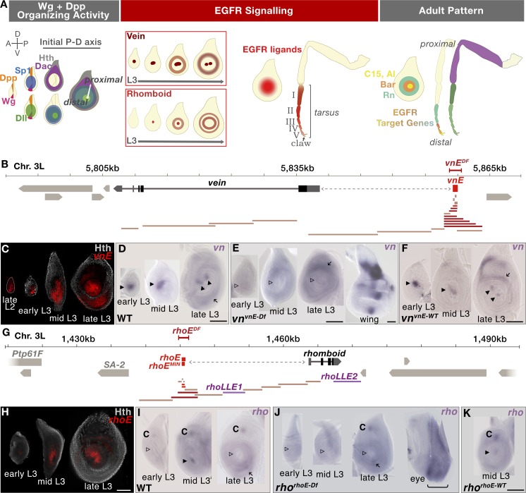
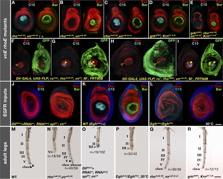
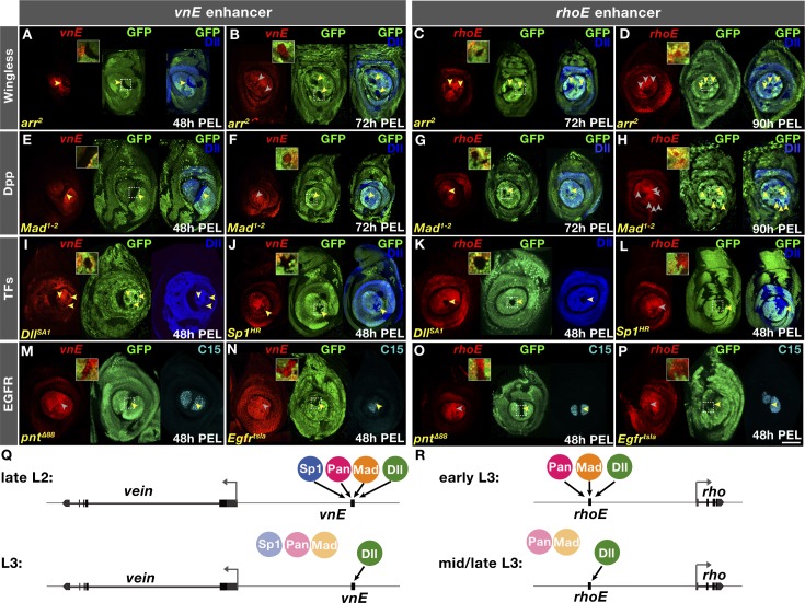
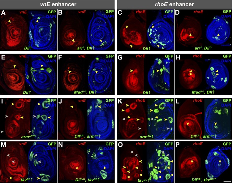
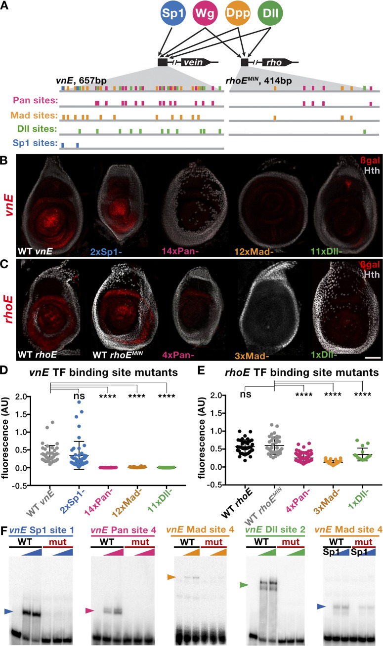
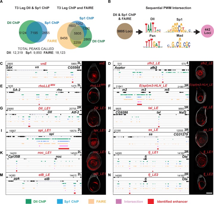
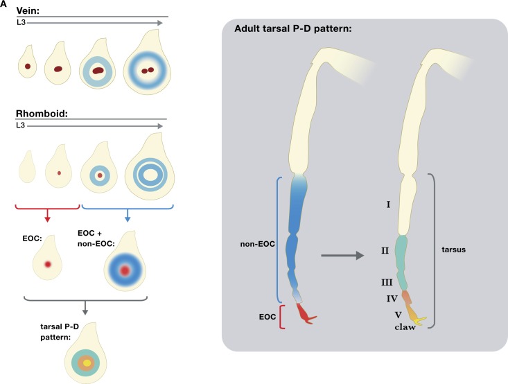
We characterized the establishment of an Epidermal Growth Factor Receptor (EGFR) organizing center (EOC) during leg development in Drosophila melanogaster. Initial EGFR activation occurs in the center of leg discs by expression of the EGFR ligand Vn and the EGFR ligand-processing protease Rho, each through single enhancers, vnE and rhoE, that integrate inputs from Wg, Dpp, Dll and Sp1. Deletion of vnE and rhoE eliminates vn and rho expression in the center of the leg imaginal discs, respectively. Animals with deletions of both vnE and rhoE (but not individually) show distal but not medial leg truncations, suggesting that the distal source of EGFR ligands acts at short-range to only specify distal-most fates, and that multiple additional 'ring' enhancers are responsible for medial fates. Further, based on the cis-regulatory logic of vnE and rhoE we identified many additional leg enhancers, suggesting that this logic is broadly used by many genes during Drosophila limb development.


Conflict of interest statement

The authors have declared that no competing interests exist.

Figures

Fig 1
Fig 1. EOC generation in third instar leg discs.
(A) Schematic representation of the establishment of an initial PD axis and EGFR signaling events in the center of leg discs (the number of rings depicted is not meant to be accurate). (B) Schematic representation of the vn genomic locus on chromosome 3L; enhancer bashing fragments for identification of vnE represented in tan did not drive expression in leg discs, dark red drove expression in leg discs and bright red designates the minimal enhancer used for further analysis; the vnvnE-Df CRISPR deletion is represented by red bracketed bar. (C) Time-course analysis of the expression pattern of vnE-lacZ reporter gene. In young discs, expression is limited to the EOC, while in late L3 additional expression appears in medial rings. (D-F) In situ analysis of vn expression in 3rd instar leg discs with genotypes: WT (D), vnvnE-Df (E; a wing disc from the same genotype serves as a positive control). vnvnE-WT, in which Recombination mediated cassette exchange (RMCE) was used to re-introduce the wild type CRM (F). Arrowheads indicate the presence (filled) or absence (open) of EOC vn expression, arrows indicate non-EOC medial expression. (G) Schematic representation of the rho genomic locus on chromosome 3L; enhancer bashing fragments for identification of rhoE represented in tan did not drive expression in leg discs, dark red drove expression in leg discs and bright red designates the minimal enhancers used for further analysis; rhoEMIN was only used for enhancer mutagenesis; rhorhoE-Df CRISPR deletion is represented by the red bracketed bar. (H) Time-course analysis of the expression pattern of rhoE-lacZ reporter gene. In young discs, expression is limited to the EOC, while in late L3 additional expression appears in medial rings. (I-K) In situ analysis of the rho expression pattern in 3rd instar leg disc with genotype: WT (I), rhorhoE-Df (J, an eye disc from the same genotype serves as a positive control), rhorhoE-WT, in which RMCE was used to re-introduce the wild type CRM (K). Arrowheads indicate presence (filled) or absence (open) of EOC rho expression, arrows indicate medial expression and “C” indicates chordotonal organ precursor expression. Scale bar = 100μm in all figures.
Fig 2
Fig 2. vnE and rhoE requirement for PD patterning of leg discs and adult legs.
(A-L) Effects on distal PD genes (C15 and Bar) in third instar leg discs in: (A) rhorhoE-Df vnvnE-Df /+; (B) rhorhoE-Df vnvnE-Df; (C) rhorhoE-Df vnvnE-D.vir; (D) grkΔFRT; Krn27-7-B; (E) grkΔFRT; rhorhoE-Df vnvnE-Df Krn27-7-B; (F,G) ruinga rhorhoE-Df vnL6 mutant clones; (H,I) ru1 rho7M43 vnvnE-Df mutant clones; (J) spi vn double RNAi; (K) Egfrtsla/+ WT leg disc at restrictive temperature; and (L) Egfrtsla mutant leg discs at restrictive temperature. (M-R) Adult leg morphology of: (M) WT; (N) rhorhoE-Df vnvnE-Df mutant; (O) spi vn double RNAi driven by Dll-Gal4 (legs with tarsal segments I-II: n = 21/102, I-III: n = 19/102, I-IV: n = 35/102, I-V: n = 27/102); (P) Egfrtsla mutant at restrictive temperature (remaining 10/42 legs less severely truncated); (Q) rhorhoE-Df vnvnE-D.vir; and (R) grkΔFRT; Krn27-7-B mutant. n refers to number of individual legs with a given number of tarsal segments present. Arrowheads indicate intact (filled) or perturbed (open) tarsal segments.
Fig 3
Fig 3. Genetic analysis of inputs into vnE and rhoE.
(A-P) lacZ reporter gene expression driven by vnE or rhoE in mutant clones as indicated. Absence of GFP marks the clone; 2X-magnified insets showcasing specific disc regions (designated with squares) are provided in each case. (A) vnE in arr2 clones generated 48h PEL; (B) vnE in arr2 clones generated 72h PEL; (C) rhoE in arr2 clones generated 72h PEL;(D) rhoE in arr2 clones generated 90h PEL; (E) vnE in Mad1-2 clones generated 48h PEL; (F) vnE in Mad1-2 clones generated 72h PEL; (G) rhoE in Mad1-2 clones generated 72h PEL; (H) rhoE in Mad1-2 clones generated 90h PEL; (I) vnE in DllSA1 clones generated 48h PEL. (J) vnE in Sp1HR clones generated 48h PEL; (K) rhoE in DllSA1 clones generated 48h PEL; (L) rhoE in Sp1HR clones generated 48h PEL; (M) vnE in pntΔ88 clones generated 48h PEL; (N) vnE in Egfrtsla Minute+ clones generated 48h PEL; (O) rhoE in pntΔ88 clones generated 48h PEL; (P) rhoE in Egfrtsla Minute+ clones generated 48h PEL. (Q, R) Schematics summarizing vnE and rhoE regulation by inputs, respectively.
Fig 4
Fig 4. Genetic interactions of inputs into vnE and rhoE.
(A-P) vnE- or rhoE-directed lacZ reporter gene expression in MARCM clones of: (A and C) Dll ectopic expression in WT background; (B and D) Dll ectopic expression in arr2 mutant background; (E and G) Dll ectopic expression in WT background; (F and H) Dll ectopic expression in Mad1-2 mutant background; (I and K) armΔN ectopic expression in WT background; (J and L) armΔN ectopic expression in DllSA1 mutant background; (M and O) tkvQD ectopic expression in WT mutant background; (N and P) tkvQD ectopic expression in DllSA1 mutant background.
Fig 5
Fig 5. Dissection of Pan, Mad, Dll and Sp1 inputs into vnE and rhoE.
(A) Schematic representation of binding sites in vnE and rhoE. Putative binding sites for each TF were mutagenized one at a time leading to progressive increase in the number of mutant binding sites until the expression driven by a mutant enhancer was lost. Sites in each TF category that were mutagenized either first or last are not sufficient for full enhancer-driven expression since fragments of vnE and rhoE (Fig 1B and 1G; S1 Table) that contain multiple such sites from each TF input cannot drive correct expression pattern. (B) Expression pattern of WT vnE-driven expression and mutant vnE enhancer-driven expression. (C) Expression pattern of WT rhoE-driven expression and mutant rhoE enhancer-driven expression. (D) Quantification of WT and mutant vnE-driven expression levels in third instar leg discs (WT vnE n = 41, 2xSp1 n = 48, 14xPan n = 24, 12xMad n = 32, 11xDll n = 49 where n indicates number of leg discs analyzed). For normalization, fluorescence was calculated as a ratio of β-gal:Dll intensity in the center of the leg disc (see Methods for details).(E) Quantification of reduction of WT and mutant rhoE-driven expression in third instar leg discs (WT rhoE n = 39, WT rhoEMIN n = 35, 4xPan n = 78, 3xMad n = 41, 1xDll n = 15 where n indicates number of leg discs analyzed). For normalization, fluorescence was calculated as a ratio of β-gal:Dll intensity in the center of the leg disc (see Methods for details).(F) EMSA analysis of selected WT vs mutant binding sites.
Fig 6
Fig 6. Genome-wide analysis of combinatorial inputs of Dll, Sp1, Wg, and Dpp in leg discs.
(A) Venn diagram representing the intersection between Dll ChIP-Seq, Sp1 ChIP-Seq and FAIRE data from third instar leg discs. (B) Schematic representation of bioinformatic intersection between Dll/Sp1 binding events and FAIRE data together with PWMs for Dll, Sp1, Pan and Mad. (C-N) Schematic representation of the binding events at selected genomic loci, the intersections and the expression pattern of tested intersection fragments for: vnE (C); zfh2_LE (D); rhoLLEMIN (E); E(spl)m3-HLH_LE (F); Dll_LE1 (G); tal_LE (H); spi_LE1 (I); ss_LE (J); noc_LE1 (K); fj_LE1 (L); elB_LE (M); fj_LE2 (N).
Fig 7
Fig 7. Summary of PD axis patterning by EGFR.
Schematic representation of EOC and non EOC sources of EGFR activation along the PD axis in leg discs (A) and adult legs (B).

References

    1. Levine M. Transcriptional enhancers in animal development and evolution. Curr Biol. 2010;20(17):R754–63. 10.1016/j.cub.2010.06.070 ; PubMed Central PMCID: PMCPMC4280268. - DOI - PMC - PubMed
    1. Estella C, Voutev R, Mann RS. A dynamic network of morphogens and transcription factors patterns the fly leg. Curr Top Dev Biol. 2012;98:173–98. 10.1016/B978-0-12-386499-4.00007-0 ; PubMed Central PMCID: PMCPMC3918458. - DOI - PMC - PubMed
    1. Cohen SM. Specification of limb development in the Drosophila embryo by positional cues from segmentation genes. Nature. 1990;343(6254):173–7. Epub 1990/01/11. 10.1038/343173a0 . - DOI - PubMed
    1. Estella C, Rieckhof G, Calleja M, Morata G. The role of buttonhead and Sp1 in the development of the ventral imaginal discs of Drosophila. Development. 2003;130(24):5929–41. Epub 2003/10/17. 10.1242/dev.00832 . - DOI - PubMed
    1. Estella C, Mann RS. Non-redundant selector and growth-promoting functions of two sister genes, buttonhead and Sp1, in Drosophila leg development. PLoS Genet. 2010;6(6):e1001001 Epub 2010/06/30. 10.1371/journal.pgen.1001001 ; PubMed Central PMCID: PMC2891808. - DOI - PMC - PubMed

Publication types

MeSH terms

LinkOut - more resources