Cortical neuronal densities and cerebral white matter demyelination in multiple sclerosis: a retrospective study
- PMID: 30143361
- PMCID: PMC6197820
- DOI: 10.1016/S1474-4422(18)30245-X
Cortical neuronal densities and cerebral white matter demyelination in multiple sclerosis: a retrospective study
Abstract
Background: Demyelination of cerebral white matter is thought to drive neuronal degeneration and permanent neurological disability in individuals with multiple sclerosis. Findings from brain MRI studies, however, support the possibility that demyelination and neuronal degeneration can occur independently. We aimed to establish whether post-mortem brains from patients with multiple sclerosis show pathological evidence of cortical neuronal loss that is independent of cerebral white-matter demyelination.
Methods: Brains and spinal cords were removed at autopsy from patients, who had died with multiple sclerosis, at the Cleveland Clinic in Cleveland, OH, USA. Visual examination of centimetre-thick slices of cerebral hemispheres was done to identify brains without areas of cerebral white-matter discoloration that were indicative of demyelinated lesions (referred to as myelocortical multiple sclerosis) and brains that had cerebral white-matter discolorations or demyelinated lesions (referred to as typical multiple sclerosis). These individuals with myelocortical multiple sclerosis were matched by age, sex, MRI protocol, multiple sclerosis disease subtype, disease duration, and Expanded Disability Status Scale, with individuals with typical multiple sclerosis. Demyelinated lesion area in tissue sections of cerebral white matter, spinal cord, and cerebral cortex from individuals classed as having myelocortical and typical multiple sclerosis were compared using myelin protein immunocytochemistry. Neuronal densities in cortical layers III, V, and VI from five cortical regions not directly connected to spinal cord (cingulate gyrus and inferior frontal cortex, superior temporal cortex, and superior insular cortex and inferior insular cortex) were also compared between the two groups and with aged-matched post-mortem brains from individuals without evidence of neurological disease.
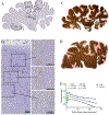
Findings: Brains and spinal cords were collected from 100 deceased patients between May, 1998, and November, 2012, and this retrospective study was done between Sept 6, 2011, and Feb 2, 2018. 12 individuals were identified as having myelocortical multiple sclerosis and were compared with 12 individuals identified as having typical multiple sclerosis. Demyelinated lesions were detected in spinal cord and cerebral cortex, but not in cerebral white matter, of people with myelocortical multiple sclerosis. Cortical demyelinated lesion area was similar between myelocortical and typical multiple sclerosis (median 4·45% [IQR 2·54-10·81] in myelocortical vs 9·74% [1·35-19·50] in typical multiple sclerosis; p=0·5512). Spinal cord demyelinated area was significantly greater in typical than in myelocortical multiple sclerosis (median 3·81% [IQR 1·72-7·42] in myelocortical vs 13·81% [6·51-29·01] in typical multiple sclerosis; p=0·0083). Despite the lack of cerebral white-matter demyelination in myelocortical multiple sclerosis, mean cortical neuronal densities were significantly decreased compared with control brains (349·8 neurons per mm2 [SD 51·9] in myelocortical multiple sclerosis vs 419·0 [43·6] in controls in layer III [p=0·0104]; 355·6 [46·5] vs 454·2 [48·3] in layer V [p=0·0006]; 366·6 [50·9] vs 458·3 [48·4] in layer VI [p=0·0049]). By contrast, mean cortical neuronal densities were decreased in typical multiple sclerosis brains compared with those from controls in layer V (392·5 [59·0] vs 454·2 [48·3]; p=0·0182) but not layers III and VI.
Interpretation: We propose that myelocortical multiple sclerosis is a subtype of multiple sclerosis that is characterised by demyelination of spinal cord and cerebral cortex but not of cerebral white matter. Cortical neuronal loss is not accompanied by cerebral white-matter demyelination and can be an independent pathological event in myelocortical multiple sclerosis. Compared with control brains, cortical neuronal loss was greater in myelocortical multiple sclerosis cortex than in typical multiple sclerosis cortex. The molecular mechanisms of primary neuronal degeneration and axonal pathology in myelocortical multiple sclerosis should be investigated in future studies.
Funding: US National Institutes of Health and National Multiple Sclerosis Society.
Copyright © 2018 Elsevier Ltd. All rights reserved.
Figures


Comment in
-
A new subtype of multiple sclerosis?Nat Rev Neurol. 2018 Oct;14(10):571. doi: 10.1038/s41582-018-0069-9. Nat Rev Neurol. 2018. PMID: 30181546 No abstract available.
-
Myelocortical multiple sclerosis: a new disease subtype?Lancet Neurol. 2018 Oct;17(10):832-834. doi: 10.1016/S1474-4422(18)30333-8. Epub 2018 Sep 18. Lancet Neurol. 2018. PMID: 30264718 No abstract available.
Similar articles
-
Axonal loss in the multiple sclerosis spinal cord revisited.Brain Pathol. 2018 May;28(3):334-348. doi: 10.1111/bpa.12516. Epub 2017 May 7. Brain Pathol. 2018. PMID: 28401686 Free PMC article.
-
High field (9.4 Tesla) magnetic resonance imaging of cortical grey matter lesions in multiple sclerosis.Brain. 2010 Mar;133(Pt 3):858-67. doi: 10.1093/brain/awp335. Epub 2010 Jan 31. Brain. 2010. PMID: 20123726
-
In vivo characterization of cortical and white matter neuroaxonal pathology in early multiple sclerosis.Brain. 2017 Nov 1;140(11):2912-2926. doi: 10.1093/brain/awx247. Brain. 2017. PMID: 29053798 Free PMC article.
-
Grey matter pathology in multiple sclerosis.Acta Neurol Scand Suppl. 2006;183:48-50. doi: 10.1111/j.1600-0404.2006.00615.x. Acta Neurol Scand Suppl. 2006. PMID: 16637929 Review.
-
The histopathology of grey matter demyelination in multiple sclerosis.Acta Neurol Scand Suppl. 2009;(189):51-7. doi: 10.1111/j.1600-0404.2009.01216.x. Acta Neurol Scand Suppl. 2009. PMID: 19566500 Review.
Cited by
-
Replication study of GWAS risk loci in Greek multiple sclerosis patients.Neurol Sci. 2019 Feb;40(2):253-260. doi: 10.1007/s10072-018-3617-6. Epub 2018 Oct 26. Neurol Sci. 2019. PMID: 30361804
-
Proceedings from the Albert Charitable Trust Inaugural Workshop on white matter and cognition in aging.Geroscience. 2020 Feb;42(1):81-96. doi: 10.1007/s11357-019-00141-8. Epub 2019 Dec 6. Geroscience. 2020. PMID: 31811528 Free PMC article. Review.
-
Cortical involvement determines impairment 30 years after a clinically isolated syndrome.Brain. 2021 Jun 22;144(5):1384-1395. doi: 10.1093/brain/awab033. Brain. 2021. PMID: 33880511 Free PMC article.
-
Nogo-A and LINGO-1: Two Important Targets for Remyelination and Regeneration.Int J Mol Sci. 2023 Feb 24;24(5):4479. doi: 10.3390/ijms24054479. Int J Mol Sci. 2023. PMID: 36901909 Free PMC article. Review.
-
Linking immune-mediated damage to neurodegeneration in multiple sclerosis: could network-based MRI help?Brain Commun. 2021 Oct 7;3(4):fcab237. doi: 10.1093/braincomms/fcab237. eCollection 2021. Brain Commun. 2021. PMID: 34729480 Free PMC article.
References
-
- Trapp BD, Nave KA. Multiple sclerosis: an immune or neurodegenerative disorder? Annu Rev Neurosci 2008; 31: 247–69. - PubMed
-
- Trapp BD, Peterson J, Ransohoff RM, et al. Axonal transection in the lesions of multiple sclerosis. N Engl J Med 1998; 338: 278–85. - PubMed
-
- Bjartmar C, Kidd G, Mörk S, et al. Neurological disability correlates with spinal cord axonal loss and reduced N-acetyl aspartate in chronic multiple sclerosis patients. Ann Neurol 2000; 48: 893–901. - PubMed
Publication types
MeSH terms
Grants and funding
LinkOut - more resources
Full Text Sources
Other Literature Sources
Medical

