. 2018 Oct 19;293(42):16142-16159.
doi: 10.1074/jbc.RA118.003525. Epub 2018 Aug 24.

The tuberous sclerosis complex subunit TBC1D7 is stabilized by Akt phosphorylation-mediated 14-3-3 binding

Affiliations

The tuberous sclerosis complex subunit TBC1D7 is stabilized by Akt phosphorylation-mediated 14-3-3 binding

James P Madigan et al. J Biol Chem. .

Abstract

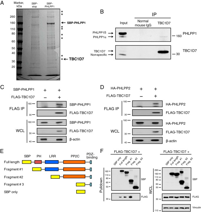
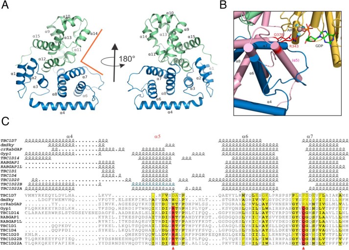
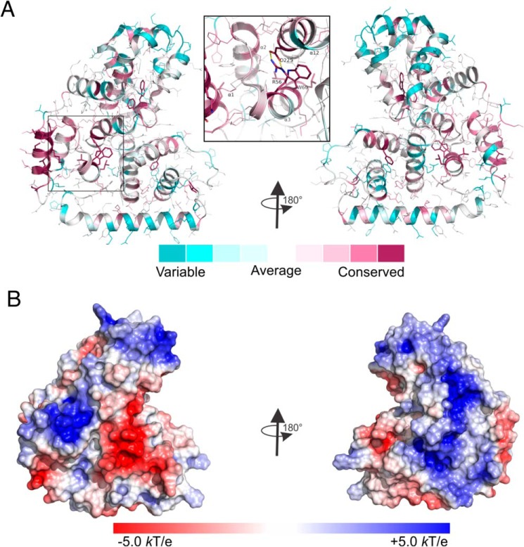
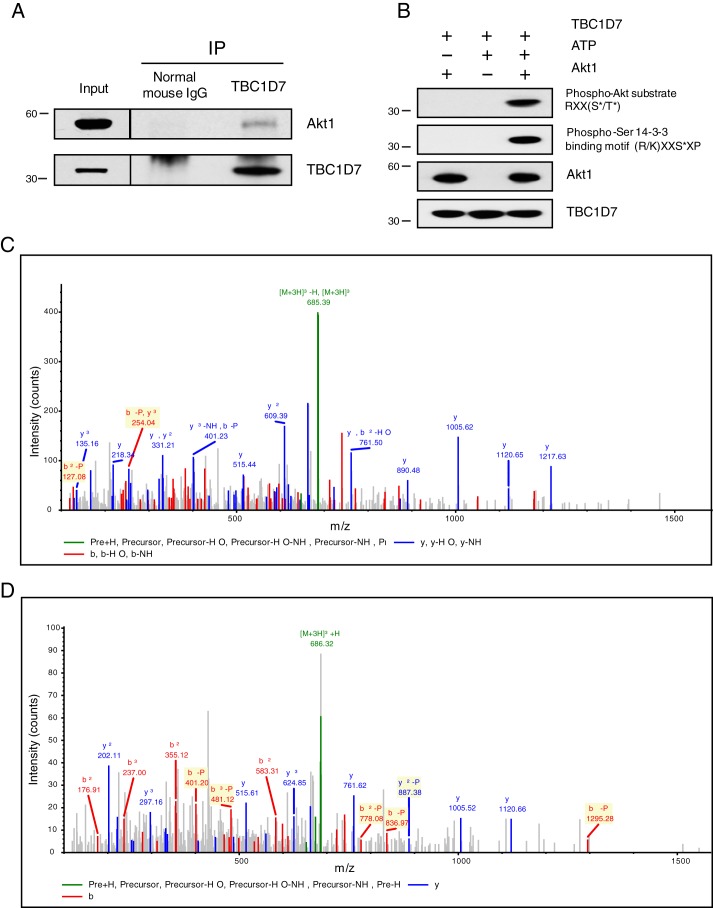
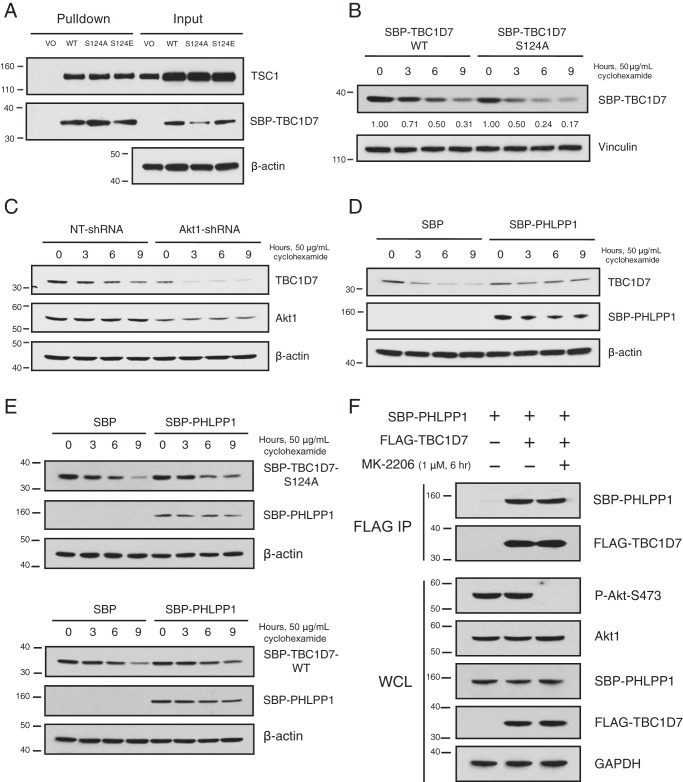
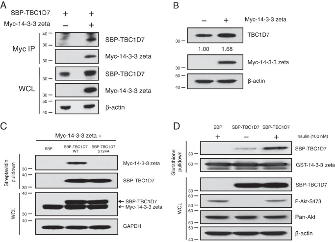
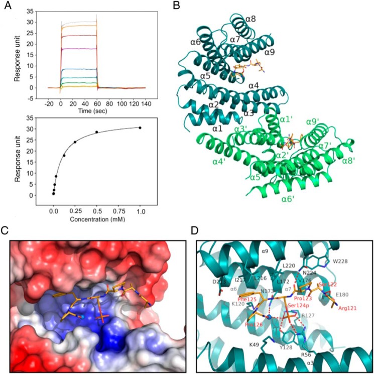
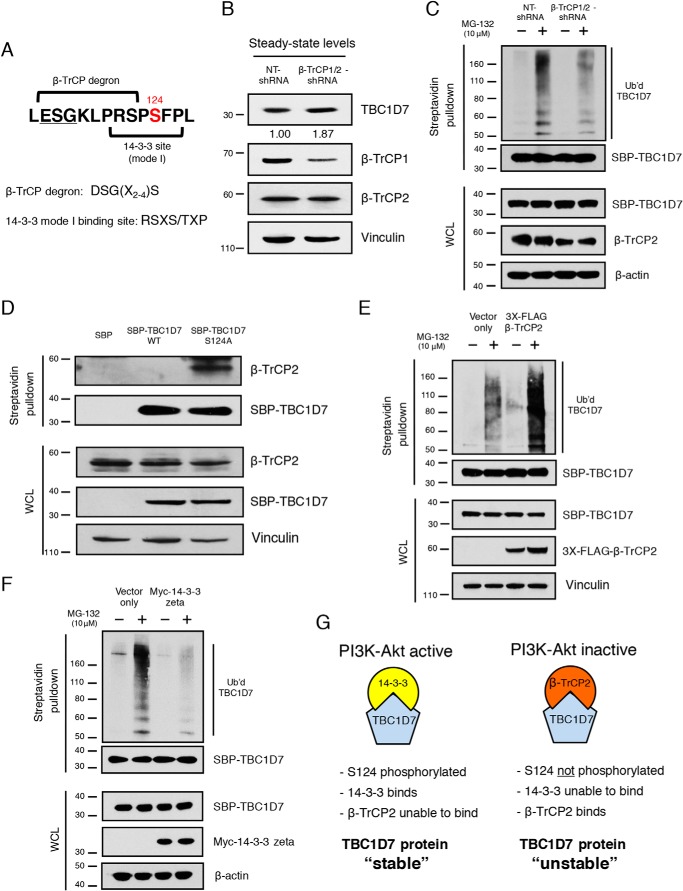
The tuberous sclerosis complex (TSC) is a negative regulator of mTOR complex 1, a signaling node promoting cellular growth in response to various nutrients and growth factors. However, several regulators in TSC signaling still await discovery and characterization. Using pulldown and MS approaches, here we identified the TSC complex member, TBC1 domain family member 7 (TBC1D7), as a binding partner for PH domain and leucine-rich repeat protein phosphatase 1 (PHLPP1), a negative regulator of Akt kinase signaling. Most TBC domain-containing proteins function as Rab GTPase-activating proteins (RabGAPs), but the crystal structure of TBC1D7 revealed that it lacks residues critical for RabGAP activity. Sequence analysis identified a putative site for both Akt-mediated phosphorylation and 14-3-3 binding at Ser-124, and we found that Akt phosphorylates TBC1D7 at Ser-124. However, this phosphorylation had no effect on the binding of TBC1D7 to TSC1, but stabilized TBC1D7. Moreover, 14-3-3 protein both bound and stabilized TBC1D7 in a growth factor-dependent manner, and a phospho-deficient substitution, S124A, prevented this interaction. The crystal structure of 14-3-3ζ in complex with a phospho-Ser-124 TBC1D7 peptide confirmed the direct interaction between 14-3-3 and TBC1D7. The sequence immediately upstream of Ser-124 aligned with a canonical β-TrCP degron, and we found that the E3 ubiquitin ligase β-TrCP2 ubiquitinates TBC1D7 and decreases its stability. Our findings reveal that Akt activity determines the phosphorylation status of TBC1D7 at the phospho-switch Ser-124, which governs binding to either 14-3-3 or β-TrCP2, resulting in increased or decreased stability of TBC1D7, respectively.

Keywords: 14-3-3 protein; Akt PKB; E3 ubiquitin ligase; mTORC1; phospho-switch; phosphorylation; protein stability; tuberous sclerosis complex (TSC); ubiquitylation (ubiquitination).


Conflict of interest statement

The authors declare that they have no conflicts of interest with the contents of this article

Figures

Figure 1.
Figure 1.
PHLPP proteins and TBC1D7 are binding partners. A, 293T cells were transfected with either SBP or SBP-PHLPP1 expression plasmids. Lysates were subject to pulldown analysis using streptavidin beads. Affinity-purified complexes were resolved on SDS-PAGE, and the gel was stained with colloidal blue. Asterisks denote bands found exclusively in the SBP-PHLPP1 lane. Bands representing SBP-PHLPP1 and TBC1D7 proteins are highlighted. B, 293T cell lysates were immunoprecipitated with either a TBC1D7 mouse mAb or a control mouse IgG. Immunoprecipitates and input whole-cell lysates were resolved on SDS-PAGE and blotted with PHLPP1 and TBC1D7 antibodies. C, 293T cells were co-transfected with either vector and SBP-PHLPP1 or FLAG-TBC1D7 and SBP-PHLPP1 expression plasmids. Lysates were immunoprecipitated with FLAG antibody–conjugated beads. Immunoprecipitates and input whole-cell lysates were resolved on SDS-PAGE and blotted with SBP, FLAG, and β-actin antibodies. D, 293T cells were co-transfected with either vector and HA-PHLPP2 or FLAG-TBC1D7 and HA-PHLPP2 expression plasmids. Lysates were immunoprecipitated with FLAG antibody–conjugated beads. Immunoprecipitates and input whole-cell lysates were resolved on SDS-PAGE and blotted with HA, FLAG, and β-actin antibodies. E, diagram representing PHLPP1 domain deletions used in Fig. 1F. F, FLAG-TBC1D7 and various SBP-PHLPP1 expression constructs were co-transfected into 293T cells. Lysates were subject to pulldown analysis using streptavidin-agarose beads. Affinity-purified complexes and input whole-cell lysates were resolved on SDS-PAGE and blotted with SBP, FLAG, and vinculin antibodies.
Figure 2.
Figure 2.
Structure of TBC1D7 and comparison with other TBC domains. A, overview of TBC1D7 structure. 15 α-helices were labeled based on the structure of yeast Gyp1 (89). The N-subdomain (α1–α8) is colored in blue, and the C-subdomain (α9–α15) is colored in light green. The rectangular groove that binds to Rab GTPase in Gyp1 is indicated using orange lines. B, an enlarged view of the region between α4 and α7 of TBC1D7 and compared with Gyp1 (salmon color) and Rab33 (yellow). Helix α5 is missing in TBC1D7. The dual-finger Arg-343/Gln-378 critical for Gyp1 RabGAP activity and the GDP molecule are shown as sticks. The AlF3 molecule is shown as a sphere. C, structure-based alignment of 12 known TBC domain structures of the region between α4 and α7. The active sites of the dual finger are indicated using red arrowheads. The structures are D. melanogaster Skywalker (dmSky, PDB code 5HJN), yeast Gyp1 (2G77), C. reinhardtii RabGAP (crRabGAP, 4P17), RABGAP1 (4NC6), RABGAP1L (3HZJ), TBC1D1 (3QYE), TBC1D4 (3QYB), TBC1D7 (3QWL), TBC1D14 (2QQ8), TBC1D20 (4HL4), TBC1D22A (2QFZ), TBC1D22B (3DZX). The α5 helix of the TBC1D22B crystal structure is presumed disordered or missing due to the use of protease and is indicated based on the high sequence identity to that of TBC1D22A.
Figure 3.
Figure 3.
Analysis of the TBC1D7 structure. A, conservation of TBC1D7 structure calculated using the ConSurf web server. Side chains of the most conserved residues are shown as sticks, whereas the rest are shown in thin lines. The enlarged insert shows the conserved salt bridge formed between residues Arg-56 and Asp-225 from helices α3 and α12 from the N- and C-subdomains, respectively. B, surface electrostatic potential of TBC1D7. Blue, positively charged residues; red, negatively charged residues.
Figure 4.
Figure 4.
Akt interacts with and phosphorylates TBC1D7 at Ser-124. A, 293T cell lysates were immunoprecipitated with either a TBC1D7 mouse mAb or a control mouse IgG. Immunoprecipitates (IP) and input whole-cell lysates were resolved on SDS-PAGE and blotted with Akt1 and TBC1D7 antibodies. B, in vitro kinase assays were performed using active Akt1 and bacterially expressed recombinant TBC1D7 protein. As negative controls, either Akt1 or ATP was absent from the reaction mixtures. Assays were resolved on SDS-PAGE and probed with either a phospho-Akt substrate, phospho-Ser 14-3-3 binding motif, or Akt1 antibodies. The phospho-Ser 14-3-3 binding motif blot was stripped and reblotted with a TBC1D7-specific antibody. C and D, tandem mass spectrum showing phosphorylation of Ser-124 of TBC1D7 (C, in vitro; D, in vivo). Shown is the MS/MS spectrum of the peptide SPpSFPLEPDDEVFLAIAK, corresponding to residues 122–139. The identified b (red) and y (blue) ions are denoted in the spectrum; fragment ions important for localization of the site of phosphorylation are highlighted in yellow.
Figure 5.
Figure 5.
Ser-124 phosphorylation stabilizes TBC1D7. A, 293T cells were transfected with either SBP vector or SBP-TBC1D7 WT, S124A, S124E expression plasmids. Lysates were subject to pulldown analysis with streptavidin beads. Affinity-purified complexes and input whole-cell lysates were resolved on SDS-PAGE and blotted with TSC1, SBP, and β-actin antibodies. B, 293T cells were transfected with either SBP-TBC1D7-WT or -S124A expression plasmids. The following day, each dish was split into four separate dishes, and the day after, they were treated with 50 μg/ml cyclohexamide for various periods of time. Whole-cell lysates were separated on SDS-PAGE and blotted with SBP and vinculin antibodies. Zero-time points for both WT and S124A SBP-TBC1D7 were set to 1; relative expression of SBP-TBC1D7 proteins is indicated, as determined by densitometric analysis. Values were normalized to vinculin loading control. C, 293T cells stably expressing either NT- or Akt1-shRNA were treated with 50 μg/ml cyclohexamide for various periods of time. Whole-cell lysates were separated on SDS-PAGE and blotted with TBC1D7, Akt1, and β-actin antibodies. D, 293T cells were transfected with either SBP vector or SBP-PHLPP1 expression plasmids. The following day, each dish was split into four separate dishes, and the day after, they were treated with 50 μg/ml cyclohexamide for various periods of time. Whole-cell lysates were separated on SDS-PAGE and blotted with TBC1D7, SBP, and β-actin antibodies. E, 293T cells were co-transfected with SBP vector or SBP-PHLPP1 and SBP-TBC1D7-S124A (top) or SBP-TBC1D7-WT (bottom). The following day, each dish was split into four separate dishes, and the day after, they were treated with 50 μg/ml cyclohexamide for various periods of time. Whole-cell lysates were separated on SDS-PAGE and blotted with SBP and β-actin antibodies. F, 293T cells were co-transfected with either vector or FLAG-TBC1D7 and SBP-PHLPP1 expression plasmids. 42 h after transfection, cells were treated with either DMSO vehicle of MK-2206 (1 μm) for 6 h. Lysates were immunoprecipitated with FLAG antibody–conjugated beads. Immunoprecipitates (IP) and input whole-cell lysates (WCL) were resolved on SDS-PAGE and blotted with SBP, FLAG, P-Akt-Ser-473, and GAPDH antibodies. The P-Akt-Ser-473 blot was stripped and reprobed with an Akt1 antibody.
Figure 6.
Figure 6.
14-3-3ζ interacts with and stabilizes TBC1D7. A, 293T cells were co-transfected with either vector and SBP-TBC1D7 or Myc-14-3-3ζ and SBP-TBC1D7 expression plasmids. Lysates were immunoprecipitated with Myc antibody. Immunoprecipitates (IP) and input lysates (WCL) were resolved on SDS-PAGE and blotted with SBP, Myc, and β-actin antibodies. B, 293T cells were transfected with either vector or Myc-14-3-3ζ. Whole-cell lysates were resolved on SDS-PAGE and blotted with TBC1D7, Myc, and β-actin antibodies. Relative expression of endogenous TBC1D7 is indicated, as determined by densitometric analysis. Values were normalized to β-actin loading control. The expression level of endogenous TBC1D7 from vector-transfected control cells was set to 1. C, 293T cells were co-transfected with Myc-14-3-3ζ and SBP vector, SBP-TBC1D7-WT, or SBP-TBC1D7-S124A. Lysates were subject to pulldown analysis with streptavidin beads. Affinity-purified complexes and input whole-cell lysates were resolved on SDS-PAGE and probed with Myc, SBP, and GAPDH antibodies. D, HeLa cells were transfected with either SBP vector or SBP-TBC1D7 expression plasmids. Cells were serum-starved (0% serum) overnight and treated for 15 min with 100 nm insulin or not. 10 μg of GSH Sepharose-conjugated recombinant GST-14-3-3ζ was incubated with cell lysates overnight. Co-purified complexes and input whole-cell lysates were resolved on SDS-PAGE and blotted with SBP, GST, P-Akt-Ser-473, and β-actin antibodies. The P-Akt-Ser-473 blot was stripped and reprobed with a pan-Akt antibody.
Figure 7.
Figure 7.
Interaction of TBC1D7 phosphopeptide with 14-3-3ζ. A, surface plasmon resonance measurement of a TBC1D7 phosphopeptide with 14-3-3. The sensorgram (top) shows the response unit when flowing peptide of increasing concentrations over a CM5 chip immobilized with 14-3-3 protein. The response was fitted to a one-site binding model (bottom) and shows a binding affinity of 102 μm. B, structure of 14-3-3 dimer with two TBC1D7 peptides. C, surface electrostatic potential map of the peptide binding groove of 14-3-3 at −5.0 kT/e to +5.0 kT/e scale. Blue, positively charged residues; red, negatively charged residues. D, stick model of the residues involved in the interaction between TBC1D7 peptide and 14-3-3 protein. The water molecule involved in binding is shown as a sphere. Salt bridges and hydrogen bonds are indicated with red dashed lines.
Figure 8.
Figure 8.
β-TrCP2 regulates TBC1D7 ubiquitination and stability. A, diagram of TBC1D7 primary sequence indicating overlapping β-TrCP degron and 14-3-3 mode I binding site. B, whole-cell lysates from 293T cells stably expressing either nontargeting (NT) or β-TrCP1/2-shRNA were resolved on SDS-PAGE and blotted with TBC1D7, β-TrCP1, β-TrCP2, and vinculin antibodies. Relative expression of endogenous TBC1D7 proteins is indicated, as determined by densitometric analysis. Values were normalized to vinculin loading control. The expression level of endogenous TBC1D7 from nontargeting shRNA-expressing control cells was set to 1. C, 293T cells stably expressing either NT- or β-TrCP1/2-shRNA were co-transfected with HA-ubiquitin and SBP-TBC1D7. Cells were treated or not with 10 μg/ml MG-132 for 4 h. Lysates were subject to pulldown analysis using streptavidin beads. Affinity-purified complexes and input whole-cell lysates (WCL) were resolved on SDS-PAGE and blotted with HA, SBP, β-TrCP2, and β-actin antibodies. D, 293T cells were transfected with either SBP, SBP-TBC1D7-WT, or SBP-TBC1D7-S124A expression plasmids. Lysates were subject to pulldown analysis using streptavidin-agarose beads. Affinity-purified complexes were resolved on SDS-PAGE and blotted with β-TrCP2, SBP, and vinculin antibodies. E, 293T cells were co-transfected with either HA-ubiquitin, SBP-TBC1D7, and empty vector or HA-ubiquitin, SBP-TBC1D7, and 3XFLAG-β-TrCP2 expression plasmids. Cells were treated, or not, with 10 μg/ml MG-132 for 4 h. Lysates were subject to pulldown analysis using streptavidin beads. Affinity-purified complexes and input whole-cell lysates were resolved on SDS-PAGE and blotted with HA, SBP, FLAG, and vinculin antibodies. F, 293T cells were co-transfected with either HA-ubiquitin, SBP-TBC1D7, and empty vector or HA-ubiquitin, SBP-TBC1D7, and Myc-14-3-3ζ expression plasmids. Cells were treated or not with 10 μg/ml MG-132 for 4 h. Lysates were subject to pulldown analysis using streptavidin beads. Affinity-purified complexes and input whole-cell lysates were resolved on SDS-PAGE and blotted with HA, SBP, Myc, and β-actin antibodies. G, diagram representing the role of TBC1D7 Ser-124 as a “phospho-switch” and controlling the stability of TBC1D7 protein. Phosphorylation of Ser-124, via PI3K-Akt signaling, determines binding of 14-3-3 and, thus, binding of β-TrCP2. Ser-124 phosphorylation-dependent binding of TBC1D7 controls the stability of TBC1D7 outside of the TSC complex.

References

    1. Hara K., Yonezawa K., Weng Q. P., Kozlowski M. T., Belham C., and Avruch J. (1998) Amino acid sufficiency and mTOR regulate p70 S6 kinase and eIF-4E BP1 through a common effector mechanism. J. Biol. Chem. 273, 14484–14494 10.1074/jbc.273.23.14484 - DOI - PubMed
    1. Manning B. D., and Cantley L. C. (2003) Rheb fills a GAP between TSC and TOR. Trends Biochem. Sci. 28, 573–576 10.1016/j.tibs.2003.09.003 - DOI - PubMed
    1. Menon S., Dibble C. C., Talbott G., Hoxhaj G., Valvezan A. J., Takahashi H., Cantley L. C., and Manning B. D. (2014) Spatial control of the TSC complex integrates insulin and nutrient regulation of mTORC1 at the lysosome. Cell 156, 771–785 10.1016/j.cell.2013.11.049 - DOI - PMC - PubMed
    1. Curatolo P., Bombardieri R., and Jozwiak S. (2008) Tuberous sclerosis. Lancet 372, 657–668 10.1016/S0140-6736(08)61279-9 - DOI - PubMed
    1. Manning B. D., and Cantley L. C. (2003) United at last: the tuberous sclerosis complex gene products connect the phosphoinositide 3-kinase/Akt pathway to mammalian target of rapamycin (mTOR) signalling. Biochem. Soc. Trans 31, 573–578 10.1042/bst0310573 - DOI - PubMed

Publication types

LinkOut - more resources