. 2018 Oct 10;38(41):8809-8821.
doi: 10.1523/JNEUROSCI.0323-18.2018. Epub 2018 Aug 24.

Presynaptic Inhibition of Primary Nociceptive Signals to Dorsal Horn Lamina I Neurons by Dopamine

Affiliations

Presynaptic Inhibition of Primary Nociceptive Signals to Dorsal Horn Lamina I Neurons by Dopamine

Yong Lu et al. J Neurosci. .

Abstract

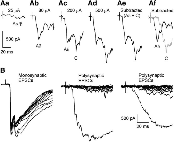
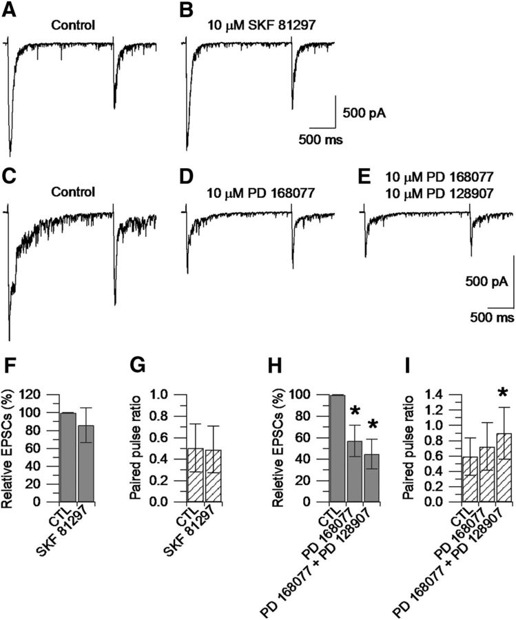
The dorsal horn of the spinal cord represents the first relay station in the pain pathway where primary nociceptive inputs are modulated by local circuits and by descending signals before being relayed to supraspinal nuclei. To determine whether dopamine can modulate primary nociceptive Aδ- and C-fiber signals, the effects of dopamine were tested on the excitatory postsynaptic currents (EPSCs) recorded from large lamina I neurons and from retrograde-labeled spinoparabrachial lamina I neurons upon stimulation of the L4/L5 dorsal root in horizontal spinal cord slices in vitro Dopamine inhibited the EPSCs in a dose-dependent manner, with substantial inhibition (33%) at 1 μm and maximum inhibition (∼70%) at 10-20 μm Dopamine reduced the frequency of miniature EPSCs recorded from large lamina I neurons, increased the paired pulse depression ratio of paired EPSCs, and induced similar inhibition of EPSCs after dialysis of large lamina I neurons with GDP-β-S, consistent with actions at presynaptic sites. Pharmacological experiments suggested that the inhibitory effects of dopamine were largely mediated by D4 receptors (53%). Similar inhibition (66%) by dopamine was observed on EPSCs recorded from ipsilateral large lamina I neurons 6 d after injection of complete Freund's adjuvant in the hindpaw, suggesting that dopamine downregulates primary nociceptive inputs to lamina I neurons during chronic inflammatory pain. We propose that presynaptic inhibition of primary nociceptive inputs to lamina I projection neurons is a mechanism whereby dopamine can inhibit incoming noxious stimuli to the dorsal horn of the spinal cord.SIGNIFICANCE STATEMENT Lamina I projection neurons represent the main output for the pain signals from the dorsal horn of the spinal cord to brainstem and thalamic nuclei. We found that dopamine inhibits the nociceptive Aδ- and C-fiber synaptic inputs to lamina I projection neurons via presynaptic actions. Similar inhibitory effects of dopamine on the EPSCs were observed in rats subjected to complete Freund's adjuvant to induce peripheral inflammation, suggesting that dopamine inhibits the synaptic inputs to lamina I neurons in the setting of injury. A better understanding of how primary nociceptive inputs to the dorsal horn of the spinal cord are modulated by descending monoaminergic signals may help in the development of new pharmacological strategies to selectively downregulate the output from lamina I projection neurons.

Keywords: D4 receptors; dopamine; dorsal horn spinal cord; lamina I neurons; nociception.


Figures

Figure 1.
Figure 1.
Horizontal spinal cord slices in vitro. A, Horizontal spinal cord slice in vitro with dorsal root (L4) attached. The dorsal root was stimulated with a suction electrode (flamed Pasteur pipette) connected to an isolated current stimulator. The dorsal root was flipped over to expose the dorsal aspect of the spinal cord to approach dorsal horn neurons with a patch pipette (recording electrode). B, Pyramidal large lamina I neuron located at the border of the white/gray matter.
Figure 2.
Figure 2.
EPSCs recorded from large lamina I neurons. Here and in the following figures, EPSCs were recorded in the presence of 10 μm bicuculline and 5 μm strychnine at 35°C unless otherwise stated. A, EPSCs were elicited by stimulation of the L4 or L5 dorsal root with increasing amount of current of 25, 80, 200, and 500 μA (0.1 ms duration). A small polysynaptic component was elicited with 25 μA of current, consistent with synaptic inputs from low-threshold Aα/β fibers (Aa). Increasing the stimulus to 80 μA elicited a component of the EPSC suggestive activation of synaptic inputs from Aδ-fibers (Ab). By increasing the stimulus to 200 μA, in addition to the Aδ component, a delayed second component of the EPSC was elicited, consistent with activation of synaptic inputs from C-fibers (Ac). Both the Aδ- and the C-fiber components were maintained when the stimulus was increased to 500 μA (Ad). The high threshold component (Aδ + C components) shown in Ae was isolated by subtracting the average of three consecutive traces elicited with 25 μA of current (Aα/β component) from the average of three consecutive traces elicited with 500 μA of current (Aα/β + Aδ + C components). The Aδ component shown in Af (black trace) was isolated by subtracting the average of three consecutive traces elicited with 25 μA of current (Aα/β component) from the average of three consecutive traces elicited with 80 μA of current (Aα/β + Aδ components); the C component shown in Af (gray trace) was isolated by subtracting the average of three consecutive traces elicited with 80 μA of current (Aα/β + Aδ components) from the average of three consecutive traces elicited with 500 μA of current (Aα/β + Aδ + C components). B, Left, Examples of monosynaptic EPSCs recorded from a large lamina I neuron. EPSCs were elicited by stimulating the dorsal root (L4) with 500 μA of current (0.1 ms). EPSCs were classified as monosynaptic based on the absence of synaptic failures and low variability (<15%) in synaptic delay during 15 consecutive stimuli at 2 Hz. Middle and right, Examples of polysynaptic EPSCs recorded from two different large lamina I neurons. EPSCs were elicited by stimulating the dorsal root (L4) with 500 μA of current (0.1 ms). In both cells, there were synaptic failures and high variability in synaptic delay during 15 consecutive stimuli at 2 Hz.
Figure 3.
Figure 3.
Dopamine inhibition of synaptic inputs to large lamina I neurons. Here and in the following figures, EPSCs were elicited by stimulating the L4 or L5 dorsal root with 400 or 500 μA of current, 0.1 ms duration, at 0.016 Hz. A, EPSC recorded in control. B, A 20 μm concentration of dopamine inhibited the EPSC by 92%. C, EPSC upon washing out dopamine. D, EPSC was completely blocked by 20 μm D-AP5 + 10 μm CNQX. E, Summary data: 20 μm dopamine inhibited the EPSCs by 71 ± 17% (n = 31), repeated-measures one-way ANOVA followed by Dunnett's post hoc test (*p < 0.05). F, Dose-dependent inhibition of EPSCs by dopamine. Each bar represents an independent experiment. For each concentration, statistical significance was assessed with a paired t test by comparing the effect of dopamine to its own control. The EPSC was reduced by 33 ± 10% (n = 10) by 1 μm dopamine (*p < 0.05); 57 ± 9% (n = 12) by 3 μm dopamine (*p < 0.05); 74 ± 16% (n = 8) by 10 μm dopamine (*p < 0.01), and 71 ± 17% (n = 31) by 20 μm dopamine (*p < 0.05). G, Effects of dopamine in males versus females. Summary data: 20 μm dopamine inhibited the EPSCs by 61 ± 15% (n = 14, from 7 rats) in males and by 63 ± 18% (n = 12, from 7 rats) in females. H, Inhibition of Aδ- and C-fiber components by dopamine. The Aδ- and the C-fiber components were isolated as described in Figure 2Af. The effect of dopamine on the Aδ- and C-fiber components was determined by comparing the EPSCs at the corresponding stimulating current before and after addition of dopamine. Summary data: 20 μm dopamine inhibited the Aδ component by 65 ± 20% and the C component by 56 ± 19% (n = 22). I, Summary data: 20 μm dopamine applied in the presence of 10 μm naloxone reduced the EPSC by 66 ± 21% (n = 7), repeated-measures one-way ANOVA followed by Dunnett's post hoc test (*p < 0.05). J, Summary data: 20 μm dopamine applied in the presence of 10 μm phentolamine reduced the EPSC by 64 ± 15% (n = 7), repeated-measures one-way ANOVA followed by Dunnett's post hoc test (*p < 0.05). K, Summary data: 20 μm dopamine applied in the presence of 10 μm methysergide reduced the EPSC by 56 ± 14% (n = 8), repeated-measures one-way ANOVA followed by Dunnett's post hoc test (*p < 0.05).
Figure 4.
Figure 4.
Dopamine inhibition of synaptic inputs to retrograde-labeled large lamina I projection neurons. A, Coronal section of a P26 rat brain illustrating the unilateral DiI injection (100 nl, 2.5 mg/ml) in the lateral PB nucleus using a Hamilton microsyringe equipped with a 32 Ga needle. The following stereotaxic coordinates were used (in millimeters relative to lambda): 0.3–0.4 posterior, 1.4–1.5 lateral, and 6.4–6.5 ventral. B, Fluorescent image showing the localization of FAST DiI in the dorsal horn of the spinal cord 4 d after injection. C, Large lamina I neuron visualized using IR-LED illumination. D, Merge. E, EPSC recorded from the retrograde-labeled lamina I neuron shown in D. F, A 20 μm concentration of dopamine inhibited the EPSC by 67%. G, EPSC upon washing out dopamine. H, Summary data: 20 μm dopamine inhibited the EPSCs by 65 ± 10% (n = 10), repeated-measures one-way ANOVA followed by Dunnett's post hoc test (*p < 0.05).
Figure 5.
Figure 5.
Dopamine inhibition of synaptic inputs to lamina I neurons in 8- to 9-week-old rats. A, EPSC recorded from a lamina I neuron in a coronal slice of the spinal cord in vitro from a 58-d-old rat. B, 20 μm dopamine inhibited the EPSC by 85%. C, EPSC upon washing out dopamine. D, Summary data from application of dopamine to coronal slices from adult (P56–P63) rats: 20 μm dopamine inhibited the EPSCs by 63 ± 11% (n = 8), repeated-measures one-way ANOVA followed by Dunnett's post hoc test (*p < 0.05).
Figure 6.
Figure 6.
Effects of dopamine on spontaneous mEPSCs recorded from large lamina I neurons. A, Spontaneous mEPSCS recorded from a large lamina I neuron in the presence of 1 μm TTX at Vh = −70 mV. B, A 20 μm concentration of dopamine reduced the median frequency of mEPSCs from 15 to 7 Hz. C, Summary data showing the effects of dopamine on the peak of mEPSCs. Values are reported as median, first quartile (25th percentile), and third quartile (75th percentile). Control (TTX): median = 25.8 pA; 25th percentile = 16.8 pA; 75th percentile = 39.9 pA. 20 μm dopamine (TTX + DA): median = 23.7 pA; 25th percentile = 19.9 pA; 75th percentile = 37.3 pA (n = 7), Wilcoxon matched-pairs test (p = 0.0938). D, Distribution of mEPSC recorded in control (TTX, shaded bars) and after application of 20 μm dopamine (TTX + DA, open bars) (n = 7). E, Cumulative probabilities of mEPSC recorded in control (TTX, solid line) and after application of 20 μm dopamine (TTX + DA, dashed line) (n = 7), Kolmogorov–Smirnov test (p < 0.0001). F, Summary data showing the effects of dopamine on the frequency of mEPSCs. Values are reported as median, first quartile (25th percentile) and third quartile (75th percentile). Control (TTX): median = 30.8 Hz; 25th percentile = 12.6 Hz; 75th percentile = 45.1 Hz. 20 μm dopamine (TTX + DA): median = 9.3 Hz; 25th percentile = 4.7 Hz; 75th percentile = 16.2 Hz (n = 7), Wilcoxon matched-pairs test *p < 0.05. G, Distribution of inter-event intervals of mEPSCs recorded in control (TTX, shaded bars) and after application of 20 μm dopamine (TTX + DA, open bars) (n = 7). H, Cumulative probabilities of interevent intervals of mEPSCs recorded in control (TTX, solid line) and after application of 20 μm dopamine (TTX + DA, dashed line) (n = 7), Kolmogorov–Smirnov test (p < 0.0001).
Figure 7.
Figure 7.
Effects of dopamine on paired EPSCs and paired pulse depression (PPD) ratio. A, Paired EPSCs recorded from a large lamina I neuron upon stimulation of the L4 dorsal root with paired stimuli (2 s apart, 500 μA, 0.1 ms duration, at 0.016 Hz). The second EPSC was smaller than the first EPSC, consistent with PPD. B, A 20 μm concentration of dopamine inhibited the first EPSC by 91% and the second EPSC by 74%. C, EPSCs upon washing out dopamine. D, In the same cell, 20 μm dopamine increased the PPR (second/first EPSC) from 0.33 to 0.85. E, Summary data: 20 μm dopamine increased the PPR from 0.55 ± 0.12 to 0.83 ± 0.22 (n = 18), repeated-measures one-way ANOVA followed by Dunnett's post hoc test (*p < 0.05).
Figure 8.
Figure 8.
Inhibitory effects of dopamine on synaptic inputs to large lamina I neurons during dialysis of postsynaptic lamina I neurons with GDP-β-S. A, Multipolar large lamina I neuron visualized with the IR-LED illumination. B, Same cell dialyzed with 1 μm Alexa Fluor-488 and GDP-β-S (0.6 mm) identified with epifluorescence after 10 min in whole-cell configuration. The cell body is completely dialyzed with the intracellular solution containing Alexa Fluor-488 and GDP-β-S, whereas the dendrites are only partially dialyzed. C, Same cell after 30 min in whole-cell configuration. Both the cell body and the dendrites are completely dialyzed with Alexa Fluor-488- and GDP-β-S-containing intracellular solution. D, Substance P-induced current recorded at Vh = − 70 mV in the presence of 1 μm TTX. A 2 μm concentration of substance P induced a transient inward current (29 ± 16 pA) in 82% (14/17) of large lamina I neurons dialyzed for 30 min with a control intracellular solution containing 0.3 mm Na-GTP. E, A 2 μm concentration of substance P failed to induce an inward current in large lamina I neurons dialyzed for 30 min with 0.6 mm GDP-β-S (n = 9). F, Paired EPSCs recorded from the same cell as in A after 30 min in whole-cell configuration. G, A 20 μm concentration of dopamine inhibited the first EPSC by 85%, the second EPSC by 67%, and increased the PPR from 0.42 to 0.93. H, Paired EPSCs upon washing out dopamine. I, Summary data using an intracellular solution containing 0.6 mm GDP-β-S: 20 μm dopamine inhibited the first EPSC by 78 ± 13% (I) and increased the PPR from 0.68 ± 0.12 to 1.04 ± 0.21 (J) (n = 16), repeated-measures one-way ANOVA followed by Dunnett's post hoc test (*p < 0.05).
Figure 9.
Figure 9.
D4 and D3 dopamine receptors mediate the inhibitory effects of dopamine on the EPSCs. A, Paired EPSCs recorded from a large lamina I neuron upon stimulation of the L4 dorsal root with paired stimuli (2 s apart, 500 μA, 0.1 ms duration, at 0.016 Hz). B, A 10 μm concentration of SKF 81297 (D1/D5 agonist) inhibited the first EPSC by 6%, the second EPSC by 1%, and increased the PPR from 0.57 to 0.60. C, Paired EPSCs recorded from a different large lamina I neuron upon stimulation of the L4 dorsal root with paired stimuli (2 s apart, 500 μA, 0.1 ms duration, at 0.016 Hz). D, A 10 μm concentration of PD 168077 (D4 agonist) inhibited the first EPSC by 53%, the second EPSC by 38%, and increased the PPR from 0.72 to 0.91. E, A 10 μm concentration of PD 128907 (D3 agonist) applied on top of PD 168077 inhibited the first EPSC by an additional 26%, the second EPSC by an additional 22%, and increased the PPR to 0.97. F, Summary data: 10 μm SKF 81297 inhibited the first EPSC by 14 ± 19% and changed the PPR (G) from 0.50 ± 0.22 to 0.49 ± 0.21 (n = 18). H, Summary data: 10 μm PD 168077 inhibited the first EPSC by 57 ± 14%. 10 μm PD 128907 applied on top of PD 168077 inhibited the first EPSC by an additional 12 ± 13% (n = 11), repeated-measures one-way ANOVA followed by Dunnett's post hoc test (*p < 0.05). I, Summary data: 10 μm PD 168077 increased the PPR from 0.59 ± 0.24 to 0.72 ± 0.30. 10 μm PD 128907 applied on top of PD 168077 increased the PPR to 0.90 ± 0.33 (n = 11), repeated-measures one-way ANOVA followed by Dunnett's post hoc test (*p < 0.05).
Figure 10.
Figure 10.
Inhibitory effects of dopamine on synaptic inputs to large lamina I neurons during chronic inflammatory pain. A, Paired EPSCs recorded from a large lamina I neuron located ipsilateral to the site of CFA injection (right hindpaw) upon stimulation of the L4 dorsal root with paired stimuli (2 s apart, 500 μA, 0.1 ms duration, at 0.016 Hz). B, A 20 μm concentration of dopamine inhibited the first EPSC by 71%, the second EPSC by 39%, and increased the PPR from 0.51 to 1.02. C, EPSCs upon washing out dopamine. D, Summary data: 20 μm dopamine inhibited the first EPSC by 66 ± 10% (n = 6), repeated-measures one-way ANOVA followed by Dunnett's post hoc test (*p < 0.05). E, Summary data: 20 μm dopamine increased the PPR from 0.50 ± 0.11 to 0.83 ± 0.10 (n = 6), repeated-measures one-way ANOVA followed by Dunnett's post hoc test (*p < 0.05).

Similar articles

Cited by

References

    1. Al Ghamdi KS, Polgár E, Todd AJ (2009) Soma size distinguishes projection neurons from neurokinin 1 receptor-expressing interneurons in lamina I of the rat lumbar spinal dorsal horn. Neuroscience 164:1794–1804. 10.1016/j.neuroscience.2009.09.071 - DOI - PMC - PubMed
    1. Al-Khater KM, Kerr R, Todd AJ (2008) A quantitative study of spinothalamic neurons in laminae I, III, and IV in lumbar and cervical segments of the rat spinal cord. J Comp Neurol 511:1–18. 10.1002/cne.21811 - DOI - PMC - PubMed
    1. Almanza A, Simón-Arceo K, Coffeen U, Fuentes-García R, Contreras B, Pellicer F, Mercado F (2015) A D2-like receptor family agonist produces analgesia in mechanonociception but not in thermonociception at the spinal cord level in rats. Pharmacol Biochem Behav 137:119–125. 10.1016/j.pbb.2015.08.013 - DOI - PubMed
    1. Bannister K, Dickenson AH (2016) What do monoamines do in pain modulation? Curr Opin Support Palliat Care 10:143–148. 10.1097/SPC.0000000000000207 - DOI - PMC - PubMed
    1. Bao J, Li JJ, Perl ER (1998) Differences in Ca2+ channels governing generation of miniature and evoked excitatory synaptic currents in spinal laminae I and II. J Neurosci 18:8740–8750. 10.1523/JNEUROSCI.18-21-08740.1998 - DOI - PMC - PubMed

Publication types

MeSH terms

LinkOut - more resources