. 2018 Oct 2;38(5):BSR20180157.
doi: 10.1042/BSR20180157. Print 2018 Oct 31.

Lactate transport facilitates neurite outgrowth

Affiliations

Lactate transport facilitates neurite outgrowth

Kun Chen et al. Biosci Rep. .

Abstract

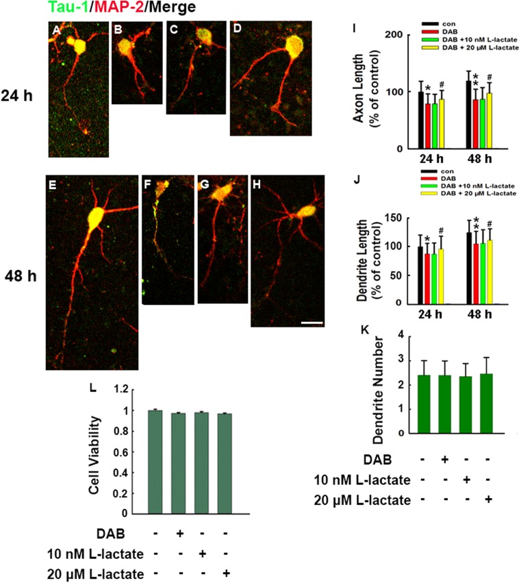
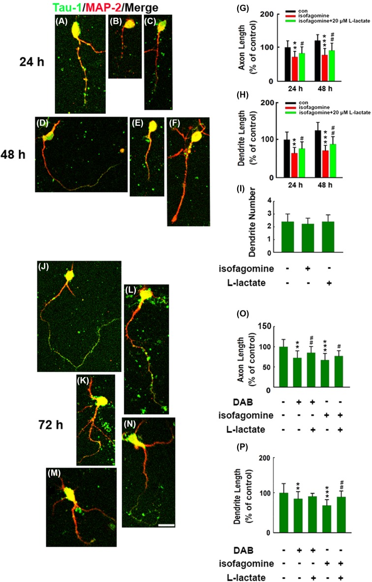
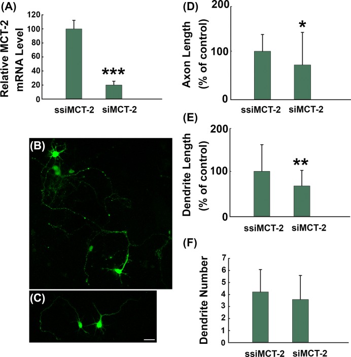
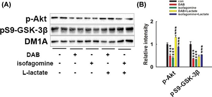
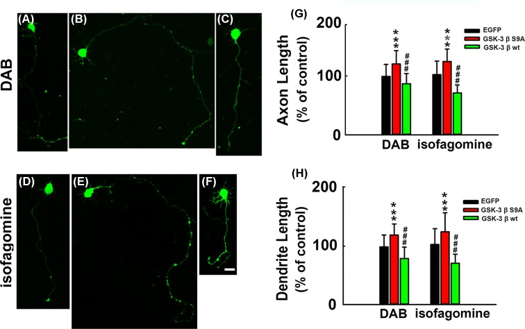
How glia affect neurite outgrowth during neural development has not been well elucidated. In the present study, we found that disruption of lactate production using 1,4-dideoxy-1,4-imino-D-arabinitol (DAB) and isofagomine significantly interfered with neurite outgrowth and that exogenous application of L-lactate rescued neurite growth failure. Monocarboxylate transporter-2-knockout, which blocked the lactate shuttle in neurons, showed a remarkable decrease in the length of axons and dendrites. We further demonstrated that Akt activity was decreased while glycogen synthase kinase 3β (GSK3β) activity was increased after astrocytic glycogen phosphorylase blockade. Additionally, GSK3βSer9 mutation reversed neurite growth failure caused by DAB and isofagomine. Our results suggested that lactate transportation played a critical role in neural development and disruption of the lactate shuttle in quiescent condition also affected neurite outgrowth in the central nervous system.

Keywords: Akt; Glycogen synthase kinase 3β (GSK-3β); L-lactate; Monocarboxylate transporters-2 (MCT-2); Neurite outgrowth.


Conflict of interest statement

The authors declare that there are no competing interests associated with the manuscript.

Figures

Figure 1
Figure 1. Disruption of astrocyte–neuron lactate transport caused by DAB-reduced neurite outgrowth
Rat primary cortical neurons were dissected from E16–18 embryonic pups. Immediately after plating, neurons were pre-treated for different concentrations of L-lactate for 30 min and were exposed with or without an inhibitor of glycogen phosphorylation DAB (100 μM) for 24 or 48 h. After treatment, neurons were fixed and double stained with MAP-2 (red) and tau-1 (green) and merged images were presented. Representative images of (A) Control neuron (con 24 h), (B) DAB-treated neuron (24 h), (C) DAB-exposed and 10 nM L-lactate pre-treated neuron (24 h), (D) DAB-exposed and 20 μM L-lactate pre-treated neuron (24 h) and (E) Control neuron (con 48 h), (F) DAB-treated neuron (48 h), (G) DAB-exposed and 10 nM L-lactate pre-treated neuron (48 h), (H) DAB-exposed and 20 μM L-lactate pre-treated neuron (48 h), and (I) quantitative analysis of the axon length for 24 and 48 h exposure in (A–H) (df = 244, F = 50.144). (J) Quantitative analysis of the dendrite length for 24 and 48 h in (A–H) (df = 244, F = 60.12). (K) Quantification of the neurite number in neurons after 48 h exposure in each treatment. (L) Cell viability was measured using a CCK-8 assay. *P<0.05, **P<0.01 versus control neurons, #P<0.05 versus DAB-treated neurons. Approximately 60–90 neurons were calculated in each group. Scale bar: 50 μm.
Figure 2
Figure 2. Disruption of astrocyte–neuron lactate transport caused by isofagomine-reduced neurite outgrowth
Rat primary cortical neurons were dissected from E16–18 embryonic pups. Immediately after plating, neurons were pre-treated with 20 μM of L-lactate for 30 min and were exposed with or without a glycogen phosphorylase inhibitor, isofagomine (8 μM), for 24 or 48 h. After treatment, neurons were fixed and double stained with MAP-2 (red) and tau-1 (green) and merged images were presented. Representative images of (A) control neuron (con 24 h), (B) isofagomine-treated neuron, (C) isofagomine-exposed and 20 µM L-lactate pre-treated neuron and for 48 h (D) control neuron (con 48 h), (E) isofagomine-treated neuron (48 h), (F) isofagomine-exposed and 20 μM L-lactate pre-treated neuron (48 h). (G) Quantitative analysis of the axon length in (A–F) (df = 281, F = 70.18). (H) Quantitative analysis of the dendrite length in (A–F) (df = 281, F = 57.36). (I) Quantification of the neurite number in neurons after 48 h exposure in each treatment. **P<0.01, ***P<0.001 versus control neurons, #P<0.05, ##P<0.01 versus isofagomine-treated neurons. Neurons were exposed with or without a glycogen phosphorylase inhibitor, DAB (100 μM) or isofagomine (8 μM), and were pre-treated with 20 μM of L-lactate for 72 h. (J) Control neuron (con, 72 h). (K) DAB-treated neurons (72 h). (L) Twenty micromolar L-lactate pre-treated before DAB-exposed neuron (72 h). (M) Isofagomine-treated neuron (72 h). (N) Twenty micromolar L-lactate pre-treated before isofagomine-exposed neuron (72 h). (O) Quantitative analysis of the axon length in (J–N) (df = 292, F = 59.16). (P) Quantitative analysis of the dendrite length in (J–N) (df = 292, F = 75.33). **P<0.01, ***P<0.001 versus con neurons, #P<0.05, ##P<0.01 versus DAB- or isofagomine-treated cells. Approximately 60–90 neurons were calculated in each group. Scale bar: 50 μm.
Figure 3
Figure 3. MCT-2 knockout induced neurite outgrowth failure
Rat cortical neurons were transfected with scrambled control LV-ssiMCT-2 or MCT-2 knockout virus LV-siMCT-2 12 h after seeding. (A) After 72 h of transfection, the total RNA was extracted from the neurons, and the MCT-2 mRNA level was detected using RT-PCR. Or the neurons were fixed and the representative micrographs of neurons were showed in (B) LV-ssiMCT-2 transfected neuron and (C) LV-siMCT-2 transfected neuron. (D) Quantitative analysis of the axon length in (B) and (C) (t = −2.5666, df = 70.367). (E) Quantitative analysis of the dendrite length in (B) and (C) (t = −2.695, df = 74.964). (F) Quantification of neurite number in neurons after transfection. *P<0.05, **P<0.01, ***P<0.001 versus LV-ssiMCT-2 transfected neurons. Approximately 60 neurons were quantified in each group. Scale bar: 50 μm.
Figure 4
Figure 4. Western blot analysis of the phosphorylated levels of Akt and Ser9 (S9) of GSK3β in DAB- or isofagomine-treated neurons
Rat primary cortical neurons were treated with DAB (100 μM) or isofagomine (8 μM) for 30 min, with or without L-lactate (20 μM) pre-treatment. After treatment, cell lysates were collected and subjected to Western blots. (A) Phosphorylated levels of Akt and Ser9 (S9) of GSK3β were detected. (B) Quantitative analysis of the blots in (A). Phosphorylated Akt and GSK3β levels were normalized with DM1A. ***P<0.001 versus con neurons, ###P<0.001 versus DAB- or isofagomine-treated neurons, df = 8, F = 95.301 in the DAB-treated group; df = 8, F = 121.839 in isofagomine-treated neurons.
Figure 5
Figure 5. GSK3β plays a key role in neurite growth failure induced by lactate shuttle disruption
Rat cortical neurons were transfected with lentivirus suspensions of empty vector, LV-GSK3βS9A or LV-GSK3βwt, 12 h after seeding; DAB (100 μM) and isofagomine (8 μM) were added to the medium, respectively. After 72 h, cells were fixed and representative images of neurons were showed in DAB-incubated neurons, (A) empty vector transfected neuron. (B) GSK3βS9A transfected neuron. (C) GSK3βwt transfected neurons. And in isofagomine-incubated neurons, (D) empty vector transfected neuron. (E) GSK3βS9A transfected neuron. (F) GSK3βwt transfected neurons. (G) Quantitative analysis of the axon length in (A–F) (df = 227, F = 48.697 in the DAB-treated group; df = 221, F = 105.641 in the isofagomine-treated group). (H) Quantitative analysis of the dendrite length in (A–F) (df = 227, F = 73.202 in the DAB-treated group; df = 221, F = 61.492 in the isofagomine-treated group). ***P<0.001 versus empty vector transfected neurons, ###P<0.01 versus GSK3βS9A transfected neurons. In each group, ~50–90 neurons were calculated. Scale bar: 50 μm.

References

    1. Brown A.M., Baltan Tekkok S. and Ransom B.R. (2004) Energy transfer from astrocytes to axons: the role of CNS glycogen. Neurochem. Inter. 45, 529–536 10.1016/j.neuint.2003.11.005 - DOI - PubMed
    1. Dringen R., Gebhardt R. and Hamprecht B. (1993) Glycogen in astrocytes: possible function as lactate supply for neighboring cells. Brain Res. 623, 208–214 10.1016/0006-8993(93)91429-V - DOI - PubMed
    1. Schipke C.G. and Kettenmann H. (2004) Astrocyte responses to neuronal activity. Glia 47, 226–232 10.1002/glia.20029 - DOI - PubMed
    1. Murphy T.H., Blatter L.A., Wier W.G. and Baraban J.M. (1993) Rapid communication between neurons and astrocytes in primary cortical cultures. J. Neurosci. 13, 2672–2679 10.1523/JNEUROSCI.13-06-02672.1993 - DOI - PMC - PubMed
    1. Henneberger C., Papouin T., Oliet S.H. and Rusakov D.A. (2010) Long-term potentiation depends on release of D-serine from astrocytes. Nature 463, 232–236 10.1038/nature08673 - DOI - PMC - PubMed

Publication types

MeSH terms

LinkOut - more resources