Review
. 2018 Nov;16(11):684-698.
doi: 10.1038/s41579-018-0064-6.

Molecular mechanisms of viral oncogenesis in humans

Affiliations
Review

Molecular mechanisms of viral oncogenesis in humans

Nathan A Krump et al. Nat Rev Microbiol. 2018 Nov.

Abstract

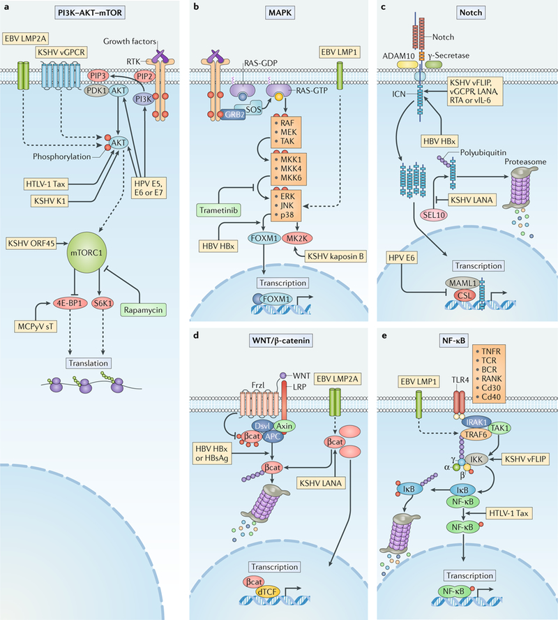
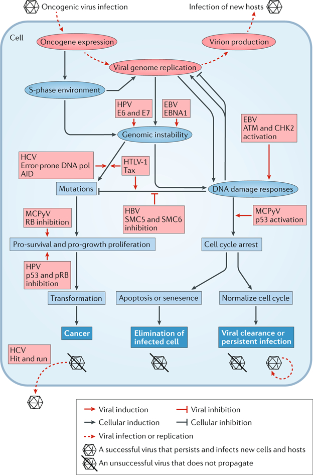
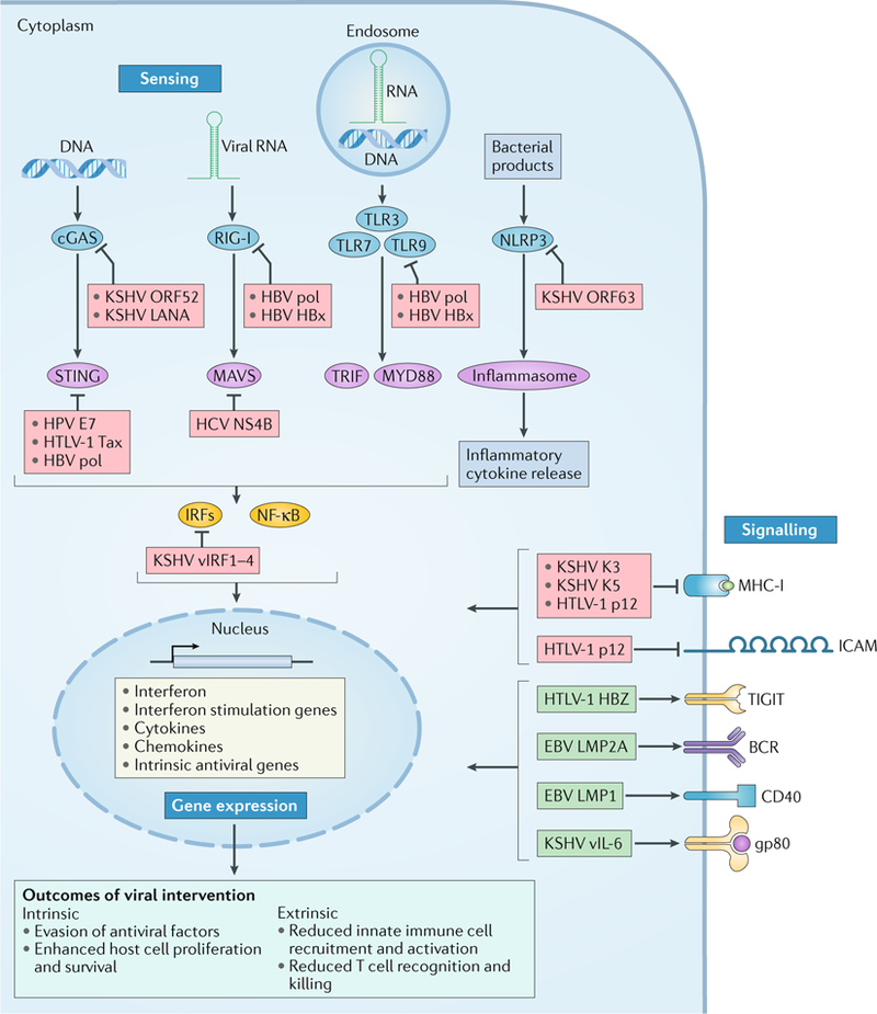
Viral infection is a major contributor to the global cancer burden. Recent advances have revealed that seven known oncogenic viruses promote tumorigenesis through shared host cell targets and pathways. A comprehensive understanding of the principles of viral oncogenesis may enable the identification of unknown infectious aetiologies of cancer and the development of therapeutic or preventive strategies for virus-associated cancers. In this Review, we discuss the molecular mechanisms of viral oncogenesis in humans. We highlight recent advances in understanding how viral manipulation of host cellular signalling, DNA damage responses, immunity and microRNA targets promotes the initiation and development of cancer.


Conflict of interest statement

Competing interests

The authors declare no competing interests.

Figures

Fig. 1 |
Fig. 1 |. Signalling pathways targeted by oncogenic viruses.
Human oncogenic viruses modulate signal transduction pathways that control cell growth, proliferation and survival to optimize cellular conditions for viral replication, virion assembly and autophagic evasion in the absence of growth or survival signals. Dysregulation of these pathways through mutation or viral factors has been implicated in many cancers. Targeting of critical axes in these pathways by human oncogenic viral factors is indicated by yellow boxes. Arrows represent activation, whereas blocking arrows represent inhibition. Dashed arrows indicate activation or promotion with multiple steps not shown. a | Mechanistic target of rapamycin (mTOR) complex 1 (mTORC1) is a master regulator that coordinates biomolecule availability and stress stimuli to yield tuned responses that promote cell growth and inhibit autophagy. Growth factor binding to receptor tyrosine kinases (RTKs) regulates mTORC1 activity through phosphatidylinositol 3-kinase (PI3K) and the serine/threonine kinase AKT. Ligand-bound RTKs autophosphorylate and recruit PI3K to the plasma membrane, where it converts phosphatidylinositol 4,5-bisphosphate (PIP2) to phosphatidylinositol 3,4,5-trisphosphate (PIP3). PIP3 recruits 3-phosphoinositide-dependent protein kinase 1 (PDK1) and AKT. Multiple viruses modulate the activity of the AKT pathway and downstream components, such as eukaryotic translation initiation factor 4E binding protein 1 (4E-BP1) and ribosomal protein S6 kinase β1 (S6K1). b | The mitogen-activated-protein kinase (MAPK) pathway is also activated by ligand-bound RTKs. Autophosphorylated tyrosine residues bind SH2 domains of growth factor receptor-bound protein 2 (GRB2), which localizes the guanine-exchange factor son-of-sevenless (SOS) to the inner membrane. SOS allows for the exchange of GDP for GTP on RAS. Activated GTP-bound RAS initiates a MAPK cascade, which activates transcription factors such as forkhead box protein M1 (FOXM1) and additional effectors such as MK2 kinase (MK2K). Together, they enhance the expression of pro-survival and pro-inflammatory genes through increased transcription and stabilization of mRNAs, respectively. c | A conformational change in Notch when bound to ligands on neighbouring cells enables sequential cleavages by a disintegrin and metalloproteinase domain-containing protein 10 (ADAM10) and γ-secretase. Cleavage releases intracellular domain of Notch (ICN) into the cytoplasm, where it can translocate to the nucleus and coordinate the transcription of proliferation and differentiation-related genes with DNA-bound CSL protein and the co-activator mastermind-like 1 (MAML1). ICN is downregulated by SEL10 polyubiquitylation-mediated proteasomal degradation. d | β-Catenin (βcat) is inactivated in a complex with adenomatous polyposis coli gene product (APC) and axin, which phosphorylates βcat and targets it for proteasomal degradation. Upon WNT glycolipoprotein binding to extracellular domains of prolow-density lipoprotein receptor related protein 1 (LRP1) and frizzled (Frzl), dishevelled (Dsvl) is recruited to the cytoplasmic domain of Frzl. Subsequent phosphorylation of LRP sequesters axin and prevents degradation of βcat. Accumulating βcat translocates to the nucleus, where it co-activates Drosophila T cell factor (dTCF)-mediated transcription of cell growth genes. e | Several immunity-related cell surface receptors, including Toll-like receptor 4 (TLR4) and tumour necrosis factor receptor (TNFR), activate the canonical nuclear factor-κB (NF-κB) pathway when bound to their respective ligands. TLR4 activation leads to phosphorylation and recruitment of interleukin-1 receptor-associated kinase 1 (IRAK1) to the adaptor protein myeloid differentiation primary response protein MYD88. A complex containing the E3-ubiquitin kinase TNF receptor-associated factor 6 (TRAF6) forms, which generates a scaffold for the polyubiquitin-binding NF-κB essential modulator (NEMO) of inhibitors of NF-κB (IκB) kinase (IKK). Orphan nuclear receptor TAK1 (also known as NR2C2) activates IKK, which then phosphorylates the inhibitory subunit (IκB) and targets it for polyubiquitylation and proteasomal degradation. A conformational change between the NF-κB subunits p50 and p65 allows activating phosphorylation and translocation to the nucleus, where it induces expression of inflammatory and pro-survival genes. BCR, B cell receptor; E5, E6, E7, early proteins 5, 6 and 7; EBV, Epstein–Barr virus; ERK1, extracellular-signal-regulated kinase 1; HBsAg, hepatitis B surface antigen; HBV, hepatitis B virus; HBx, HBVX protein; HPV, human papilloma virus; HTLV-1, human T-lymphotropic virus 1; JNK, JUN N-terminal kinase; KSHV, Kaposi sarcoma-associated virus; LANA, latency-associated nuclear antigen; LMP, latent membrane protein; MCPyV, Merkel cell polyomavirus; MEK, MAPK/ERK kinase; MKK, mitogen-activated protein kinase kinase; RAF, RAF proto-oncogene serine/threonine-protein kinase; RANK, receptor activator of NF-κB (also known as TNFRSF11A); RTA, replication and transcription activator; sT, small tumour antigen; Tax, transactivator from X-gene region; TCR, T cell receptor; vFLIP, viral FLICE inhibitory protein; vGPCR, viral G protein-coupled receptor.
Fig. 2 |
Fig. 2 |. Viral oncoproteins and DNA damage responses influence the fate of the host cell.
The schematic depicts changes to the cellular environment as a result of oncogenic virus infection. Red ellipses represent stages of the life cycle that are shared by oncogenic viruses; red boxes represent effects caused by the indicated viral effector. Blue ellipses represent the immediate changes to the cellular environment resulting from virus infection; blue boxes represent subsequent effects on the cell; blue boxes with white text are the possible fates of the infected cell. Arrows signify that the factor or status promotes the effect it points to, whereas blocking arrows signify inhibition. For example, genomic instability and viral genome replication can both induce DNA damage responses, which in turn support or hinder viral replication, depending on the viral infection context. Successful viruses avoid abortive fates (virion with a line through it), such as programmed cell death or cancer, to persist and infect new hosts. AID, activation-induced cytidine deaminase; ATM, ataxia telangiectasia mutated; CHK2, checkpoint kinase 2; E6, E7, early proteins 6 and 7; EBNA1, Epstein–Barr virus nuclear antigen 1; EBV, Epstein–Barr virus; HBV, hepatitis B virus; HCV, hepatitis Cvirus; HPV, human papilloma virus; HTLV-1, human T-lymphotropic virus 1; MCPyV, Merkel cell polyomavirus; p53, cellular tumour antigen p53; pol, polymerase; pRB, retinoblastoma protein; SMC5/6, structural maintenance of chromosomes complex 5/6.
Fig. 3 |
Fig. 3 |. Modulation of host immune responses by oncogenic viruses.
Proteins encoded by oncogenic viruses can target the host immune response (blue boxes with white text), including sensing of pathogen-associated molecular patterns, immune gene expression profiles and intercellular signalling. Arrows indicate activation, whereas blocking arrows indicate inhibition. Viral DNA and RNA structures are detected by pattern recognition receptors (blue ellipses), including cyclic GMP–AMP synthase (cGAS), retinoic acid-inducible gene I (RIG-I) and endosomal Toll-like receptors (TLRs). Activation is transduced through intermediates or adaptors (purple ellipses), such as stimulator of interferon genes protein (STING), mitochondrial antiviral-signalling protein (MAVS), TIR domain-containing adaptor molecule 1 (TRIF; also known as TICAM1) and myeloid differentiation primary response 88 (MYD88). Activated transcription factors, such as interferon regulatory factors (IRFs) and nuclear factor-κB (NF-κB), upregulate expression of immune genes (yellow box). Alternatively, inflammasome activation by NOD-, LRR- and pyrin domains-containing 3 (NLRP3) can mediate proteolytic activation of inflammatory cytokines and inflammatory cell death in response to bacterial effectors or cell damage signals. Oncogenic viruses undermine inflammatory responses at the level of pathogen sensing and signal transduction (red boxes). They also limit recruitment of leukocytes to infected cells by reducing immune modulator intercellular adhesion molecule (ICAM) expression and downregulating the display of viral peptides on major histocompatibility complex I (MHC-I). Oncogenic viruses that infect adaptive immune cells can induce or simulate pro-expansion signals and promote a state that is unresponsive to antigen and endogenous cytokines (green boxes). BCR, B cell receptor; E7, early protein 7; gp80, glycoprotein 80; HBV, hepatitis B virus; HBx, HBV-X protein; HBZ, HTLV-1 basic zipper factor; HCV, hepatitis C virus; HPV, human papilloma virus; HTLV-1, human T-lymphotropic virus 1; KSHV, Kaposi sarcoma-associated virus; LANA, latency-associated nuclear antigen; LMP, latent membrane protein; NS4B, non-structural protein 4B; pol, polymerase; TIGIT, T cell immunoglobulin and ITIM domain; vIL-6, viral interleukin-6; vIRF1–4, viral interferon regulatory factors 1–4.

References

    1. zur Hausen H & de Villiers EM Cancer “causation” by infections — individual contributions and synergistic networks. Semin. Oncol 41, 860–875 (2014). - PubMed
    1. Moore PS & Chang Y Why do viruses cause cancer? Highlights of the first century of human tumour virology. Nat. Rev. Cancer 10, 878–889 (2010). - PMC - PubMed
    1. Epstein MA, Achong BG & Barr YM Virus particles in cultured lymphoblasts from Burkitt’s lymphoma. Lancet 1, 702–703 (1964). - PubMed
    1. Chang Y et al. Identification of herpesvirus-like DNA sequences in AIDS-associated Kaposi’s sarcoma. Science 266, 1865–1869 (1994).

      In this study, representational difference analysis identifies KSHV as a new human herpesvirus associated with Kaposi sarcoma.

    1. Doorbar J Molecular biology of human papillomavirus infection and cervical cancer. Clin. Sci 110, 525–541 (2006). - PubMed

Publication types

LinkOut - more resources